
Recommended
More Related Content
What's hot
What's hot (20)
Similar to Cardio vascular system (cvs) examination
Similar to Cardio vascular system (cvs) examination (20)
Recently uploaded
Recently uploaded (20)
Cardio vascular system (cvs) examination
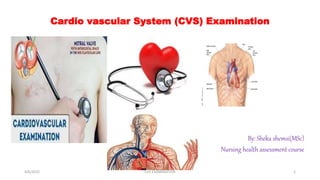
- 1. Cardio vascular System (CVS) Examination By: Sheka shemsi(MSc) Nursing health assessment course 4/6/2022 CVS EXAMINATION 1
- 2. CVS……. Circulation through the heart is shown in the diagram, which identifies the cardiac chambers, valves, and direction of blood flow. Because of their positions; o The tricuspid and mitral valves are often called atrioventricular valves. o The aortic and pulmonic valves are called semilunar valves. 4/6/2022 CVS EXAMINATION 2
- 3. An overview of the CVS Examination The evaluation of the CVS includes a thorough health history, a detailed examination of:- o the heart o the peripheral arterial and venous circulations an appropriate laboratory studies. 4/6/2022 CVS EXAMINATION 3
- 4. Health history Common or concerning symptoms of Cardiovascular Disease Dyspnoea:-shortness of breath (SOB) on exertion Chest pain:-which radiates to the left neck, shoulder and left upper arm Body swelling:-Which usually starts from the leg Palpitation:- Is subjective unpleasant perception of one’s own heartbeat Cough:- Which usually occurs at night (nocturnal) Syncope:-Is sudden episode of fainting related to hemodynamic derangement Dyspnea: This is a state of shortness of breath (SOB) on exertion. o But, it may occur at rest as the heart failure progresses. o Also called difficult or labored breathing. 4/6/2022 CVS EXAMINATION 4
- 5. The degree of dyspnea is graded based on the NHA Class (NHAC): Tab.1 New York Heart Association Functional Classification Class I No limitation of physical activity No symptoms with ordinary exertion Class II Slight limitation of physical activity Ordinary activity causes symptoms Class III Marked limitation of physical activity Less than ordinary activity causes symptoms Asymptomatic at rest Class IV Inability to carry out any physical activity without discomfort Symptoms occur at rest SOURCE: Modified from The Criteria Committee of the NHAC. 4/6/2022 CVS EXAMINATION 5
- 6. o Paroxysmal Nocturnal Dyspnea (PND): Is type SOB that occurs during sleep. The patient suddenly wakes up due the SOB and then sits up or rushes to open window/door to get fresh air. o Orthopnea: Is type SOB that occurs during recumbent position. It is evaluated by the number of pillows that are used to relieve the symptom. 4/6/2022 CVS EXAMINATION 6
- 7. concerning symptoms of Cardiovascular Disease (CVD)…………….. Chest pain: Angina pectoris is a cardiac pain. It arises in the precordial area usually on the retrosternal region It radiates to the left neck, shoulder and left upper arm It has piercing, or squeezing character It is aggravated by exertion & relieved by rest 4/6/2022 CVS EXAMINATION 7
- 8. Physical Examination Instruments needed for CVS examination include: Stethoscope Penlight Centimeter ruler and tape measure Sphygmomanometer General Considerations The patient must be properly undressed above the waist. The examination room must be quiet to perform adequate auscultation. 4/6/2022 CVS EXAMINATION 8
- 9. General signs of Cardiovascular Disease (CVD) • Observe the patient for general signs of cardiovascular disease. Breathing pattern; for respiratory distress Signs of respiratory distress, such as tachypnea, flaring, and retractions, may be associated with the client attempts to compensate for hypoxemia caused by a chronic heart disease. Skin color: for cyanosis; the mucous membranes are usually pink. Lips: for cyanosis. Mouth and tongue: for central cyanosis. Eyes for pallor of the conjunctiva: indicates anemia 4/6/2022 CVS EXAMINATION 9
- 10. Hands for edema: indicates poor venous return. Color of the palm: for cyanosis. Shape of the finger nail Clubbing of finger nail (concavity of finger nail means the angle is > 180o) suggests chronic cyanosis or infective endocarditis. Capillary refilling time Checked by pressing tip of finger for a brief period then release the pressure. Normally the color returns immediately when the pressure is released. Capillary refill is normally < 2 seconds, indicating good circulation and perfusion of the tissues. Sluggish return of color up on releasing pressure suggests decreased peripheral circulation. 4/6/2022 CVS EXAMINATION 10
- 11. Temperature and moisture Asses the temperature and moisture by using dorsum of the hand. Normally on palpation there is no temperature difference & it is warm and dry. Moist and cool hand may occur in decreased peripheral circulation. Legs for edema: indentation remains > 5 seconds after brief pressing • + 1 (mild) +2 & +3 (moderate) +4 (severe edema) • 2 mm 4-6mm > 8mm • for varicose veins and ulcer 4/6/2022 CVS EXAMINATION 11
- 12. Components of Cardiovascular System Examination A.Arterial System Examination B.Venous System Examination C.Precordial/ Cardiac Examination 1. Arterial System Examination The important part of arterial system examination includes palpation of the pulses of the major arteries and measuring of the blood pressure. 4/6/2022 CVS EXAMINATION 12
- 13. Location of the major arteries Major arteries are temporal, carotid, brachial, radial, femoral, popliteal, posterior tibial, and dorsalispedis. ☼ Pulse Rate: the radial artery is preferred. Pulse classification based on the rate: Normal 60-100 beats/min Bradycardia < 60 beats/min Tachycardia > 100 beats/min ☼ Pulse Rhythm: Pulse classification based on rhythm: Regular - Regular Regular - Irregular Irregular - Irregular ☼ Pulse Volume (amplitude): Best checked on carotid arteries. Observe for carotid pulsation and volume. Pulse classification based on volume: Feeble or weak, Normal ,Rebound 4/6/2022 CVS EXAMINATION 13
- 14. ☼ Radio femoral delay Radial artery & femoral artery are palpated symmetrically at the same time. Normally they are synchronize/symmetrical. In some obstructions there will be delay of femoral artery. • E.g. Coarctation of aorta, occlusive aortic disease • Measuring the Blood Pressure Assessment of blood pressure is important to detect conditions of hypertension or hypovolemic shock. The patient should be seated and quiet for 3 to 5 minutes before the BP is taken. 4/6/2022 CVS EXAMINATION 14
- 15. 2. Venous System Examination • Jugular Venous Pressure (JVP) Estimating the JVP is the most important part of venous system examination. It is a reflection of right atria pressure & known as central venous pressure (CVP). JVP examination is used to estimate CVP . • Distinguish the internal jugular vein from the carotid artery pulsation. 4/6/2022 CVS EXAMINATION 15
- 16. Steps for assessing the Jugular Venous Pressure (JVP) 1. Position the patient supine with the head of the table elevated 30O. 2. Adjust the angle of table elevation to bring out the venous pulsation. 3. Use tangential side lighting & look for rapid and double wave with each heart beat 4. With elevation of the head identify the highest point of pulsation (HPP) in the neck. 5. Using a horizontal line from HPP then measure vertically from the sternal angle 4/6/2022 CVS EXAMINATION 16
- 17. Interpretation of JVP Normally: o JVP measurement should be < 3 cm above the sternal angle in a health adult. o JV is distended in supine position and loud voice. o But in shock patient JV is not seen even in supine position. o Distention with elevation of the head or JVP measured at >4 cm above the sternal angle is considered elevated JVP . o Indicates raised right atria pressure which is most often found in right ventricular failure (RHF), fluid over load, tricuspid stenosis, or obstruction of superior vena cava. The way to report: o JVP is 5 cm at 450 inclination with reference at sternal angle or an angle of louis. 4/6/2022 CVS EXAMINATION 17
- 18. 3. Precordial or Cardiac Examination Precordium is the part of the anterior chest wall which overlies the heart. The examiner should stand at the patient’s right side. For most of the cardiac examination, three positions are needed: 1. supine with the head of the bed or table elevated to about 30°, 2. turning to the left side, and 3. sitting & leaning forward 4/6/2022 CVS EXAMINATION 18
- 19. Techniques of Precordial Examination 1. Inspection of the Precordium Position the patient supine with the head of the table slightly elevated. Begin the heart examination by inspecting the precordium, or anterior chest. A. Inspect the shape and symmetry of the anterior chest from the front & side views. o The rib cage is normally symmetric. Precordial bulge which may indicate long standing cardiac disease. Bulging of the left side of the chest wall may indicate an enlarged heart. Previous scars over the chest wall 4/6/2022 CVS EXAMINATION 19
- 20. A. Observe for any chest/ precordial movement associated with the heart’s contraction. o Normally there is no an observable pulsation and heave or lift. Abnormal pulsations Multiple pulsation e.g. multi valvular lesions Quiet pulsation e.g. Pericardial effusion Heave, an obvious lifting of the chest wall during contraction, may indicate an enlarged heart. 4/6/2022 CVS EXAMINATION 20
- 21. Location of the apical impulse A. Location of the apical impulse, sometimes called the point of maximum intensity, is located where the left ventricle taps the chest wall during contraction. When visible the apical impulse is normally seen or located in the left 5th ICS at or 1 cm medially to the LMCL or 7-9 cm laterally from MSL. • In about half of the patients the apical impulse is invisible but it can normally be seen in thin patient. 4/6/2022 CVS EXAMINATION 21
- 22. Palpation of the Precordium Place the entire palmar surface of your fingers together or the ball of your hand on the chest wall to palpate the precordium. Systematically palpate the entire precordium to detect any parasternal pulsations, heaves, or vibrations. to Identify the PMI (Point of maximal impulse) a. Parasternal impulses Generally palpate the entire precordium using the ball of your hand to detect parasternal impulses at the right & left interspaces, along the left sterna borders, & at the apical area. In normal individuals parasternal impulse is not palpable & no any abnormal sensations like vibrations, heaves or lifts. 4/6/2022 CVS EXAMINATION 22
- 23. a. Abnormal sensations Palpate the parasternal region at the same locations for abnormal sensations. Thrills (a palpable murmur) A thrill is a rushing vibration that feels like a cat’s purr. It is caused by turbulent blood flow from a defective heart valve and a heart murmur. Parasternal heave (lifting the ball of your hand or a pen when we put on the left parasternal area) It is the sensation of the heart lifting up against the chest wall. It may be associated with an enlarged heart or a heart contracting with extra force. 4/6/2022 CVS EXAMINATION 23
- 24. a. Identify the PMI (Point of maximal impulse): PMI is usually located at the same area of the apical impulse. Place the patient in the supine position elevating the trunk approximately 30-45 degrees. Using the palmar surface of your finger tips palpate the apex over the left precordium in the 4th, 5th, and 6th ICSs near the MCL to confirm the characteristics of the apical impulse for location and diameter. • Diameter Usually measures < 2.5 cm (1cm by 2cm) and occupies only one interspace. In the left lateral position, a diameter > 3 cm indicates left ventricular enlargement. 4/6/2022 CVS EXAMINATION 24
- 25. Displacement of PMI: Upward and to the left from pregnancy, Lateral displacement from cardiac enlargement in Congestive heart failure & Cardiomyopathy , Downward and lateral displacement of the apex below the 5th interspace in Left ventricular dilation, Mediastinal displacement from deformities of the thorax 4/6/2022 CVS EXAMINATION 25
- 26. Percussion of the Precordium: Percussion of the heart borders is rarely performed during PE. It has little significance in precordial examination. The borders of the heart are better identified by radiologic examination. (Note: often done by chest X-ray) Normally dullness in the 3rd, 4th, 5th, and possibly 6th left ICSs. To estimate cardiac size like cardiomegaly. Percussion is also done when one suspects dextro-cardia. 4/6/2022 CVS EXAMINATION 26
- 27. Auscultation of the Precordium: The stethoscope has two parts:- o Diaphragm: Preferred to auscultate high pitched sounds e.g. S1, S2, murmurs of aortic & mitral regurgitation. Should be pressed firmly against the chest. o Bell: Preferred to auscultate low pitched sounds e.g. S3, S4, and the murmur of mitral stenosis. Applied lightly, with just enough. 4/6/2022 CVS EXAMINATION 27
- 28. Areas of auscultation o These areas are named for the valve producing the sound. A. Aortic area— is the 2nd ICS to the right of the sternum. B. Pulmonic area— is the 2nd ICS to the left of the sternum. C. Erb’s point— 3rd ICS to the left of the sternum. D. Tricuspid area (right ventricular or septal area) — is the 4th & 5th ICS to the left of the sternum. E. Mitral area (left ventricular or apical area) — is at the apex 4/6/2022 CVS EXAMINATION 28
- 29. Sequences of auscultation o The auscultation sequence is started using the diaphragm of stethoscope to listen at all the auscultatory areas (progress from the base of heart moving from right 2nd ICS to the left 2nd ICS then down to the left lower border to the apex). • Then with the bell of the stethoscope listen along the lower left sterna border in the left 4th & 5th ICS then listen at the apex Position of the patient for heart auscultation a. Position the patient supine with the head of the table slightly elevated(to auscultalteby diaphragm). b. Ask the patient to roll partly onto the left side while you listen in the left 4th & 5th ICS and at the apex with bell bell of the stethoscope. Bring the left ventricle close to the chest wall Accentuates (brings out) a left-sided S3, S4 and murmur of mitral stenosis. 4/6/2022 CVS EXAMINATION 29
- 30. a. Patient to sitting up, leaning forward, exhaling completely, and stop breathing in expiration while you listen for the murmur of aortic regurgitation. With the diaphragm of the stethoscope, listening along the left sternal border and at the apex, pausing periodically so the patient may breathe Accentuates (brings out) murmur of aortic regurgitation and pericardial friction rub. 4/6/2022 CVS EXAMINATION 30
- 31. Heart sounds During auscultation focus on: normal, abnormal, and extra or additional heart sounds 1. Normal heart sounds • The sound resembles the pronunciation of lub-dub. a. S1 (the first heart sound): S1 results from closer of atrioventricular valves (tricuspid/mitral valves). Normally S1 is louder at the apex (in the tricuspid & mitral areas) and soft at the base of the heart. This signal the onset of systole. Key findings: S1 is loud in: mitral stenosis, tachycardia and hyper-dynamic circulation like e.g. anemia S1 is soft (muffled) in: mitral regurgitation, bradycardia and LVF 4/6/2022 CVS EXAMINATION 31
- 32. a. S2 (the 2nd heart sound): S2 results from closer of the semilunar valves (aortic and pulmonic valves). Normally S2 is louder at the base (aortic & pulmonic area) but soft at the apex. Key findings: S2 is loud: in systemic and pulmonary hypertension. S2 is soft: in aortic and pulmonic regurgitation. Extra or Additional heart sounds (S3 and S4) These extra or additional heart sounds are low pitched sounds. They occurs during diastole. If either S3 or S4 is very loud it is often heard as gallop/triple rhythm. 4/6/2022 CVS EXAMINATION 32
- 33. S3 (3rd heart sound or ventricular gallop): S3 is auscultate using bell of stethoscope because it is low pitched sound. S3 occurs during diastole as result of rapid ventricular filling. It is also caused by overload. S3 may be heard in the tricuspid & mitral areas following S2 sound. S3 is best heard when the client is in the left lateral position, and the sound resembles the pronunciation of lub- dub-by. It is normal in children and young adult. It occurs abnormally in: patients with heart failure - left sided heart failure - S3 heard best in mitral area - right sided heart failure - S3 heard best in tricuspid area 4/6/2022 CVS EXAMINATION 33
- 34. S4 (4th heart Sound or atrial gallop): S4 is auscultate using bell of stethoscope because it is low pitched sound. S4 occurs during forceful atrial contraction against a stiffened ventricle e.g. due to aortic stenosis or hypertensive heart disease. It results also from resistance to ventricular feeling in a condition such as Cardiomyopathy, and Systemic and pulmonary hypertension. S4 may be heard in the tricuspid and mitral areas before S1 sound. S4 is best heard when the client is in the supine position, and the sound resembles the pronunciation of le-lub-dub. S4 can be a normal sound in some older patients and trained athletes especially after exercise. 4/6/2022 CVS EXAMINATION 34
- 35. Summation Gallop:. • simultaneous occurrence of S3 & S4 is called summation gallop Other abnormal sounds heard on auscultation • Murmur: Murmur is abnormal sound (swishing or blowing sounds) heard in systole or diastole, which occur due to “turbulence” of blood flow through a valve. Murmur is distinguishable from heart sounds by their longer duration. Murmurs are prolonged series of auditory vibrations. Position to be used to auscultate murmur Sitting, leaning forward to listen along the left sterna border down to apex Left side lying position to listen at the apex. 4/6/2022 CVS EXAMINATION 35
- 36. Murmur may occurs B/c of: 1. Increased velocity of blood (e.g. Exercise, thyrotoxicosis) 2. Decreases velocity of blood (e.g. Anemia) 3. Structural defect in the valves or an unusual opening occurs in the chambers (e.g. valvular lesions) 4/6/2022 CVS EXAMINATION 36
- 37. The intensity of a murmur is graded I to VI. Grade Volume Thrill 1/6 Very faint (weak); barely heard in a quiet room No 2/6 Quiet, but heard clearly and immediately after placing the stethoscope on the chest No 3/6 Moderately loud No 4/6 Loud (Louder than grade 3) Yes 5/6 Heard with the stethoscope partially off the chest Yes 6/6 Heard with the stethoscope completely off the chest Yes 4/6/2022 CVS EXAMINATION 38
- 38. Pericardial friction rub It is a leathery (rubbing) sound heard in systole or diastole, which suggests pericardial inflammation (Pericarditis). Caused by abrasion of the pericardial surfaces during the cardiac cycle Can be heard best using the diaphragm of the stethoscope, with the patient sitting up and leaning forward 4/6/2022 CVS EXAMINATION 39
- 39. THANK YOU 4/6/2022 CVS EXAMINATION 41
Editor's Notes
- : Although this diagram shows all valves in an open position, they are not all open simultaneously in the living heart.
- Retrosternal means behind the breastbone, or sternum. Retrosternal chest pain, therefore, is a pain that occurs inside the chest.
- for exophthalmus (protrude eye ball: may be seen in thyrotoxicosis for yellowish discoloration of sclera or Icterus (jaundice): may be found in acutely congested liver
- Palpate the characteristics of the pulses in the extremities to assess the circulation. Evaluate the pulsation in each extremity and compare your findings bilaterally. All arteries should be palpated symmetrically at the same time except carotid arteries as this could cut off the blood supply to the brain and cause syncope
- Diameter Usually measures < 2.5 cm (1cm by 2cm) and occupies only one interspace. In the left lateral position, a diameter > 3 cm indicates left ventricular enlargement.
- yperdynamic circulation is abnormally increased circulatory volume. Systemic vasodilation and the associated decrease in peripheral vascular resistance ...
- Crescendo-decrescendo (diamond-shaped), or Plateau (even).