Understanding Brain Activity Through EEG
•Download as PPTX, PDF•
5 likes•412 views

This case study describes a 54-year-old woman with recurrent epithelial ovarian cancer who was undergoing chemotherapy. After her third round of ifosfamide treatment, she developed neurological symptoms. An electroencephalogram (EEG) detected frequent low-frequency periodic waves across both hemispheres, consistent with ifosfamide-induced encephalopathy. Her chemotherapy was stopped but her condition deteriorated further with worsening encephalopathy and acute kidney injury, and she ultimately passed away within a month.

Recommended
More Related Content
What's hot
What's hot (20)
Similar to Understanding Brain Activity Through EEG
Similar to Understanding Brain Activity Through EEG (20)
Recently uploaded
Recently uploaded (20)
Understanding Brain Activity Through EEG
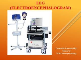
- 1. EEG (ELECTROENCEPHALOGRAM) Created & Presented By: Shashvat M.Sc. Neuropsychology
- 2. An electroencephalogram (EEG) is a test that detects electrical activity in your brain using electrodes attached to your scalp. Your brain cells communicate via electrical impulses and are active all the time, even when you're asleep. This activity shows up as wavy lines on an EEG recording Richard Caton (1875) –localization of sensory functions with monkeys and rabbits Hans Berger (1924) – first EEG recording done on humans
- 3. Why is an EEG performed? An EEG is used to detect problems in the electrical activity of the brain that may be associated with certain brain disorders. The measurements given by an EEG are used to confirm or rule out various conditions, including: • seizure disorders (such as epilepsy) • head injury • encephalitis (inflammation of the brain) • brain tumor • encephalopathy (disease that causes brain dysfunction) • memory problems • sleep disorders • stroke • Dementia When someone is in a coma, an EEG may be performed to determine the level of brain activity
- 4. EEG COMPONENTS Active electrodes Amplifier and ADC (Analog to Digital Converter) USB converter Analog Input Box Computer storage and display
- 5. BASIC ACQUISITION • EEG signals are a measure the potential difference between two electrodes. • Action Potentials are caused by an exchange of ions across the neuron membrane and an AP is a temporary change in the membrane potential that is transmitted along the axon. • The conduction velocity of action potentials lies between 1 and 100 m/s. APs are initiated by many different types of stimuli; sensory nerves respond to many types of stimuli, such as chemical, light, electricity, pressure, touch, and stretching.
- 6. Brainwaves At the root of all our thoughts, emotions and behaviours is the communication between neurons within our brains. Brainwaves are produced by synchronised electrical pulses from masses of neurons communicating with each other. Types of Brainwaves: • Delta waves (.5 to 4 Hz) • Theta waves (4 to 8 Hz) • Alpha waves (8 to 12 Hz) • Beta waves (12 to 38 Hz) • Gamma waves (38 to 42 Hz)
- 7. Delta Waves Characteristics: -frequency: .5-4 Hz -amplitude: 20-200µV Meditative waves (slowest and high amplitude), Found during periods of deep sleep in most people, Characterized by very irregular and slow wave patterns,Also useful in detecting tumors and abnormal brain behaviors STUDIES • Sleep and sleep disorders • Alcoholism and sleep
- 8. Theta Waves Characteristics: -frequency: 4-7Hz -amplitude: 20-100µV Cognitive high level thinking (correlates to mental operations, attention , info uptake , processing and learning or memory recall ,Frequency increases with task difficulty ,Can be recorded from all over cortex, Carry frequency of cognitive processing , Believed to be more common in children than adults Walter Study (1952) found these waves to be related to displeasure, pleasure, and drowsiness. STUDIES • N-Back task • Spatial Navigation • Brain monitoring in operational environment
- 9. Characteristics: - frequency: 8-12 Hz -amplitude: 20-60 µV Easily produced when quietly sitting in relaxed position with eyes closed (few people have trouble producing alpha waves) STUDIES • Meditation • Biofeedback • Attention Alpha Wave
- 10. Beta Waves Characteristics: -frequency: 12-30 Hz -amplitude: 2-20 µV Active , busy , anxious thinking and active concentration. Increases with motor movements that have focused attention The most common form of brain waves. Are present during mental thought and activity. STUDIES • Motor Control • Stimulant induced alertness
- 11. Gamma Waves Characteristics: -frequency: 30-44Hz -amplitude: 3-5µV Occur with sudden sensory stimuli, Gamma waves are important for learning, memory and information processing.
- 12. ELECTRODE PLACEMENT • Typically adopt an accepted placement scheme for applying electrodes to the scalp. • The International 10-20 placement system is the most widely adopted. • It uses a set of measurements relative to landmarks on the head. • Name reflects the fact that electrodes are placed at intervals that are 10% or 20% of the distance between landmarks.
- 13. • Requires distance from front to back of head and distance from left to right. • Front to back defined as distance from nasion to inion. • Nasion - intersection of the frontal bone and two nasal bones • Inion - the most prominent projection of the occipital bone at the posterioinferior (lower rear) part of the skull ELECTRODE PLACEMENT
- 14. • Requires distance from front to back of head and distance from left to right. • Left right defined as distance between pre- auricular points. • Pre-auricular point- root of the zygomatic arch anterior to the tragus ELECTRODE PLACEMENT
- 15. EEG Recording
- 16. EEG AS A TIME SERIES • The EEG signal is recorded together with noise that stems from a number of sources. • Essentially anything that is not the signal of interest is considered noise. • Noise amplitude is usually larger than the signal of interest.
- 17. SOURCES OF NOISE IN EEG • Capacitive coupling – the electrodes and cables are coupled to signals such as lights, computers, cell phones, etc. can induce voltage in the leads. – Theoretically this is the same for all leads so should be removed by common mode rejection. – In practice, however, this is not always the case so it is best to keep distance between leads and electrical sources. • Induction – Loop created between body and equipment allows for the formation of a magnetic field that can induce current flow in wires. – The best solution is to wrap the cables around each other so that opposing magnetic fields will cancel each other.
- 18. CASE STUDY A 54-year-old, gravid 2, para 2, married woman presented with recurrent epithelial ovarian cancer (endometrioid adenocarcinoma). She had noninsulin dependent diabetes mellitus, diabetic retinopathy, and hypertension. Seven years earlier, she underwent coronary artery bypass grafting due to coronary artery disease. Three years ago, she was transferred to our center after laparoscopic bilateral salpingo-oophorectomy which led to a diagnosis of epithelial ovarian cancer. She underwent a debulking operation, including total abdominal hysterectomy and pelvic and paraaortic lymph node dissection, total omentectomy, appendectomy, peritoneal mass excision, and left ureter resection and anastomosis. The tumor was categorized as International Federation of Obstetrics and Gynecology (FIGO) stage IIIc and cytoreductive surgery was suboptimal. Following the surgery, she received 6 cycles of adjuvant chemotherapy with paclitaxel/carboplatin. However, because there was disease progression in this patient, she was further treated with 3 cycles of topotecan, followed by 3 cycles of docetaxel. An abdomino-pelvic computed tomography (CT) scan showed aggravation of disease and her serum carbohydrate antigen 125 (CA-125) concentration was elevated from 534 to 1,270 U/mL . In addition, brain magnetic resonance imaging (MRI) findings were normal. An electroencephalogram showed frequent 1-1.5 Hz periodic or single triphasic waves on both hemispheres (Fig. 1). She was diagnosed with ifosfamide -induced encephalopathy, and her chemotherapy was discontinued immediately. Her neurologic symptoms and signs did not improve. Despite the supportive care, her encephalopathy became aggravated, and she developed ifosfamide-induced acute tubular necrosis. She was discharged without any hope for recovery and died within 1 month.
- 19. CASE STUDY Fig.1