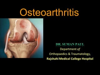
2. Osteoarthritis (OA)
• OA is the most common
form of arthritis and the most
common joint disease
• Most of the people who have
OA are older than age 45,
and women are more
than
commonly affected
men.
3. Definition
• Osteoarthritis is an idiopathic disease
characterized by degeneration of articular
cartilage leads to fibrillation, fissures, gross
ulceration and finally disappearance of the
full thickness of articular cartilage.
4. • As defined by the American College of
Rheumatology (ACR), OA is a
heterogeneous group of condition that leads
to joint signs and symptoms which are
associated with defective integrity of
articular cartilage, in addition to related
changes in the underlying bone at the joint
margins.
5. •The repair mechanisms of tissue absorption and
synthesis get out of balance and result in
osteophyte formation (bone spurs) and bone cysts
6. OA
OA is a disease of
joints that affects all
of the weight-bearing
components of the
joint:
•Articular
cartilage
•Menisci
•Bone
7. OA – Articular Cartilage
Articular cartilage is the main tissue affected
OA results in:
•Increased tissue swelling
•Change in color
•Cartilage fibrillation
•Cartilage erosion down to subchondral bone
8. OA – Articular Cartilage
A)Normal articular
cartilage from 21-year
old adult (3000X)
B)Osteoarthritic
cartilage (3000X)
22. OA – Symptoms
•
•
OA usually occurs slowly -
It may be many years before
the damage to the joint
becomes noticeable
Only a third of people
whose X-rays show
OA report pain or
other symptoms:
– Steady or intermittent pain in a joint
– Stiffness that tends to follow periods of inactivity, such as sleep
or sitting
– Swelling or tenderness in one or more joints [not necessarily
occurring on both sides of the body at the same time]
– Crunching feeling or sound of bone rubbing on bone (called
crepitus) when the joint is used
23. Two Major Types of OA
• Primary or Idiopathic
– Most common type
– Diagnosed when there is no known cause for
the symptoms
• Secondary
– Diagnosed when there is an identifiable cause
• Trauma or Underlying joint disorder
• Each of these major types has subtypes
24.
25. OA vs. Aging
Unlike aging, OA is progressive and a significantly
more active process
26. Osteoarthritis with lateral osteophyte, loss of articular cartilage and
some subchondral bony sclerosis- X-ray shows loss of joint space
OA – Overall Changes
27.
28. Laboratory findings in OA
• THERE ARE NO DIAGNOSTIC LAB
TESTS FOR OSTEOARTHRITIS
• OA is not a systemic disease, therefore:
– ESR, Chem 7, CBC, and UA all WNL
• Synovial fluid
• Mild leukocytosis (<2000 WBC/microliter)
• Can be used to exclude gout, CPPD, or septic
arthritis if diagnosis is in doubt
29. Synovial fluid analysis
• Severe, acute joint pain is an
uncommon manifestation of OA
• Clear fluidWBC <2000/mm3
• Normal viscosity
31. Asymmetrical joint space narrowing from loss of
articular cartilage
The medial (inside) part of the knee is most commonly affected by osteoarthritis.
OA – Radiographic Diagnosis
32. OA – Radiographic Diagnosis
•Asymmetrical
joint space
narrowing
•Periarticular
sclerosis
•Osteophytes
•Sub-chrondral
bone cysts
33.
34. OA – Arthroscopic Diagnosis
Normal Articular Cartilage
Ostearthritic degenerated cartilage
with exposed subchondral bone
Arthroscopy allows earlier
diagnosis by demonstrating the
more subtle cartilage changes
that are not visible on x-ray
35. •OA is a condition which progresses slowly over a
period of many years and cannot be cured
•Treatment is directed at decreasing the symptoms of
the condition, and slowing the progress of the
condition
•Functional treatment goals:
•Limit pain
•Increase range of motion
•Increase muscle strength
OA – Disease Management
39. • Pharmacologic
– Glucosamine 20 studies with >2500 patients
• If only high quality studies evaluated:
– No benefit over placebo on pain
• If all studies included:
– Pain may improve by as much as 13 points
• 2 RCT’s using Rotta preparation:
– Demonstrated slowing of radiological progression of OA
over a 3 year period
40. • Pharmacologic
– Diacerein
• Pain improved 5 points compared to placebo
• Over 3 years,
– Slowed progress of OA in the hip compared to placebo
– Did not slow progress of OA in the knee
• Diarrhea is most common side effect
– 42 out of 100 had diarrhea in the first 2 weeks
– 18 discontinued because of side effects (13 in placebo)
44. •Osteoarthritis usually
affects the inside half
(medial compartment) of
the knee more often than
the outside (lateral
compartment).
•This can lead to the lower
extremity becoming
slightly bowlegged, or in
medical terms, a genu
varum deformity
Proximal Tibial Osteotomy
45. Proximal Tibial Osteotomy
•The result is that the weight bearing line of the lower
extremity moves more medially (towards the medial
compartment of the knee).
•The end result is that there is more pressure on the medial
joint surfaces, which leads to more pain and faster
degeneration.
•In some cases, re-aligning the angles in the lower extremity
can result in shifting the weight-bearing line to the lateral
compartment of the knee. This, presumably, places the
majority of the weight-bearing force into a healthier
compartment. The result is to reduce the pain and delay the
progression of the degeneration of the medial compartment.
46. Proximal Tibial Osteotomy
•In the procedure to realign the
angles, a wedge of bone is
removed from the lateral side
of the upper tibia.
•A staple or plate and screws
are used to hold the bone in
place until it heals.
•This converts the extremity
from being bow-legged to
knock-kneed.
•The Proximal Tibial Osteotomy buys some time before ultimately
needing to perform a total knee replacement. The operation
probably lasts for 5-7 years if successful.
47. The ultimate solution for osteoarthritis of the knee is to
replace the joint surfaces with an artificial knee joint:
•Usually only considered in people over the age of 60
•Artificial knee joints last about 12 years in an elderly population
•Not recommended in younger patients because:
•The younger the patient, the more likely the artificial joint will fail
•Replacing the knee the second and third time is much harder and much
less likely to succeed.
•Younger patients are more active and place more stress on the artificial
joint, that can lead to loosening and failure earlier
•Younger patients are also more likely to outlive their artificial joint, and
will almost surely require a revision at some point down the road.
•Younger patients sometimes require the surgery (simply because
no other acceptable solution is available to treat their condition)
Total Knee Replacement
48. •The ends of the femur, tibia, and patella are shaped to accept
the artificial surfaces.
•The end result is that all moving surfaces of the knee are
metal against plastic
Total Knee Replacement