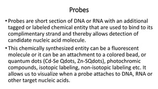
1. Probes
•Probes are short section of DNA or RNA with an additional
tagged or labeled chemical entity that are used to bind to its
complimentary strand and thereby allows detection of
candidate nucleic acid molecule.
•This chemically synthesized entity can be a fluorescent
molecule or it can be an attachment to a colored bead, or
quantum dots (Cd-Se Qdots, Zn-SQdots), photochromic
compounds, isotopic labeling, non-isotopic labeling etc. It
allows us to visualize when a probe attaches to DNA, RNA or
other target nucleic acids.
2.
3. Labeling Of Probes:
• Probes can be labeled at specific location within the
oligonucleotides or internally at multiple sites. Some probes are
of defined length and some are heterogeneous populations of
labeled molecules.
• In vivo labeling: DNA and RNA can be directly labeled inside
tissue culture cells by adding labeled deoxynucleotides in culture
plate in vivo. This method is restricted only to prepare labeled
viral DNA from virus-infected cells and to study RNA processing
events.
• In vitro labeling: It is a more versatile method involving in
vitro labeling of purified RNA, DNA or oligonucleotide using DNA
polymerases for incorporation of labeled nucleotides.
4. • 3-5.5.1 L abeling during synthesis:
• In vitro labeling of DNA can be done by various methods as
follows:
• a) Nick-translation
• b) Random primed labeling
• c) PCR-mediated labeling- Labeling of RNA is generally
accomplished by an in vitro transcription system. These
procedures require DNA or RNA polymerases to add labeled
nucleotides to synthesize in vitro probes. It requires at least
one labeled nucleotides among four nucleotides.
5. DNA Nick translation:
• It involves insertion of random single-strand breaks
called nicks in one of the strands of double stranded
target DNA which exposes 3′- OH termini and 5′-
PO4 termini. The nicks are introduced by
endonuclease like pancreatic deoxyribonuclease I
(DNase I).
6.
7. • Addition of DNase I and multi-subunit enzyme E. coli DNA polymerase I is
used for nick translation which contributes both activities like:
• (i) 5′ → 3′ exonuclease that attacks the exposed 5′ termini of a nick and
sequentially removes the nucleotides in 5′ → 3′ direction;
• (ii) DNA polymerase adds nucleotides to the free 3′-OH group, in 5′ → 3′
direction replacing the nucleotides removed by exonuclease and causing
lateral displacement (translation) of the nick.
• This method requires 100-fold less radioactive precursor than in-
vivo labeling method. The amount of radiolabel incorporated depends on
number of nicks created by DNase I. Too much or too little nicking must be
avoided. At low temperatures (about 15°C), the disadvantage is that only
one complete regeneration of existing nucleotide sequence takes place and
reaction does not proceed further.
8. Random primed DNA labeling:
• This method known as oligo-labeling is based upon
hybridization of a mixture of all possible hexanucleotides.
The template DNA is initially denatured and then cooled
slowly so as to allow random hexanucleotides to bind at
complementary sequences at which extension takes place
through PCR.
9.
10. PCR mediated DNA labeling:
• This method has several advantages over other methods.
• Defined segments of target DNA can be amplified and
labeled independently of restriction sites.
• Amount of template DNA is required very small and
• No need to isolate fragments of DNA or to sub-clone into
vectors containing bacteriophage promoters.
11. • The standard PCR reaction can be modified to incorporate labeled
nucleotides. The methods commonly used are:
• Standard PCR-based DNA labeling- The probe generation reaction is
modified to incorporate one or more labeled nucleotide precursors at a
concentration same as oligonucleotide conc.(Km) or slightly above Km and
others at concentrations exceeding Km, which become incorporated into the
PCR product throughout its length.
• Primer-mediated 5′ end labeling. This method uses a 5' end-labeled primer,
which is incorporated during PCR reaction. It is utilized in DNA sequencing,
PCR based mutagenesis etc.
• Radiolabeled probes can be generated for both strands using equal
concentrations of primers or biased heavily in favor of one strand of DNA
using higher concentration of one primer.
12.
13. Preparation of Labeled Nucleotides
• Nucleotides can be labeled by isotopic and non-isotopic methods.
• 3-5.7.1 Isotopic labeling:
• Isotopes generally used for labeling nucleotides are 32P, 33P, 35S or
3H. They can be detected directly in solution or on X-ray film using
autoradiography.
14. Properties of radioisotopes used for labeling DNA and RNA probes:
• The strength of autoradiography signal depends on intensity of radiation
emitted by radioisotope and duration of exposure.
• 32P emits high energy β-particles which offer high detection sensitivity.
Thus, it is widely used in Southern blot hybridization, dot-blot
hybridization, colony hybridization.
• But it is relatively unstable and when fine resolution is required to
interpret results, the image is unambiguous due to its high energy β-
particle emission.
• Due to this, 35S-labeled, 33P-labeled (moderate half-lives) and 3H-
labeled nucleotides are used which emit less energetic β- radiation.
They are used in DNA sequencing and in-situ hybridization. 3H requires
long exposure time due to low energy β-particle emission.
15. Non-isotopic labeling:
• Non-isotopic labeling systems involve the use of nonradioactive
probes. These methods are developed recently as compared to
radioisotope labeling methods, but are finding wide variety of
applications in different ways. Two types of non-radioactive labeling
are conducted: direct and indirect.
• 3-5.7 .2.1 Direct non-isotopic labeling, where a nucleotide containing
label such as Fluorescein, Texas Red, Rhodamine that will be detected
when incorporated with the help of spacer molecule. These modified
nucleotides having tag, fluoresce when excited by light of certain
wavelength.
16. An example of fluorescein conjugated dUTP . The fluorescein group is linked to the 5′
carbon atom of the uridine by a spacer group. Similarly, Rhodamine can also be used
in place of fluorescein.
17. • Indirect non-isotopic labeling involves chemical linkage of reporter
molecule to a nucleotide. When this modified nucleotide is
incorporated into DNA, then it is specifically bound to a protein or
other ligand which has high affinity against the reporter group. Long
spacer is introduced between nucleotide and reporter so as to reduce
steric hindrances for binding of affinity molecule.
18.
19. • Two widely used non-isotopic labeling methods are:
• Biotin-Streptavidin Method: This method uses two ligands which has high
affinity towards each other: Biotin works as the reporter and the bacterial
protein streptavidin is used as the affinity molecule. Biotinylated nucleotides
like bio-11-dUTP are used as labeling agents with a spacer of 4-16 C atoms
long between biotin and dNTP. However, Biotin is a ubiquitous constituent of
mammalian tissues and tends to stick easily to certain type of nylon
membranes which leads to high levels of background during in situ, northern
and southern hybridization. To overcome this background problem,
digoxigenin is used.
• Digoxigenin, a plant steroid obtained from Digitalis plant and is used as a
reporter and an affinity molecule. Digoxigenin is thus an all-purpose
immuno-tag, and in particular a standard immune histochemical marker
for in situ hybridization.
20. Detection of non-radioactively labeled probes after
hybridization:
• Affinity molecules (streptavidin or digoxigenin-specific antibody) are
conjugated with a variety of marker groups or molecules. They include
various fluorophores or enzymes such as alkaline phosphatase and
peroxidase which can permit detection via colorimetric assays, chemical
luminescence assays or fluorescent assay.
• In colorimetric assays, alkaline phosphatase catalyzes removal of
phosphate group from BCIP (5-bromo-4-chloro-3-indolyl phosphate),
generating a product that dimerizes to di-bromo-di-chloro indigo, which
reduces NBT (Nitrobluetetrazolium) to insoluble purple dye, diformazan
that becomes visible at sites where probe has hybridized.
21. • Fluorescent assays make use of HNPP (2-hydroxy-3-naphthoic acid 2'-
phenylanilide phosphate). After de-phosphorylation by alkaline
phosphatase, HNPP generates fluorescent precipitate on membranes
that can be excited by irradiation at 290nm. The response signal
emitted at 509nm are captured by CCD cameras.
• Chemiluminescence is the fastest and most sensitive assay using HRP
(Horseradish peroxidase) – luminal detection system. HRP catalyzes the
oxidation of luminal in the presence of H2 O2 , generating reactive
peroxide that emits light at 425nm during decomposition to its ground
state.
22. Agarose gel electrophoresis
• There are several physical methods available for separating nucleic
acid (DNA and RNA) based on its size. Gel electrophoresis is a
separation technique which is purely based on charge and size. This is
eventually one of the traditional methods of separating and analyzing
nucleic acid. In this method, gel made from agarose acts as a
separating medium. Agarose which is linear polymer agarose is a
polysaccharide, whose monomeric unit is a disaccharide of D-
galactose and 3,6-anhydro-L-galactopyranose.
23. • Agarose is in powdered form, and is insoluble in water at room temperature.
It gets dissolved in boiling water and when it starts to cool, it undergoes
cross-linking (H-bonding) and results in polymerization (agarose gel matrix).
Extent of cross-linking depends on percentage of agarose (higher percentage
results in higher cross linking thus more sieving effect due to small pore
size).
• Charge of the DNA is negative and therefore it migrates to the anode
(positively charged electrode), if a voltage is applied.
• Nucleic acid molecules are separated by applying an electric field to move
the DNA through an agarose gel matrix.
• The migration rate of the DNA is mainly affected by the factors such as size
of the DNA, agarose concentration used and conformation of the DNA.
24. • The migration of the molecules in gel electrophoresis is directly proportional
to the size of the molecule.
• The gel sieves the movement of the molecules based on their size. Small
molecules migrate faster and than bigger ones as small molecules can move
more easily through gel pores.
• Due to difference in the migration rate of various size DNA molecules in gel
DNA fragments are separated based on sizes.
• The size of the fragments can be determined by running standard DNA ladder
run in parallel.
• DNA fragments of length ranging from 50 base pair to several million base
pair can be separated using agarose gel electrophoresis.
• Migration rate of the fragments also depends on the concentration of agarose
used to prepare gel.
25. • The concentration of agarose is inversely proportional to the rate of
migration of the DNA fragments, i.e. lower the concentration, the faster is
the DNA migration rate and vice versa.
• Generally used agarose concentration is 0.7% to separate DNA fragments of
range 2- 10 kb and 2% agarose for separation of small fragments such as
0.1- 1 kb.
• Low percent gels are weak and high percent gels are often brittle. Standard
1 % agarose gels are common for many applications, which can resolve DNA
fragments from 0.5- 30 kb in length.
• Agarose gels are horizontally laid and are completely covered by running
buffer; hence they are referred to as submarine gels. The agarose gels are
prepared in a gel casting trays which are of different sizes. The small gel slab
are used for quick check in which the resolution is not great and runs for 30
to 40 minutes.
26. Electrophoresis buffer
• For the electrophoresis of DNA many buffers have been
recommended. But the most commonly used are TAE (Tris-acetate-
EDTA) and TEB (Tris-botate-EDTA).
• Due to the difference in ionic strength of these two buffers, DNA
fragments will migrate at different rates.
• The gels must be prepared in the same buffer in which the gels are
run i.e. either TAE or TBE buffer.
• A buffer not only provides ions to support the conductivity, but also
establish a pH. If you use concentrated buffer, enough heat may
generate to melt the gel. The working concentration of the buffer is
1X and stock is prepared for 50X.
27. • Agarose gels are prepared as percentage weight/volume solutions. Thus to prepare
standard 1% agarose gel 1 gram of agarose is dissolve in 100 ml of buffer.
• For bigger gels just scale up the volume accordingly.
• Agarose do not dissolve in the buffer, rather it has to be melted by boiling which is
typically done in a microwave oven.
• While melting agarose, care should be taken to prevent boil over.
• To visualize the DNA on agarose gel UV-fluorescent dye is required. Ethidium
bromide is a florescent dye that intercalates between bases of nucleic acids and
allows convenient detection of DNA fragments in agarose under UV light.
• After agarose has cooled to about 60°C, a final concentration of 0.5 µg/ml ethidium
bromide is added to agarose solution before pouring the agarose into the gel tray
which is sealed and come is positioned. The agarose solution is allowed to cool at
room temperature for an hour to solidify gel. Comb is carefully removed from the
solidified gel and seal of the gel tray is removed. The gel is placed in the buffer tank
carefully with buffer completely submerge the gel
28.
29. • Load your DNA samples in corresponding wells in the gel.
• Remember that DNA is negatively charged and runs towards the positive
electrode.
• The gel wells should be nearer towards black electrode and farther from
red electrode (DNA should run to the red end).
• Turn on the power to run the gel with the voltage set at 60-80 volts for
40 minutes for small gels and 90-100 volts for 1-2 hours for larger gels.
• Agarose gels run at high voltage may result in melting the gel and
distortion of bands.
• One can confirm that the gel is running by checking for bubbles from the
electrodes. Switch off the power at the end of the run. The next step is
visualization of gel for bands
30.
31. Visualization
• Ethidium bromide (EtBr) is traditionally used as a dye that binds to
DNA and fluoresces under ultraviolet light.
• EtBr causes mutation and must be handled as hazardous waste. Due
to the hazardous nature of the EtBr, recently non-toxic dyes have
been introduced in which the gels have to be stained first and then
destained to visualize the bands. But, if you are using EtBr as a dye to
visualize DNA bands you need to be more careful in handling as to
prevent the hazardous effect of EtBr. The EtBr gels have to be
disposed separately in hazard wastes disposal bags.