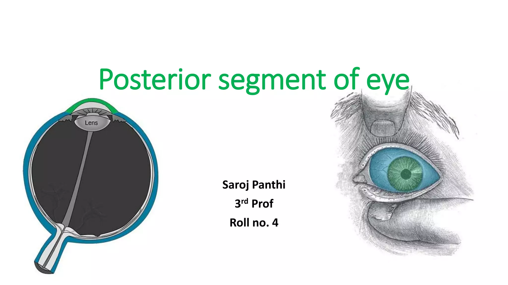
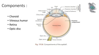
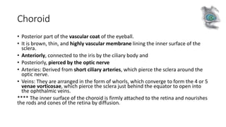
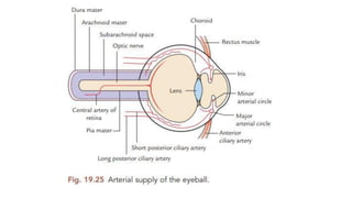
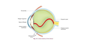
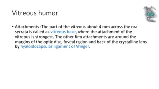
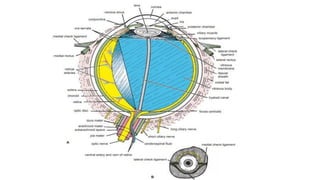
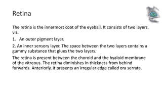
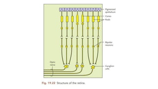
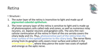
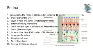
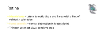
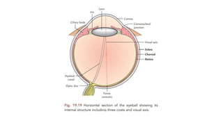
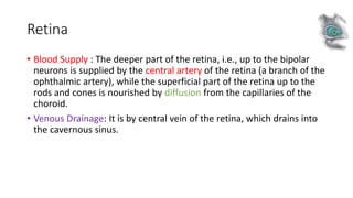
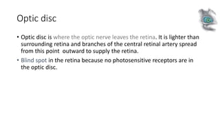
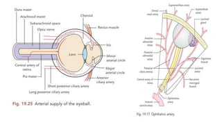
Posterior segment of eye
Saroj Panthi
3rd Prof
Roll no. 4
Components :
• Choroid
• Vitreous humor
• Retina
• Optic disc
Choroid
• Posterior part of the vascular coat of the eyeball.
• It is brown, thin, and highly vascular membrane lining the inner surface of the
sclera.
• Anteriorly, connected to the iris by the ciliary body and
• Posteriorly, pierced by the optic nerve
• Arteries: Derived from short ciliary arteries, which pierce the sclera around the
optic nerve.
• Veins: They are arranged in the form of whorls, which converge to form the 4 or 5
venae vorticosae, which pierce the sclera just behind the equator to open into
the ophthalmic veins.
**** The inner surface of the choroid is firmly attached to the retina and nourishes
the rods and cones of the retina by diffusion.
Choroid
• Structure : It consists of four layers (from outside inwards), viz.
1. Suprachoroid lamina (lamina fusca): It consists of loose network of
elastic and collagen fibres and is traversed by long posterior ciliary
vessels and nerves.
2. Vascular lamina: It consists of loose areolar tissue and pigment
cells. It contains branches of short posterior ciliary arteries and
veins, which converge in whorls to form 4 or 5 venae vorticosae,
which pierce the sclera and drain into ophthalmic veins.
Choroid
3. Capillary lamina (capillary layer of choroid): It consists of fine
network of capillaries, which nourish photoreceptors of the retina (rods
and cones) by diffusion.
4. Basal lamina (membrane of Bruch): It is a thin transparent
membrane, which is firmly attached to the pigment cell layer of the
retina. The last three layers form the choroid proper, which is
separated from sclera by suprachoroid lamina.
****In some animals such as cat, tiger, lion, etc. the specialized cells of
choroid form a reflecting media called tapetum, which produces
greenish glare in the eyes of these animals in the night.
Choroid
Choroiditis
Vitreous humor
• It is a transparent, gelatinous substance. This can’t be replaced unlike
aqueous humor.
• The vitreous body is 99% water but, vitally, also contains collagen
fibrils and hyaluronic acid, which impart cohesion and a gel-like
consistency.
• The vitreous is adherent to the retina at certain points, particularly at
the optic disc and at the ora serrata.
Vitreous humor
• The vitreous body can be divided into two parts: the cortex and the medulla or nucleus
(the main vitreous body)
1. Cortical vitreous: It lies adjacent to the retina posteriorly and ciliary body, zonules and
lens anteriorly. The density of collagen fibrils is greater in this peripheral part. The
condensation of these fibrils form a false anatomic membrane which is called as
anterior hyaloid membrane anterior to ora serrata and posterior hyaloid membrane
posterior to ora. The attachment of the anterior hyaloid membrane to the posterior
lens surface is firm in young and weak in elders, whereas, posterior hyaloid membrane
remains loosely attached to the internal limiting membrane of the retina throughout
life.
2. Vitreous body (medulla or nucleus): It has a less dense fibrillar structure and is a true
biological gel. It is here where liquefactions of the vitreous gel start first.
Microscopically, the vitreous body is homogenous, but exhibits wavy lines as of
watered silk in the slit-lamp beams. Running down the centre of the vitreous body
from the optic disc to the posterior pole of the lens is the hyaloid canal (Cloquet’s
canal) of doubtful existence in adults. Down this canal ran the hyaloid artery of the
foetus.
Vitreous humor
• Attachments :The part of the vitreous about 4 mm across the ora
serrata is called as vitreous base, where the attachment of the
vitreous is strongest. The other firm attachments are around the
margins of the optic disc, foveal region and back of the crystalline lens
by hyaloidocapsular ligament of Wieger.
Vitreous humor
Functions of vitreous
• Screen out UV and IR light.
• Provide a clear media for optical transmission.
D/s of Vitreous humor
Vitreous liquefaction
Vitreous detachments
Inflammatory vitreous opacities
Vitreous haemorrhage
Retina
The retina is the innermost coat of the eyeball. It consists of two layers,
viz.
1. An outer pigment layer.
2. An inner sensory layer. The space between the two layers contains a
gummy substance that glues the two layers.
The retina is present between the choroid and the hyaloid membrane
of the vitreous. The retina diminishes in thickness from behind
forwards. Anteriorly, it presents an irregular edge called ora serrata.
Retina
• Structure :
1. The outer layer of the retina is insensitive to light and made up of
pigmented cuboidal epithelium.
2. The inner sensory layer of the retina is sensitive to light and is made up
of photoreceptors cells called rods and cones; as well as numerous relay
neurons, viz. bipolar neurons and ganglion cells. The very thin non-
cellular continuation of the retina in front of the ora serrata covers the
ciliary body and iris. Thus, photosensitive part of the retina lines the
inner surface of the eyeball posterior to the ciliary body. The nerve fibres
arising from its ganglion cells covers inner surface and collect in the
inferomedial region, where they pierce the outer two coats of eyeball
and emerge as the optic nerve.
Retina
• Histologically, the retina is composed of following 10 layers:
1. Outer pigmented layer
2. Layer of rods and cones (photoreceptor cells)
3. External limiting membrane
4. Outer nuclear layer (Cell bodies of rods and cones)
5. Outer plexiform layer
6. Inner nuclear layer (Cell bodies of bipolar neurons)
7. Inner plexiform layer
8. Ganglion cell layer
9. Nerve fibre layer
10. Internal limiting membrane
Retina
• Macula lutea - Lateral to optic disc a small area with a hint of
yellowish coloration
• Fovea centralis – central depression in Macula lutea
• Thinnest yet most visual sensitive area
Retina
• Blood Supply : The deeper part of the retina, i.e., up to the bipolar
neurons is supplied by the central artery of the retina (a branch of the
ophthalmic artery), while the superficial part of the retina up to the
rods and cones is nourished by diffusion from the capillaries of the
choroid.
• Venous Drainage: It is by central vein of the retina, which drains into
the cavernous sinus.
D/s of Retina
Retinitis
Eale’s d/s
Retinal artery occlusions
Hypertensive retinopathy
Optic disc
• Optic disc is where the optic nerve leaves the retina. It is lighter than
surrounding retina and branches of the central retinal artery spread
from this point outward to supply the retina.
• Blind spot in the retina because no photosensitive receptors are in
the optic disc.
D/s of Optic disc
Coloboma
Drusen
Hypoplasia
Arterial Supply of Eyeball
• The eyeball is supplied by the following arteries:
1. Central artery of the retina
2. Long and short posterior ciliary arteries
3. Anterior ciliary arteries
In the region of ciliary body, a major arterial circle is formed by the
anastomosis between long posterior ciliary artery and anterior ciliary
artery. From major arterial circle the vessel passes centripetally and
anastomoses close to the pupillary margin to form the minor arterial
circle.
Reference
• Snell Clinical Anatomy
• Gray’s Anatomy
• Textbook of Human anatomy Vishram Singh
• Comprehensive ophthalmology A.K Khurana
सर्वे जन ाः सुखिनो भर्वतु
Thank You

Posterior segment of eye.pdf

  • 1.
    Posterior segment ofeye Saroj Panthi 3rd Prof Roll no. 4
  • 2.
    Components : • Choroid •Vitreous humor • Retina • Optic disc
  • 3.
    Choroid • Posterior partof the vascular coat of the eyeball. • It is brown, thin, and highly vascular membrane lining the inner surface of the sclera. • Anteriorly, connected to the iris by the ciliary body and • Posteriorly, pierced by the optic nerve • Arteries: Derived from short ciliary arteries, which pierce the sclera around the optic nerve. • Veins: They are arranged in the form of whorls, which converge to form the 4 or 5 venae vorticosae, which pierce the sclera just behind the equator to open into the ophthalmic veins. **** The inner surface of the choroid is firmly attached to the retina and nourishes the rods and cones of the retina by diffusion.
  • 5.
    Choroid • Structure :It consists of four layers (from outside inwards), viz. 1. Suprachoroid lamina (lamina fusca): It consists of loose network of elastic and collagen fibres and is traversed by long posterior ciliary vessels and nerves. 2. Vascular lamina: It consists of loose areolar tissue and pigment cells. It contains branches of short posterior ciliary arteries and veins, which converge in whorls to form 4 or 5 venae vorticosae, which pierce the sclera and drain into ophthalmic veins.
  • 6.
    Choroid 3. Capillary lamina(capillary layer of choroid): It consists of fine network of capillaries, which nourish photoreceptors of the retina (rods and cones) by diffusion. 4. Basal lamina (membrane of Bruch): It is a thin transparent membrane, which is firmly attached to the pigment cell layer of the retina. The last three layers form the choroid proper, which is separated from sclera by suprachoroid lamina. ****In some animals such as cat, tiger, lion, etc. the specialized cells of choroid form a reflecting media called tapetum, which produces greenish glare in the eyes of these animals in the night.
  • 7.
  • 8.
    Vitreous humor • Itis a transparent, gelatinous substance. This can’t be replaced unlike aqueous humor. • The vitreous body is 99% water but, vitally, also contains collagen fibrils and hyaluronic acid, which impart cohesion and a gel-like consistency. • The vitreous is adherent to the retina at certain points, particularly at the optic disc and at the ora serrata.
  • 9.
    Vitreous humor • Thevitreous body can be divided into two parts: the cortex and the medulla or nucleus (the main vitreous body) 1. Cortical vitreous: It lies adjacent to the retina posteriorly and ciliary body, zonules and lens anteriorly. The density of collagen fibrils is greater in this peripheral part. The condensation of these fibrils form a false anatomic membrane which is called as anterior hyaloid membrane anterior to ora serrata and posterior hyaloid membrane posterior to ora. The attachment of the anterior hyaloid membrane to the posterior lens surface is firm in young and weak in elders, whereas, posterior hyaloid membrane remains loosely attached to the internal limiting membrane of the retina throughout life. 2. Vitreous body (medulla or nucleus): It has a less dense fibrillar structure and is a true biological gel. It is here where liquefactions of the vitreous gel start first. Microscopically, the vitreous body is homogenous, but exhibits wavy lines as of watered silk in the slit-lamp beams. Running down the centre of the vitreous body from the optic disc to the posterior pole of the lens is the hyaloid canal (Cloquet’s canal) of doubtful existence in adults. Down this canal ran the hyaloid artery of the foetus.
  • 11.
    Vitreous humor • Attachments:The part of the vitreous about 4 mm across the ora serrata is called as vitreous base, where the attachment of the vitreous is strongest. The other firm attachments are around the margins of the optic disc, foveal region and back of the crystalline lens by hyaloidocapsular ligament of Wieger.
  • 12.
    Vitreous humor Functions ofvitreous • Screen out UV and IR light. • Provide a clear media for optical transmission.
  • 13.
    D/s of Vitreoushumor Vitreous liquefaction Vitreous detachments Inflammatory vitreous opacities Vitreous haemorrhage
  • 15.
    Retina The retina isthe innermost coat of the eyeball. It consists of two layers, viz. 1. An outer pigment layer. 2. An inner sensory layer. The space between the two layers contains a gummy substance that glues the two layers. The retina is present between the choroid and the hyaloid membrane of the vitreous. The retina diminishes in thickness from behind forwards. Anteriorly, it presents an irregular edge called ora serrata.
  • 17.
    Retina • Structure : 1.The outer layer of the retina is insensitive to light and made up of pigmented cuboidal epithelium. 2. The inner sensory layer of the retina is sensitive to light and is made up of photoreceptors cells called rods and cones; as well as numerous relay neurons, viz. bipolar neurons and ganglion cells. The very thin non- cellular continuation of the retina in front of the ora serrata covers the ciliary body and iris. Thus, photosensitive part of the retina lines the inner surface of the eyeball posterior to the ciliary body. The nerve fibres arising from its ganglion cells covers inner surface and collect in the inferomedial region, where they pierce the outer two coats of eyeball and emerge as the optic nerve.
  • 18.
    Retina • Histologically, theretina is composed of following 10 layers: 1. Outer pigmented layer 2. Layer of rods and cones (photoreceptor cells) 3. External limiting membrane 4. Outer nuclear layer (Cell bodies of rods and cones) 5. Outer plexiform layer 6. Inner nuclear layer (Cell bodies of bipolar neurons) 7. Inner plexiform layer 8. Ganglion cell layer 9. Nerve fibre layer 10. Internal limiting membrane
  • 19.
    Retina • Macula lutea- Lateral to optic disc a small area with a hint of yellowish coloration • Fovea centralis – central depression in Macula lutea • Thinnest yet most visual sensitive area
  • 21.
    Retina • Blood Supply: The deeper part of the retina, i.e., up to the bipolar neurons is supplied by the central artery of the retina (a branch of the ophthalmic artery), while the superficial part of the retina up to the rods and cones is nourished by diffusion from the capillaries of the choroid. • Venous Drainage: It is by central vein of the retina, which drains into the cavernous sinus.
  • 22.
    D/s of Retina Retinitis Eale’sd/s Retinal artery occlusions Hypertensive retinopathy
  • 23.
    Optic disc • Opticdisc is where the optic nerve leaves the retina. It is lighter than surrounding retina and branches of the central retinal artery spread from this point outward to supply the retina. • Blind spot in the retina because no photosensitive receptors are in the optic disc.
  • 24.
    D/s of Opticdisc Coloboma Drusen Hypoplasia
  • 25.
    Arterial Supply ofEyeball • The eyeball is supplied by the following arteries: 1. Central artery of the retina 2. Long and short posterior ciliary arteries 3. Anterior ciliary arteries In the region of ciliary body, a major arterial circle is formed by the anastomosis between long posterior ciliary artery and anterior ciliary artery. From major arterial circle the vessel passes centripetally and anastomoses close to the pupillary margin to form the minor arterial circle.
  • 28.
    Reference • Snell ClinicalAnatomy • Gray’s Anatomy • Textbook of Human anatomy Vishram Singh • Comprehensive ophthalmology A.K Khurana
  • 29.
    सर्वे जन ाःसुखिनो भर्वतु Thank You