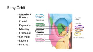
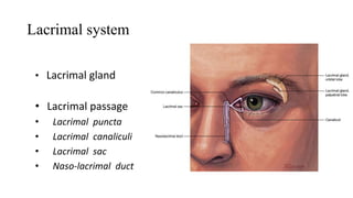
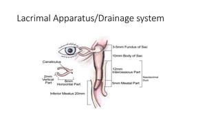
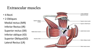
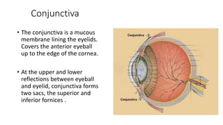
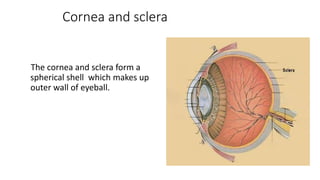
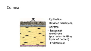
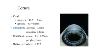
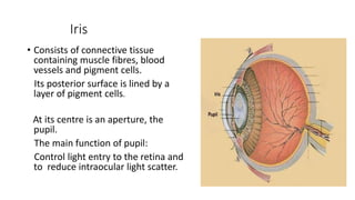
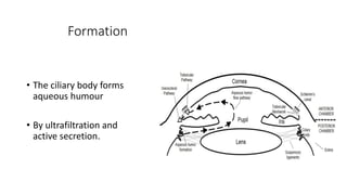
Ocular Anatomy
Rajkumar Bera 613 (6)
Bony Orbit
• Made by 7
Bones:-
• Frontal
• Zygomatic
• Maxillary
• Ethmoidal
• Sphenoid
• Lacrimal
• Palatine
Blood supply
Network of blood vessels which form an anastomosis
between branches derived from the external carotid artery
via the face and from the internal carotid artery via the orbit.
Lymphatics
• Lymphatic fluid drains into the preauricular and submandibular
nodes.
• Preauricular lymphadenopathy is a useful sign of infective eyelid
swelling (especially viral).
Lacrimal system
• Lacrimal gland
• Lacrimal passage
• Lacrimal puncta
• Lacrimal canaliculi
• Lacrimal sac
• Naso-lacrimal duct
Lacrimal Apparatus/Drainage system
Extraocular muscles
• 4 Recti
• 2 Obliques
Medial rectus (MR)
Inferior Rectus (IR)
Superior rectus (SR)
Inferior oblique (IO)
Superior Oblique(SO)
Lateral Rectus (LR)
Conjunctiva
• The conjunctiva is a mucous
membrane lining the eyelids.
Covers the anterior eyeball
up to the edge of the cornea.
• At the upper and lower
reflections between eyeball
and eyelid, conjunctiva forms
two sacs, the superior and
inferior fornices .
Cornea and sclera
The cornea and sclera form a
spherical shell which makes up
outer wall of eyeball.
Cornea and sclera
• The sclera is :
- principally collagenous,
- avascular (apart from some vessels on its surface)
- relatively acellular.
It is perforated posteriorly by the optic nerve, and by sensory and
motor nerves and blood vessels to the eyeball.
The cornea and sclera merge at the corneal edge (the limbus).
Corneoscleral limbus
• Transition zone:
• anterior:bowman`s terminal-Descemet’s terminal
• posterior:scleral spur/iris root
• 1.5~2.0mm width
• Appearance
• Semitransparant zone
• White sclera
• Angle of AC
Cornea
• Made up of 5 layers
• Specialized Transparent Tissue
• No blood vessels
• Primarily responsible for refracting light (43-44 diopters)
• More nerve endings than anywhere else in the body
• Protection to the eye
• The only part of the eye that is transplanted from one person to
another
Cornea
• Epithelium
• Bowman membrane
• Stroma
• Descemet
membrane
(posterior limiting
layer of cornea)
• Endothelium
Cornea
• Oval
• transverse:11.5~12mm
• vertical:10.5~11mm
• curvature:anterior: 7.8mm
posterior:6.8mm
• thickness:centre:0.5~0.55mm
periphery:1mm
• Refractive index:1.377
• The uvea comprises the
• iris and ciliary body anteriorly
• choroid posteriorly
UVEA
Iris
• Consists of connective tissue
containing muscle fibres, blood
vessels and pigment cells.
Its posterior surface is lined by a
layer of pigment cells.
At its centre is an aperture, the
pupil.
The main function of pupil:
Control light entry to the retina and
to reduce intraocular light scatter.
Ciliary body
• The ciliary body is a specialised structure uniting the iris with the
choroid.
• Secretes aqueous humour
• Anchors the lens via the zonules, through which it modulates lens
convexity.
• The posterior part of the ciliary body merges into the retina at the ora
serrata.
Choroid
• The choroid, consisting of blood vessels, connective tissue and
pigment cells, is sandwiched between the retina and the sclera.
• It provides oxygen and nutrition to the outer retinal layers.
Lens
• The discus-like lens comprises
a mass of long cells known as
fibres. Has a hard nucleus
surrounded by less dense
fibres, the cortex.
• Relatively dehydrated
• Transparent.
Aqueous humour
• Fills the anterior and posterior chambers.
• The anterior chamber is the space between the cornea and the iris.
• Behind the iris and in front of the lens is the posterior chamber.
They are connected by the pupil.
Formation
• The ciliary body forms
aqueous humour
• By ultrafiltration and
active secretion.
Vitreous
• A thick, transparent gel like substance that fills the center of
eyeball, giving it form and shape
• The vitreous body is 99% water but, vitally, also contains collagen
fibrils and hyaluronan, which impart cohesion and a gel-like
consistency.
• The vitreous is adherent to the retina at certain points,
particularly at the optic disc and at the ora serrata.
• Volume - 4.5ml
• RI:1.3360≈aqueous humor
• Transparent ,thin
• Anterior rim:ora serrata
• 3 parts:-
• Peripheral Retina
• Macula/fovea
• Optic disc
Retina
Histology of retina
1 RPE
2 Layers of Rods & Cones
3 External Limiting Membrane
4 Outer Nuclear layer
5 Outer Plexiform layer
6 Inner Nuclear layer
7 Inner Plexiform Layer
8 Ganglion Cell layer
9 Nerve Fibre layer
10 Internal limiting membrane
Thank You

ocular_anatomy.pptx

  • 1.
  • 2.
    Bony Orbit • Madeby 7 Bones:- • Frontal • Zygomatic • Maxillary • Ethmoidal • Sphenoid • Lacrimal • Palatine
  • 3.
    Blood supply Network ofblood vessels which form an anastomosis between branches derived from the external carotid artery via the face and from the internal carotid artery via the orbit.
  • 4.
    Lymphatics • Lymphatic fluiddrains into the preauricular and submandibular nodes. • Preauricular lymphadenopathy is a useful sign of infective eyelid swelling (especially viral).
  • 5.
    Lacrimal system • Lacrimalgland • Lacrimal passage • Lacrimal puncta • Lacrimal canaliculi • Lacrimal sac • Naso-lacrimal duct
  • 6.
  • 7.
    Extraocular muscles • 4Recti • 2 Obliques Medial rectus (MR) Inferior Rectus (IR) Superior rectus (SR) Inferior oblique (IO) Superior Oblique(SO) Lateral Rectus (LR)
  • 8.
    Conjunctiva • The conjunctivais a mucous membrane lining the eyelids. Covers the anterior eyeball up to the edge of the cornea. • At the upper and lower reflections between eyeball and eyelid, conjunctiva forms two sacs, the superior and inferior fornices .
  • 9.
    Cornea and sclera Thecornea and sclera form a spherical shell which makes up outer wall of eyeball.
  • 10.
    Cornea and sclera •The sclera is : - principally collagenous, - avascular (apart from some vessels on its surface) - relatively acellular. It is perforated posteriorly by the optic nerve, and by sensory and motor nerves and blood vessels to the eyeball. The cornea and sclera merge at the corneal edge (the limbus).
  • 11.
    Corneoscleral limbus • Transitionzone: • anterior:bowman`s terminal-Descemet’s terminal • posterior:scleral spur/iris root • 1.5~2.0mm width • Appearance • Semitransparant zone • White sclera • Angle of AC
  • 12.
    Cornea • Made upof 5 layers • Specialized Transparent Tissue • No blood vessels • Primarily responsible for refracting light (43-44 diopters) • More nerve endings than anywhere else in the body • Protection to the eye • The only part of the eye that is transplanted from one person to another
  • 13.
    Cornea • Epithelium • Bowmanmembrane • Stroma • Descemet membrane (posterior limiting layer of cornea) • Endothelium
  • 14.
    Cornea • Oval • transverse:11.5~12mm •vertical:10.5~11mm • curvature:anterior: 7.8mm posterior:6.8mm • thickness:centre:0.5~0.55mm periphery:1mm • Refractive index:1.377
  • 15.
    • The uveacomprises the • iris and ciliary body anteriorly • choroid posteriorly UVEA
  • 16.
    Iris • Consists ofconnective tissue containing muscle fibres, blood vessels and pigment cells. Its posterior surface is lined by a layer of pigment cells. At its centre is an aperture, the pupil. The main function of pupil: Control light entry to the retina and to reduce intraocular light scatter.
  • 17.
    Ciliary body • Theciliary body is a specialised structure uniting the iris with the choroid. • Secretes aqueous humour • Anchors the lens via the zonules, through which it modulates lens convexity. • The posterior part of the ciliary body merges into the retina at the ora serrata.
  • 18.
    Choroid • The choroid,consisting of blood vessels, connective tissue and pigment cells, is sandwiched between the retina and the sclera. • It provides oxygen and nutrition to the outer retinal layers.
  • 19.
    Lens • The discus-likelens comprises a mass of long cells known as fibres. Has a hard nucleus surrounded by less dense fibres, the cortex. • Relatively dehydrated • Transparent.
  • 20.
    Aqueous humour • Fillsthe anterior and posterior chambers. • The anterior chamber is the space between the cornea and the iris. • Behind the iris and in front of the lens is the posterior chamber. They are connected by the pupil.
  • 21.
    Formation • The ciliarybody forms aqueous humour • By ultrafiltration and active secretion.
  • 22.
    Vitreous • A thick,transparent gel like substance that fills the center of eyeball, giving it form and shape • The vitreous body is 99% water but, vitally, also contains collagen fibrils and hyaluronan, which impart cohesion and a gel-like consistency. • The vitreous is adherent to the retina at certain points, particularly at the optic disc and at the ora serrata. • Volume - 4.5ml • RI:1.3360≈aqueous humor
  • 23.
    • Transparent ,thin •Anterior rim:ora serrata • 3 parts:- • Peripheral Retina • Macula/fovea • Optic disc Retina
  • 24.
    Histology of retina 1RPE 2 Layers of Rods & Cones 3 External Limiting Membrane 4 Outer Nuclear layer 5 Outer Plexiform layer 6 Inner Nuclear layer 7 Inner Plexiform Layer 8 Ganglion Cell layer 9 Nerve Fibre layer 10 Internal limiting membrane
  • 25.