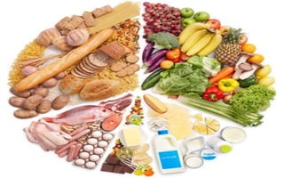
2. Introduction
•The breakdown of large food molecules to
chemical building blocks (monomers)
•These are the types of substances that the
GIT walls can absorb
•The break down is accomplished by enzymes
from glands of the GIT
•There are 4 main food substances that are
required for life- carbohydrates, proteins,
lipids and nucleic acids
3. Hydrolysis as a means of breakdown
•Carbohydrates break down from polysaccharides to
disaccharides and the final break down is monosaccharides
•Proteins are multiple amino acids bound by peptide bonds -
polypeptides
• They breakdown to form tri, di peptides and the smallest units are
the amino acids
•Lipids are made of 3 fatty acids and glycerol (triglyceride)
•Bonds that bind these units are broken down by specific
enzymes
4. Carbohydrates
•Largest polysaccharides include starch, glycogen and
cellulose
•Other smaller forms include disaccharides (sucrose,
lactose, maltose) and the smallest are monosaccharides
(fructose, galactose and glucose)
•Mouth; Ptyalin (alpha amylase) enzyme secreted by salivary
glands breaks down polysaccharides to maltose (di) by 5%
•This break down continues in the middle of the bolus in the
stomach and conversion of a further 30 to 40% is noted
5. •Small intestine –
•Pancreatic amylase (alpha amylase) breaks
down any remaining polysaccharides to
maltose and other smaller polymers
•Epithelial enzymes are present to
breakdown these smaller polymers;
•Lactase – lactose to galactose and glucose
•Sucrase – sucrose to fructose and glucose
•Maltase – maltose to molecules of glucose
6. Principles of absorption
• Fluid absorbed in a day = that ingested (2L) plus secreted (7L)
• Stomach has poor absorption ability and only absorbs alcohol
and certain drugs like aspirin
• This is due to lack of villi and presence of tight junctions in
epithelium
• Small intestines – have vulvulae connivients (x3) that have villi
(x10) on them
• The villi have microvilli (x20) that make the brush boarder
• Water movement obeys the law of osmosis, it is absorbed
when the blood is more hypertonic
• Ions - different mechanisms are noted for ion absorption
7. Carbohydrate absorption
• Mostly absorbed as monosacchs by secondary active transport
• Sodium from cytosol of epithelial cell is actively taken into blood,
depleting the cytosolic sodium content
• Sodium from lumen moves into cytosol due to the depleted cytosol
sodium
• In order for sodium to move into cytosol, a symporter is used that
has to carry glucose as well
• Therefore glucose is shuttled into the cell due to sodium pull caused
by active uptake of sodium into blood
• Once inside the cell, the glucose is taken up into paracellular space by
another transporter
8.
9.
10. Proteins
•Characteristics of proteins are a sequence of amino acids
attached by peptide bonds
•Stomach
• Pepsin activated at 2-3 pH breaks down proteins and especially
works on collagen
•Small intestines –
• Trypsin and chymotrypsin break down proteins into small
polymers
• Carboxypeptidase cleaves proteins to amino acids
• Elastase breaks down elastin fibers
• Few proteins break down to amino acids by pancreatic juice
• Final products are di and tripeptides
11. •Enterocytes release peptidase on the surface
and inside the cytosol
•These include aminopeptidases and dipeptidases
•They split the polypeptides to amino acids and
those that are absorbed in di or tripeptide form
are broken down further in the cytosol
•Proteins are absorbed as amino acids into the
blood
14. Fats
• Dietary lipids are primarily (90%) triacylglycerols but also
include cholesterolesters, phospholipids, essential unsaturated
fatty acids and fat-soluble vitamins (A, D, E, K).
• Essentially all (98%) of the fat consumed in the diet is
absorbed, and most is transported to adipose tissues for
storage
• Mouth – lingual lipase breaks down 10% of the fat
• Lingual lipase followed by gastric lipase start minor digestion of
triglycerides
• Remove one of the three fatty acids leaving diacylglycerol and
free fatty acid as a product.
• triacylglycerol → fatty acid + diacylglycerol (lingual or gastric lipase)
• Small Intestine- breaks down fats into smaller globules
15. •This is a process of emulsification
•Made possible by bile salts that have polar and non
polar ends
•The polar ends are soluble in water and mix with it,
while the non polar dissolves with the fat
•Agitation of the GIT causes them to split and thus
increase the surface area on which enzymes can
work on
16.
17. •Lipase is water soluble and can only digest the part that is
exposed
• The emulsification is therefore imperative for complete digestion
•Both pancreatic and enteric lipase split the fats into free
fatty acids and 2-monoglycerides
•These need to be ferried into the cell as quickly as possible
as they can easily bind to form fats again
•The bile salts form micelles by binding to these monomers
and ferrying them to the cells
• At the brush boarder of the epithelium, the fat monomers are
absorbed as they are lipid soluble and the bile salts return to chyme
in the intestine
18. Steps of lipid digestion/absorption
• The major digestion of all lipids occurs in the lumen of the small
intestines
• Pancreatic lipase removes the fatty acids from carbons one and
three of the glycerol backbone.
• Requires colipase(a small protein) for its activity.
• cholesterol esterase, removes the ester portion of dietary
cholesterol ester to facilitate its uptake across the intestinal wall
• Phospholipase A2 removes a fatty acid from carbon 2 of the
glycerol backbone leaving a fatty acid on carbon 1 and the polar
headgroup on carbon 3 (lysophospholipid).
• triacylglycerol → 2 fatty acids + monoacylglycerol (pancreatic lipase)
• cholesterol ester → cholesterol + ester (cholesterol esterase)
• phospholipids → fatty acid + lysophospholipid or lisophosphoglycerides
(phospholipase A2)
20. Fats
•Micelles carry the fat globules to epithelium
•Soluble fat enters cells and the monomers are taken
up by ER to form new triglycerides
21. • TGs are water insoluble and lipase is water soluble.
• Digestion of TGs takes place at lipid-water interfaces.
• Rate of digestion depends on the surface area of the interface.
• Bile salts are amphipathic, they act as detergent emulsifying the
lipid drops and increasing the surface area of the interface.
22. • Bile salts also activates the lipase.
• Inadequate production of bile salts results in steatorrhea.
23. ABSORPTION OF DIETARY LIPIDS
2-monoacylglycerols, fatty acids, lysophosphoglycerides,
free cholesterol form micelles with bile salts.
Lipid absorption – passive diffusion process.
24. • Micelles migrate to the microvilli and lipids diffuse into the cells.
• Bile acids are actively absorbed and transferred to the liver via
portal vein.
• Bile salts can circulate through intestine and liver several time
per day.
25. • Solubilization of lipids by a biological detergent occurs as follows;
• bile acid is produced by the liver from cholesterol and converted to bile
salts by intestinal bacteria
• Bile salts act in the absorption of lipids by reversibly forming micelles
• products of triacylglycerol hydrolysis are continuously transferred from
emulsion droplets to the mixed micelles
• Uptake of lipids by the epithelial cells lining the intestines occurs by
passive diffusion from the mixed micelles through the membrane
• Cholesterol is converted back to its cholesterol ester form and fatty acids
are reattached to glycerol backbones to form new molecules of
triacylglycerol
• lipid products are then incorporated into a lipoprotein known as a
chylomicron
• exported into the lymphatic system
• delivered to tissues such as adipose tissue and muscle
26. • These lipids assemble with
phospholipids and apoproteins
(apolipoproteins) to form
spherical particles called
lipoprotein
Structure: Hydrophobic core:
-TGs, -
cholesteryl esters
Hydrophilic surfaces: -
cholesterol, -
phospholipids, -
apolipoproteins
TRANSPORT FORMS OF LIPIDS
• TGs, cholesterol and cholesterol esters are insoluble in water and cannot
be transported in blood or lymph as free molecules
28. Water Soluble Vitamins
• Most water-soluble vitamins can be absorbed by simple diffusion
if taken in sufficiently high doses
• Water-soluble vitamins—thiamin, riboflavin, niacin, pyridoxine,
pantothenate, biotin, and ascorbic acid—are absorbed by carriers
that are Na+ cotransporters.
• They are taken up across the brush border membrane from the
lumen.
• Most vitamins are absorbed in the upper small intestine, but
vitamin B12 is absorbed in the ileum.
• This vitamin binds to intrinsic factor, a protein secreted by the
stomach, and the complex is absorbed across the ileal mucosa
34. •The parietal cells of the gastric glands secrete a glycoprotein
called intrinsic factor, which combines with vitamin B12 in
food and makes the B12 available for absorption by the gut.
•Intrinsic factor binds tightly with the vitamin B12.
•In this bound state, the B12 is protected from digestion by
the gastrointestinal secretions.
•Still in the bound state, intrinsic factor binds to specific
receptor sites on the brush border membranes of the
mucosal cells in the ileum.
•Then, vitamin B12 is transported into the blood during the
next few hours by the process of pinocytosis, carrying
intrinsic factor and the vitamin together through the
membrane.
35. Large intestine
•Most of the absorption occurs in the proximal half of the
colon
•Active absorption of sodium occurs causing a cl- drag and this
creates an osmotic pull
•Water leaves the cell to blood by simple diffusion (osmosis)
38. • The net absorption or net secretion of water in the intestine is the result of
bidirectional movements of water from mucosa to serosa (m s flux
or absorption) and from serosa to mucosa (s m flux or secretion).
• Electrolytes and H20 may cross intestinal epithelial cells by either cellular or
paracellular (between cells) routes.
• Tight junctions attach the epithelial cells to one another at the luminal
membrane.
• The permeability of the tight junctions varies with the type of epithelium,
where a "tight" (impermeable) epithelium is in the colon and a
“leaky" (permeable) epithelia are in the small intestine and gallbladder.
• The intestinal mucosal surface consists of a bimolecular lipid membrane, which
(presumably) contains small pores or channels.
• Water and water-soluble substances can hypothetically enter the cell through
these pores only, while lipid-soluble substrates can directly cross the lipid cell
membrane.
• Specialized protein pores, referred to as aquaporins (AQP) have been identified
in many tissues, including colon epithelium.
39. Secretion of water
• Crypt cells actively secrete electrolytes, leading
to water secretion: The apical or lumenal
membrane of crypt epithelial cells contain an ion
channel- a cyclic AMP-dependent chloride channel
known also as the cystic fibrosis transmembrane
conductance regulator or CFTR.
• This channel is responsible for secretion of water
by the following steps:
• Chloride ions enter the crypt epithelial cell by
cotransport with sodium and potassium; sodium is
pumped back out via sodium pumps, and potassium
is exported via a number of channels.
• Activation of adenylyl cyclase leads to generation
of cyclic AMP.
• Elevated intracellular concentrations of cAMP in
crypt cells activate the CFTR, resulting in secretion
of chloride ions into the lumen.
H2
0
Accumulation of negatively-
charged chloride anions in the
crypt creates an electric potential
that attracts sodium, pulling it into
the lumen, apparently across tight
junctions - the net result is
secretion of NaCl.
Secretion of NaCl into the crypt
creates an osmotic gradient across
the tight junction and water is
drawn into the lumen.
40. • Several types of bacteria produce toxins that
strongly, often permanently, activate the
adenylate cyclase in crypt enterocytes.
• This leads to elevated levels of cAMP,
causing the chloride channels to essentially
become stuck in the "open" position".
• The result is massive secretion of water that
is manifest as severe diarrhea.
• Cholera toxin, produced by cholera bacteria,
is the best known example of this
phenomenon, but several other bacteria
produce toxins that act similarly.
41. Absorption of water
• Intestinal absorption of water is a passive process and requires movement of
solutes.
• Water accompanies solute and moves across the intestinal mucosa in response to
osmotic gradients.
• The rate of water uptake in any region of the intestine is a function of solute
absorption in this region.
• All areas of the intestines (including small bowel and colon) absorb water, the
relative amounts absorbed depending on the presence of solutes, and the types
of solutes present.
• In the jejunum, the active transport of sugars and amino acids causes passive
movement of salt and water, which accounts for most of the water uptake in this
area.
42. Pathways of water absorption across the intestinal epithelium
• An osmotic gradient is set up across
the lateral membrane by means of the
Na+, K+-ATPase.
• Water may be drawn osmotically
through four pathways:
• diffusion through the lipid bilayer of
the enterocyte;
• paracellular diffusion through the TJ;
• symport with Na+ and glucose by the
SGLT1;
• diffusion through aquaporins apically
as well as laterally.