2. CONCLUSIONS .........................................................................................................................................................169
REFERENCES ............................................................................................................................................................170
INTRODUCTION
Antiseptics and disinfectants are used extensively in hospi-
tals and other health care settings for a variety of topical and
hard-surface applications. In particular, they are an essential
part of infection control practices and aid in the prevention of
nosocomial infections (277, 454). Mounting concerns over the
potential for microbial contamination and infection risks in the
food and general consumer markets have also led to increased
use of antiseptics and disinfectants by the general public. A
wide variety of active chemical agents (or “biocides”) are
found in these products, many of which have been used for
hundreds of years for antisepsis, disinfection, and preservation
(39). Despite this, less is known about the mode of action of
these active agents than about antibiotics. In general, biocides
have a broader spectrum of activity than antibiotics, and, while
antibiotics tend to have specific intracellular targets, biocides
may have multiple targets. The widespread use of antiseptic
and disinfectant products has prompted some speculation on
the development of microbial resistance, in particular cross-
resistance to antibiotics. This review considers what is known
about the mode of action of, and mechanisms of microbial
resistance to, antiseptics and disinfectants and attempts, wher-
ever possible, to relate current knowledge to the clinical envi-
ronment.
A summary of the various types of biocides used in antisep-
tics and disinfectants, their chemical structures, and their clin-
ical uses is shown in Table 1. It is important to note that many
of these biocides may be used singly or in combination in a
variety of products which vary considerably in activity against
microorganisms. Antimicrobial activity can be influenced by
many factors such as formulation effects, presence of an or-
ganic load, synergy, temperature, dilution, and test method.
These issues are beyond the scope of this review and are
discussed elsewhere (123, 425, 444, 446, 451).
DEFINITIONS
“Biocide” is a general term describing a chemical agent,
usually broad spectrum, that inactivates microorganisms. Be-
cause biocides range in antimicrobial activity, other terms may
be more specific, including “-static,” referring to agents which
inhibit growth (e.g., bacteriostatic, fungistatic, and sporistatic)
and “-cidal,” referring to agents which kill the target organism
(e.g., sporicidal, virucidal, and bactericidal). For the purpose of
this review, antibiotics are defined as naturally occurring or
synthetic organic substances which inhibit or destroy selective
bacteria or other microorganisms, generally at low concentra-
tions; antiseptics are biocides or products that destroy or in-
hibit the growth of microorganisms in or on living tissue (e.g.
health care personnel handwashes and surgical scrubs); and
disinfectants are similar but generally are products or biocides
that are used on inanimate objects or surfaces. Disinfectants
can be sporostatic but are not necessarily sporicidal.
Sterilization refers to a physical or chemical process that
completely destroys or removes all microbial life, including
spores. Preservation is the prevention of multiplication of mi-
croorganisms in formulated products, including pharmaceuti-
cals and foods. A number of biocides are also used for cleaning
purposes; cleaning in these cases refers to the physical removal
of foreign material from a surface (40).
MECHANISMS OF ACTION
Introduction
Considerable progress has been made in understanding the
mechanisms of the antibacterial action of antiseptics and dis-
infectants (215, 428, 437). By contrast, studies on their modes
of action against fungi (426, 436), viruses (298, 307), and pro-
tozoa (163) have been rather sparse. Furthermore, little is
known about the means whereby these agents inactivate prions
(503).
Whatever the type of microbial cell (or entity), it is probable
that there is a common sequence of events. This can be envis-
aged as interaction of the antiseptic or disinfectant with the cell
surface followed by penetration into the cell and action at the
target site(s). The nature and composition of the surface vary
from one cell type (or entity) to another but can also alter as
a result of changes in the environment (57, 59). Interaction at
the cell surface can produce a significant effect on viability (e.g.
with glutaraldehyde) (374, 421), but most antimicrobial agents
appear to be active intracellularly (428, 451). The outermost
layers of microbial cells can thus have a significant effect on
their susceptibility (or insusceptibility) to antiseptics and dis-
infectants; it is disappointing how little is known about the
passage of these antimicrobial agents into different types of
microorganisms. Potentiation of activity of most biocides may
be achieved by the use of various additives, as shown in later
parts of this review.
In this section, the mechanisms of antimicrobial action of a
range of chemical agents that are used as antiseptics or disin-
fectants or both are discussed. Different types of microorgan-
isms are considered, and similarities or differences in the na-
ture of the effect are emphasized. The mechanisms of action
are summarized in Table 2.
General Methodology
A battery of techniques are available for studying the mech-
anisms of action of antiseptics and disinfectants on microor-
ganisms, especially bacteria (448). These include examination
of uptake (215, 428, 459), lysis and leakage of intracellular
constituents (122), perturbation of cell homeostasis (266,
445), effects on model membranes (170), inhibition of en-
zymes, electron transport, and oxidative phosphorylation (162,
272), interaction with macromolecules (448, 523), effects on
macromolecular biosynthetic processes (133), and microscopic
examination of biocide-exposed cells (35). Additional and use-
ful information can be obtained by calculating concentration
exponents (n values [219, 489]) and relating these to mem-
brane activity (219). Many of these procedures are valuable for
detecting and evaluating antiseptics or disinfectants used in
combination (146, 147, 202, 210).
Similar techniques have been used to study the activity of
antiseptics and disinfectants against fungi, in particular yeasts.
Additionally, studies on cell wall porosity (117–119) may pro-
vide useful information about intracellular entry of disinfec-
tants and antiseptics (204–208).
Mechanisms of antiprotozoal action have not been widely
investigated. One reason for this is the difficulty in cultur-
ing some protozoa (e.g., Cryptosporidium) under laboratory
conditions. However, the different life stages (trophozoites
and cysts) do provide a fascinating example of the problem
148 MCDONNELL AND RUSSELL CLIN. MICROBIOL. REV.
3. of how changes in cytology and physiology can modify re-
sponses to antiseptics and disinfectants. Khunkitti et al. (251–
255) have explored this aspect by using indices of viability,
leakage, uptake, and electron microscopy as experimental tools.
Some of these procedures can also be modified for study-
ing effects on viruses and phages (e.g., uptake to whole cells
and viral or phage components, effects on nucleic acids and
proteins, and electron microscopy) (401). Viral targets are
TABLE 1. Chemical structures and uses of biocides in antiseptics and disinfectants
Continued on following page
VOL. 12, 1999 ANTISEPTICS AND DISINFECTANTS 149
4. predominantly the viral envelope (if present), derived from
the host cell cytoplasmic or nuclear membrane; the capsid,
which is responsible for the shape of virus particles and for
the protection of viral nucleic acid; and the viral genome.
Release of an intact viral nucleic acid into the environment
following capsid destruction is of potential concern since
some nucleic acids are infective when liberated from the cap-
sid (317), an aspect that must be considered in viral disin-
fection. Important considerations in viral inactivation are
dealt with by Klein and Deforest (259) and Prince et al.
TABLE 1—Continued
Continued on following page
150 MCDONNELL AND RUSSELL CLIN. MICROBIOL. REV.
5. (384), while an earlier paper by Grossgebauer is highly rec-
ommended (189).
Alcohols
Although several alcohols have been shown to be effective
antimicrobials, ethyl alcohol (ethanol, alcohol), isopropyl alco-
hol (isopropanol, propan-2-ol) and n-propanol (in particular in
Europe) are the most widely used (337). Alcohols exhibit rapid
broad-spectrum antimicrobial activity against vegetative bacte-
ria (including mycobacteria), viruses, and fungi but are not
sporicidal. They are, however, known to inhibit sporulation
and spore germination (545), but this effect is reversible (513).
Because of the lack of sporicidal activity, alcohols are not
recommended for sterilization but are widely used for both
hard-surface disinfection and skin antisepsis. Lower concen-
trations may also be used as preservatives and to potentiate the
activity of other biocides. Many alcohol products include low
levels of other biocides (in particular chlorhexidine), which
remain on the skin following evaporation of the alcohol, or
excipients (including emollients), which decrease the evapora-
tion time of the alcohol and can significantly increase product
efficacy (68). In general, isopropyl alcohol is considered slightly
more efficacious against bacteria (95) and ethyl alcohol is more
potent against viruses (259); however, this is dependent on the
concentrations of both the active agent and the test microor-
ganism. For example, isopropyl alcohol has greater lipophilic
properties than ethyl alcohol and is less active against hydro-
philic viruses (e.g., poliovirus) (259). Generally, the antimicro-
bial activity of alcohols is significantly lower at concentrations
below 50% and is optimal in the 60 to 90% range.
Little is known about the specific mode of action of alcohols,
but based on the increased efficacy in the presence of water, it
is generally believed that they cause membrane damage and
rapid denaturation of proteins, with subsequent interference
with metabolism and cell lysis (278, 337). This is supported by
specific reports of denaturation of Escherichia coli dehydroge-
nases (499) and an increased lag phase in Enterobacter aero-
genes, speculated to be due to inhibition of metabolism re-
quired for rapid cell division (101).
Aldehydes
Glutaraldehyde. Glutaraldehyde is an important dialdehyde
that has found usage as a disinfectant and sterilant, in partic-
ular for low-temperature disinfection and sterilization of en-
doscopes and surgical equipment and as a fixative in electron
TABLE 1—Continued
TABLE 2. Summary of mechanisms of antibacterial action of antiseptics and disinfectants
Target Antiseptic or disinfectant Mechanism of action
Cell envelope (cell wall, outer membrane) Glutaraldehyde Cross-linking of proteins
EDTA, other permeabilizers Gram-negative bacteria: removal of Mg21
, release of some LPS
Cytoplasmic (inner) membrane QACs Generalized membrane damage involving phospholipid bilayers
Chlorhexidine Low concentrations affect membrane integrity, high concentrations
cause congealing of cytoplasm
Diamines Induction of leakage of amino acids
PHMB, alexidine Phase separation and domain formation of membrane lipids
Phenols Leakage; some cause uncoupling
Cross-linking of macromolecules Formaldehyde Cross-linking of proteins, RNA, and DNA
Glutaraldehyde Cross-linking of proteins in cell envelope and elsewhere in the cell
DNA intercalation Acridines Intercalation of an acridine molecule between two layers of base
pairs in DNA
Interaction with thiol groups Silver compounds Membrane-bound enzymes (interaction with thiol groups)
Effects on DNA Halogens Inhibition of DNA synthesis
Hydrogen peroxide, silver ions DNA strand breakage
Oxidizing agents Halogens Oxidation of thiol groups to disulfides, sulfoxides, or disulfoxides
Peroxygens Hydrogen peroxide: activity due to from formation of free hydroxy
radicals (zOH), which oxidize thiol groups in enzymes and pro-
teins; PAA: disruption of thiol groups in proteins and enzymes
VOL. 12, 1999 ANTISEPTICS AND DISINFECTANTS 151
6. icroscopy. Glutaraldehyde has a broad spectrum of activity
against bacteria and their spores, fungi, and viruses, and a
considerable amount of information is now available about the
ways whereby these different organisms are inactivated (Tables
2 and 3). Earlier reviews of its mechanisms of action have been
published (179, 182, 374, 482).
The first reports in 1964 and 1965 (182) demonstrated that
glutaraldehyde possessed high antimicrobial activity. Subse-
quently, research was undertaken to evaluate the nature of its
bactericidal (339–344, 450) and sporicidal (180, 181, 507, 508)
action. These bactericidal studies demonstrated (374) a strong
binding of glutaraldehyde to outer layers of organisms such as
E. coli and Staphylococcus aureus (179, 212, 339–341, 343, 344),
inhibition of transport in gram-negative bacteria (179), inhibi-
tion of dehydrogenase activity (343, 344) and of periplasmic
enzymes (179), prevention of lysostaphin-induced lysis in S. au-
reus (453) and of sodium lauryl sulfate-induced lysis in E. coli
(340, 344), inhibition of spheroplast and protoplast lysis in
hypotonic media (340, 344), and inhibition of RNA, DNA, and
protein synthesis (320). Strong interaction of glutaraldehyde
with lysine and other amino acids has been demonstrated (450).
Clearly, the mechanism of action of glutaraldehyde involves
a strong association with the outer layers of bacterial cells,
specifically with unprotonated amines on the cell surface, pos-
sibly representing the reactive sites (65). Such an effect could
explain its inhibitory action on transport and on enzyme sys-
tems, where access of substrate to enzyme is prohibited. Partial
or entire removal of the cell wall in hypertonic medium, lead-
ing to the production of spheroplasts or protoplasts and the
subsequent prevention of lysis by glutaraldehyde when these
forms are diluted in a hypotonic environment, suggests an ad-
ditional effect on the inner membrane, a finding substantiated
by the fact that the dialdehyde prevents the selective release of
some membrane-bound enzymes of Micrococcus lysodeikticus
(138). Glutaraldehyde is more active at alkaline than at acidic
pHs. As the external pH is altered from acidic to alkaline,
more reactive sites will be formed at the cell surface, leading to
a more rapid bactericidal effect. The cross-links thus obtained
mean that the cell is then unable to undertake most, if not all,
of its essential functions. Glutaraldehyde is also mycobacteri-
cidal. Unfortunately, no critical studies have as yet been un-
dertaken to evaluate the nature of this action (419).
The bacterial spore presents several sites at which interac-
tion with glutaraldehyde is possible, although interaction with
a particular site does not necessarily mean that this is associ-
ated with spore inactivation. E. coli, S. aureus, and vegetative
cells of Bacillus subtilis bind more glutaraldehyde than do rest-
ing spores of B. subtilis (377, 378); uptake of glutaraldehyde is
greater during germination and outgrowth than with mature
spores but still lower than with vegetative cells. Low concen-
trations of the dialdehyde (0.1%) inhibit germination, whereas
much higher concentrations (2%) are sporicidal. The alde-
hyde, at both acidic and alkaline pHs, interacts strongly with
the outer spore layers (508, 509); this interaction reduces the
release of dipicolinic acid (DPA) from heated spores and the
lysis induced by mercaptoethanol (or thioglycolate)-peroxide
combinations. Low concentrations of both acidic and alkaline
glutaraldehyde increase the surface hydrophobicity of spores,
again indicating an effect at the outermost regions of the cell.
It has been observed by various authors (182, 374, 376, 380)
that the greater sporicidal activity of glutaraldehyde at alkaline
pH is not reflected by differences in uptake; however, uptake
per se reflects binding and not necessarily penetration into the
spore. It is conceivable that acidic glutaraldehyde interacts
with and remains at the cell surface whereas alkaline glutaral-
dehyde penetrates more deeply into the spore. This contention
is at odds with the hypothesis of Bruch (65), who envisaged the
acidic form penetrating the coat and reacting with the cortex
while the alkaline form attacked the coat, thereby destroying
the ability of the spore to function solely as a result of this
surface phenomenon. There is, as yet, no evidence to support
this theory. Novel glutaraldehyde formulations based on acidic
rather than alkaline glutaraldehyde, which benefit from the
greater inherent stability of the aldehyde at lower pH, have
been produced. The improved sporicidal activity claimed for
these products may be obtained by agents that potentiate the
activity of the dialdehyde (414, 421).
During sporulation, the cell eventually becomes less suscep-
tible to glutaraldehyde (see “Intrinsic resistance of bacterial
spores”). By contrast, germinating and outgrowing cells reac-
quire sensitivity. Germination may be defined as an irreversible
process in which there is a change of an activated spore from
a dormant to a metabolically active state within a short period.
Glutaraldehyde exerts an early effect on the germination pro-
cess. L-Alanine is considered to act by binding to a specific
receptor on the spore coat, and once spores are triggered to
germinate, they are committed irreversibly to losing their dor-
mant properties (491). Glutaraldehyde at high concentrations
inhibits the uptake of L-[14
C]alanine by B. subtilis spores, albeit
by an unknown mechanism (379, 414). Glutaraldehyde-treated
spores retain their refractivity, having the same appearance
under the phase-contrast microscope as normal, untreated
spores even when the spores are subsequently incubated in
germination medium. Glutaraldehyde is normally used as a 2%
solution to achieve a sporicidal effect (16, 316); low concen-
trations (,0.1%) prevent phase darkening of spores and also
prevent the decrease in optical density associated with a late
event in germination. By contrast, higher concentrations (0.1
to 1%) significantly reduce the uptake of L-alanine, possibly as
a result of a sealing effect of the aldehyde on the cell surface.
Mechanisms involved in the revival of glutaraldehyde-treated
spores are discussed below (see “Intrinsic resistance of bacte-
rial spores”).
There are no recent studies of the mechanisms of fungicidal
action of glutaraldehyde. Earlier work had suggested that the
fungal cell wall was a major target site (179, 182, 352), espe-
cially the major wall component, chitin, which is analogous to
the peptidoglycan found in bacterial cell walls.
Glutaraldehyde is a potent virucidal agent (143, 260). It
reduces the activity of hepatitis B surface antigen (HBsAg) and
especially hepatitis B core antigen ([HBcAg] in hepatitis B
virus [HBV]) (3) and interacts with lysine residues on the
surface of hepatitis A virus (HAV) (362). Low concentrations
TABLE 3. Mechanism of antimicrobial action of glutaraldehyde
Target
microorganism
Glutaraldehyde action
Bacterial spores ..........Low concentrations inhibit germination; high con-
centrations are sporicidal, probably as a conse-
quence of strong interaction with outer cell layers
Mycobacteria...............Action unknown, but probably involves mycobacte-
rial cell wall
Other nonsporulat-
ing bacteria..............Strong association with outer layers of gram-positive
and gram-negative bacteria; cross-linking of
amino groups in protein; inhibition of transport
processes into cell
Fungi............................Fungal cell wall appears to be a primary target site,
with postulated interaction with chitin
Viruses.........................Actual mechanisms unknown, but involve protein-
DNA cross-links and capsid changes
Protozoa ......................Mechanism of action not known
152 MCDONNELL AND RUSSELL CLIN. MICROBIOL. REV.
7. (,0.1%) of alkaline glutaraldehyde are effective against puri-
fied poliovirus, whereas poliovirus RNA is highly resistant to
aldehyde concentrations up to 1% at pH 7.2 and is only slowly
inactivated at pH 8.3 (21). In other words, the complete po-
liovirus particle is much more sensitive than poliovirus RNA.
In light of this, it has been inferred that glutaraldehyde-in-
duced loss of infectivity is associated with capsid changes (21).
Glutaraldehyde at the low concentrations of 0.05 and 0.005%
interacts with the capsid proteins of poliovirus and echovirus,
respectively; the differences in sensitivity probably reflect ma-
jor structural variations in the two viruses (75).
Bacteriophages were recently studied to obtain information
about mechanisms of virucidal action (298–304, 306, 307). Many
glutaraldehyde-treated P. aeruginosa F116 phage particles had
empty heads, implying that the phage genome had been eject-
ed. The aldehyde was possibly bound to F116 double-stranded
DNA but without affecting the molecule; glutaraldehyde also
interacted with phage F116 proteins, which were postulated to
be involved in the ejection of the nucleic acid. Concentrations
of glutaraldehyde greater than 0.1 to 0.25% significantly af-
fected the transduction of this phage; the transduction process
was more sensitive to the aldehyde than was the phage itself.
Glutaraldehyde and other aldehydes were tested for their
ability to form protein-DNA cross-links in simian virus 40
(SV40); aldehydes (i.e., glyoxal, furfural, prionaldehyde, acet-
aldehyde, and benzylaldehyde) without detectable cross-link-
ing ability had no effect on SV40 DNA synthesis, whereas
acrolein, glutaraldehyde, and formaldehyde, which formed
such cross-links (144, 271, 297), inhibited DNA synthesis (369).
Formaldehyde. Formaldehyde (methanal, CH2O) is a mono-
aldehyde that exists as a freely water-soluble gas. Formalde-
hyde solution (formalin) is an aqueous solution containing ca.
34 to 38% (wt/wt) CH2O with methanol to delay polymeriza-
tion. Its clinical use is generally as a disinfectant and sterilant
in liquid or in combination with low-temperature steam. Form-
aldehyde is bactericidal, sporicidal, and virucidal, but it works
more slowly than glutaraldehyde (374, 482).
Formaldehyde is an extremely reactive chemical (374, 442)
that interacts with protein (156, 157), DNA (155), and RNA
(155) in vitro. It has long been considered to be sporicidal by
virtue of its ability to penetrate into the interior of bacterial
spores (500). The interaction with protein results from a com-
bination with the primary amide as well as with the amino
groups, although phenol groups bind little formaldehyde (155).
It has been proposed that formaldehyde acts as a mutagenic
agent (291) and as an alkylating agent by reaction with car-
boxyl, sulfhydryl, and hydroxyl groups (371). Formaldehyde
also reacts extensively with nucleic acid (489) (e.g., the DNA of
bacteriophage T2) (190). As pointed out above, it forms pro-
tein-DNA cross-links in SV40, thereby inhibiting DNA synthe-
sis (369). Low concentrations of formaldehyde are sporostatic
and inhibit germination (512). Formaldehyde alters HBsAg
and HBcAg of HBV (3).
Itisdifficulttopinpointaccuratelythemechanism(s)respon-
sible for formaldehyde-induced microbial inactivation. Clearly,
its interactive, and cross-linking properties must play a consid-
erable role in this activity. Most of the other aldehydes (glutar-
aldehyde, glyoxyl, succinaldehyde, and o-phthalaldehyde [OPA])
that have sporicidal activity are dialdehydes (and of these, gly-
oxyl and succinaldehyde are weakly active). The distance be-
tween the two aldehyde groups in glutaraldehyde (and possibly
in OPA) may be optimal for interaction of these-CHO groups
in nucleic acids and especially in proteins and enzymes (428).
Formaldehyde-releasing agents. Several formaldehyde-re-
leasing agents have been used in the treatment of peritonitis
(226, 273). They include noxythiolin (oxymethylenethiourea),
tauroline (a condensate of two molecules of the aminosulponic
acid taurine with three molecules of formaldehyde), hexamine
(hexamethylenetetramine, methenamine), the resins melamine
and urea formaldehydes, and imidazolone derivatives such as
dantoin. All of these agents are claimed to be microbicidal on
account of the release of formaldehyde. However, because the
antibacterial activity of taurolin is greater than that of free
formaldehyde, the activity of taurolin is not entirely the result
of formaldehyde action (247).
o-Phthalaldehyde. OPA is a new type of disinfectant that is
claimed to have potent bactericidal and sporicidal activity and
has been suggested as a replacement for glutaraldehyde in
endoscope disinfection (7). OPA is an aromatic compound
with two aldehyde groups. To date, the mechanism of its an-
timicrobial action has been little studied, but preliminary evi-
dence (526) suggests an action similar to that of glutaralde-
hyde. Further investigations are needed to corroborate this
opinion.
Anilides
The anilides have been investigated primarily for use as
antiseptics, but they are rarely used in the clinic. Triclocarban
(TCC; 3,4,49-triclorocarbanilide) is the most extensively stud-
ied in this series and is used mostly in consumer soaps and
deodorants. TCC is particularly active against gram-positive
bacteria but significantly less active against gram-negative bac-
teria and fungi (30) and lacks appreciable substantivity (per-
sistency) for the skin (37). The anilides are thought to act by
adsorbing to and destroying the semipermeable character of
the cytoplasmic membrane, leading to cell death (194).
Biguanides
Chlorhexidine. Chlorhexidine is probably the most widely
used biocide in antiseptic products, in particular in handwash-
ing and oral products but also as a disinfectant and preserva-
tive. This is due in particular to its broad-spectrum efficacy,
substantivity for the skin, and low irritation. Of note, irritability
has been described and in many cases may be product specific
(167, 403). Despite the advantages of chlorhexidine, its activity
is pH dependent and is greatly reduced in the presence of or-
ganic matter (430). A considerable amount of research has
been undertaken on the mechanism of the antimicrobial action
of this important bisbiguanide (389) (Tables 2 and 4), although
most of the attention has been devoted to the way in which it
TABLE 4. Mechanisms of antimicrobial action of chlorhexidine
Type of
microorganism
Chlorhexidine action
Bacterial spores ..........Not sporicidal but prevents development of spores;
inhibits spore outgrowth but not germination
Mycobacteria...............Mycobacteristatic (mechanism unknown) but not
mycobactericidal
Other nonsporulat-
ing bacteria..............Membrane-active agent, causing protoplast and
spheroplast lysis; high concentrations cause pre-
cipitation of proteins and nucleic acids
Yeasts...........................Membrane-active agent, causing protoplast lysis and
intracellular leakage; high concentrations cause
intracellular coagulation
Viruses.........................Low activity against many viruses; lipid-enveloped
viruses more sensitive than nonenveloped viruses;
effect possibly on viral envelope, perhaps the lipid
moieties
Protozoa ......................Recent studies against A. castellanii demonstrate
membrane activity (leakage) toward trophozoites,
less toward cysts
VOL. 12, 1999 ANTISEPTICS AND DISINFECTANTS 153
8. inactivates nonsporulating bacteria (215, 428, 430, 431, 451).
Nevertheless, sufficient data are now available to examine its
sporostatic and mycobacteriostatic action, its effects on yeasts
and protozoa, and its antiviral activity.
Chlorhexidine is a bactericidal agent (120, 215). Its interac-
tion and uptake by bacteria were studied initially by Hugo et
al. (222–224), who found that the uptake of chlorhexidine by
E. coli and S. aureus was very rapid and depended on the
chlorhexidine concentration and pH. More recently, by using
[14
C]chlorhexidine gluconate, the uptake by bacteria (145) and
yeasts (204) was shown to be extremely rapid, with a maximum
effect occurring within 20 s. Damage to the outer cell layers
takes place (139) but is insufficient to induce lysis or cell death.
The agent then crosses the cell wall or outer membrane, pre-
sumably by passive diffusion, and subsequently attacks the bac-
terial cytoplasmic or inner membrane or the yeast plasma
membrane. In yeasts, chlorhexidine “partitions” into the cell
wall, plasma membrane, and cytoplasm of cells (205). Damage
to the delicate semipermeable membrane is followed by leak-
age of intracellular constituents, which can be measured by
appropriate techniques. Leakage is not per se responsible for
cellular inactivation but is a consequence of cell death (445).
High concentrations of chlorhexidine cause coagulation of in-
tracellular constituents. As a result, the cytoplasm becomes
congealed, with a consequent reduction in leakage (222–224,
290), so that there is a biphasic effect on membrane perme-
ability. An initial high rate of leakage rises as the concentration
of chlorhexidine increases, but leakage is reduced at higher
biocide concentrations because of the coagulation of the cy-
tosol.
Chlorhexidine was claimed by Harold et al. (199) to be an
inhibitor of both membrane-bound and soluble ATPase as well
as of net K1
uptake in Enterococcus faecalis. However, only
high biguanide concentrations inhibit membrane-bound ATPase
(83), which suggests that the enzyme is not a primary target for
chlorhexidine action. Although chlorhexidine collapses the mem-
brane potential, it is membrane disruption rather than ATPase
inactivation that is associated with its lethal effects (24, 272).
The effects of chlorhexidine on yeast cells are probably sim-
ilar to those previously described for bacteria (204–207). Chlor-
hexidine has a biphasic effect on protoplast lysis, with reduced
lysis at higher biguanide concentrations. Furthermore, in whole
cells, the yeast cell wall may have some effect in limiting the
uptake of the biguanide (208). The findings presented here and
elsewhere (47, 136, 137, 527) demonstrate an effect on the
fungal plasma membrane but with significant actions elsewhere
in the cell (47). Increasing concentrations of chlorhexidine (up
to 25 mg/ml) induce progressive lysis of Saccharomyces cerevi-
siae protoplasts, but higher biguanide concentrations result in
reduced lysis (205).
Work to date suggests that chlorhexidine has a similar effect
on the trophozoites of Acanthameoba castellanii, with the cysts
being less sensitive (251–255). Furr (163) reviewed the effects
of chlorhexidine and other biocides on Acanthameoba and
showed that membrane damage in these protozoa is a signifi-
cant factor in their inactivation.
Mycobacteria are generally highly resistant to chlorhexidine
(419). Little is known about the uptake of chlorhexidine (and
other antiseptics and disinfectants) by mycobacteria and on the
biochemical changes that occur in the treated cells. Since the
MICs for some mycobacteria are on the order of those for
chlorhexidine-sensitive, gram-positive cocci (48), the inhibitory
effects of chlorhexidine on mycobacteria may not be dissimilar
to those on susceptible bacteria. Mycobacterium avium-intra-
cellulare is considerably more resistant than other mycobacte-
ria (48).
Chlorhexidine is not sporicidal (discussed in “Mechanisms
of resistance”). Even high concentrations of the bisbiguanide
do not affect the viability of Bacillus spores at ambient tem-
peratures (473, 474), although a marked sporicidal effect is
achieved at elevated temperatures (475). Presumably, suffi-
cient changes occur in the spore structure to permit an in-
creased uptake of the biguanide, although this has yet to be
shown experimentally. Little is known about the uptake of
chlorhexidine by bacterial spores, although coatless forms take
up more of the compound than do “normal” spores (474).
Chlorhexidine has little effect on the germination of bacte-
rial spores (414, 422, 432, 447) but inhibits outgrowth (447).
The reason for its lack of effect on the former process but its
significant activity against the latter is unclear. It could, how-
ever, be reflected in the relative uptake of chlorhexidine, since
germinating cells take up much less of the bisbiguanide than do
outgrowing forms (474). Binding sites could thus be reduced in
number or masked in germinating cells.
The antiviral activity of chlorhexidine is variable. Studies
with different types of bacteriophages have shown that chlor-
hexidine has no effect on MS2 or K coliphages (300). High
concentrations also failed to inactivate Pseudomonas aerugi-
nosa phage F116 and had no effect on phage DNA within the
capsid or on phage proteins (301); the transduction process
was more sensitive to chlorhexidine and other biocides than
was the phage itself. This substantiated an earlier finding (306)
that chlorhexidine bound poorly to F116 particles. Chlorhexi-
dine is not always considered a particularly effective antiviral
agent, and its activity is restricted to the lipid-enveloped viruses
(361). Chlorhexidine does not inactivate nonenveloped viruses
such as rotavirus (485), HAV (315), or poliovirus (34). Its
activity was found by Ranganathan (389) to be restricted to the
nucleic acid core or the outer coat, although it is likely that the
latter would be a more important target site.
Alexidine. Alexidine differs chemically from chlorhexidine in
possessing ethylhexyl end groups. Alexidine is more rapidly
bactericidal and produces a significantly faster alteration in
bactericidal permeability (79, 80). Studies with mixed-lipid and
pure phospholipid vesicles demonstrate that, unlike chlorhex-
idine, alexidine produces lipid phase separation and domain
formation (Table 2). It has been proposed (80) that the nature
of the ethylhexyl end group in alexidine, as opposed to the
chlorophenol one in chlorhexidine, might influence the ability
of a biguanide to produce lipid domains in the cytoplasmic
membrane.
Polymeric biguanides. Vantocil is a heterodisperse mixture
of polyhexamethylene biguanides (PHMB) with a molecular
weight of approximately 3,000. Polymeric biguanides have
found use as general disinfecting agents in the food industry
and, very successfully, for the disinfection of swimming pools.
Vantocil is active against gram-positive and gram-negative bac-
teria, although P. aeruginosa and Proteus vulgaris are less sen-
sitive. Vantocil is not sporicidal. PHMB is a membrane-active
agent that also impairs the integrity of the outer membrane of
gram-negative bacteria, although the membrane may also act
as a permeability barrier (64, 172). Activity of PHMB increases
on a weight basis with increasing levels of polymerization,
which has been linked to enhanced inner membrane perturba-
tion (173, 174).
Unlike chlorhexidine but similar to alexidine (Table 2),
PHMB causes domain formation of the acidic phospholipids of
the cytoplasmic membrane (61–64, 172, 173, 227). Permeability
changes ensue, and there is believed to be an altered function
of some membrane-associated enzymes. The proposed se-
quence of events during its interaction with the cell enve-
lope of E. coli is as follows: (i) there is rapid attraction of
154 MCDONNELL AND RUSSELL CLIN. MICROBIOL. REV.
9. PHMB toward the negatively charged bacterial cell surface,
with strong and specific adsorption to phosphate-containing
compounds; (ii) the integrity of the outer membrane is im-
paired, and PHMB is attracted to the inner membrane; (iii)
binding of PHMB to phospholipids occurs, with an increase in
inner membrane permeability (K1
loss) accompanied by bac-
teriostasis; and (iv) complete loss of membrane function fol-
lows, with precipitation of intracellular constituents and a bac-
tericidal effect.
Diamidines
The diamidines are characterized chemically as described in
Table 1. The isethionate salts of two compounds, propamidine
(4,4-diaminodiphenoxypropane) and dibromopropamidine
(2,2-dibromo-4,4-diamidinodiphenoxypropane), have been
used as antibacterial agents. Their antibacterial properties and
uses were reviewed by Hugo (213) and Hugo and Russell (226).
Clinically, diamidines are used for the topical treatment of
wounds.
The exact mechanism of action of diamidines is unknown,
but they have been shown to inhibit oxygen uptake and induce
leakage of amino acids (Table 2), as would be expected if they
are considered as cationic surface-active agents. Damage to
the cell surface of P. aeruginosa and Enterobacter cloacae has
been described (400).
Halogen-Releasing Agents
Chlorine- and iodine-based compounds are the most signif-
icant microbicidal halogens used in the clinic and have been
traditionally used for both antiseptic and disinfectant purposes.
Chlorine-releasing agents. Excellent reviews that deal with
the chemical, physical, and microbiological properties of chlo-
rine-releasing agents (CRAs) are available (42, 130). The most
important types of CRAs are sodium hypochlorite, chlorine
dioxide, and the N-chloro compounds such as sodium di-
chloroisocyanurate (NaDCC), with chloramine-T being used
to some extent. Sodium hypochlorite solutions are widely used
for hard-surface disinfection (household bleach) and can be
used for disinfecting spillages of blood containing human im-
munodeficiency virus or HBV. NaDCC can also be used for
this purpose and has the advantages of providing a higher
concentration of available chlorine and being less susceptible
to inactivation by organic matter. In water, sodium hypochlo-
rite ionizes to produce Na1
and the hypochlorite ion, OCl2
,
which establishes an equilibrium with hypochlorous acid,
HOCl (42). Between pH 4 and 7, chlorine exists predominantly
as HClO, the active moiety, whereas above pH9, OCl2
pre-
dominates. Although CRAs have been predominantly used as
hard-surface disinfectants, novel acidified sodium chlorite (a
two-component system of sodium chlorite and mandelic acid)
has been described as an effective antiseptic (248).
Surprisingly, despite being widely studied, the actual mech-
anism of action of CRAs is not fully known (Table 2). CRAs
are highly active oxidizing agents and thereby destroy the cel-
lular activity of proteins (42); potentiation of oxidation may
occur at low pH, where the activity of CRAs is maximal,
although increased penetration of outer cell layers may be
achieved with CRAs in the unionized state. Hypochlorous acid
has long been considered the active moiety responsible for
bacterial inactivation by CRAs, the OCl2
ion having a minute
effect compared to undissolved HOCl (130). This correlates
with the observation that CRA activity is greatest when the
percentage of undissolved HOCl is highest. This concept ap-
plies to hypochlorites, NaDCC, and chloramine-T.
Deleterious effects of CRAs on bacterial DNA that involve
the formation of chlorinated derivatives of nucleotide bases
have been described (115, 128, 477). Hypochlorous acid has
also been found to disrupt oxidative phosphorylation (26) and
other membrane-associated activity (70). In a particularly in-
teresting paper, McKenna and Davies (321) described the in-
hibition of bacterial growth by hypochlorous acid. At 50 mM
(2.6 ppm), HOCl completely inhibited the growth of E. coli
within 5 min, and DNA synthesis was inhibited by 96% but
protein synthesis was inhibited by only 10 to 30%. Because
concentrations below 5 mM (260 ppm) did not induce bacterial
membrane disruption or extensive protein degradation, it was
inferred that DNA synthesis was the sensitive target. In con-
trast, chlorine dioxide inhibited bacterial protein synthesis (33).
CRAs at higher concentrations are sporicidal (44, 421, 431);
this depends on the pH and concentration of available chlorine
(408, 412). During treatment, the spores lose refractivity, the
spore coat separates from the cortex, and lysis occurs (268). In
addition, a number of studies have concluded that CRA-treat-
ed spores exhibit increased permeability of the spore coat (131,
268, 412).
CRAs also possess virucidal activity (34, 46, 116, 315, 394,
407, 467, 485, 486). Olivieri et al. (359) showed that chlorine
inactivated naked f2 RNA at the same rate as RNA in intact
phage, whereas f2 capsid proteins could still adsorb to the host.
Taylor and Butler (504) found that the RNA of poliovirus type
1 was degraded into fragments by chlorine but that poliovirus
inactivation preceded any severe morphological changes. By
contrast, Floyd et al. (149) and O’Brien and Newman (357)
demonstrated that the capsid of poliovirus type 1 was broken
down. Clearly, further studies are needed to explain the anti-
viral action of CRAs.
Iodine and iodophors. Although less reactive than chlorine,
iodine is rapidly bactericidal, fungicidal, tuberculocidal, viru-
cidal, and sporicidal (184). Although aqueous or alcoholic (tinc-
ture) solutions of iodine have been used for 150 years as an-
tiseptics, they are associated with irritation and excessive
staining. In addition, aqueous solutions are generally unstable;
in solution, at least seven iodine species are present in a com-
plex equilibrium, with molecular iodine (I2) being primarily
responsible for antimictrobial efficacy (184). These problems
were overcome by the development of iodophors (“iodine car-
riers” or “iodine-releasing agents”); the most widely used are
povidone-iodine and poloxamer-iodine in both antiseptics and
disinfectants. Iodophors are complexes of iodine and a solubi-
lizing agent or carrier, which acts as a reservoir of the active
“free” iodine (184). Although germicidal activity is maintained,
iodophors are considered less active against certain fungi and
spores than are tinctures (454).
Similar to chlorine, the antimicrobial action of iodine is
rapid, even at low concentrations, but the exact mode of action
is unknown. Iodine rapidly penetrates into microorganisms
(76) and attacks key groups of proteins (in particular the free-
sulfur amino acids cysteine and methionine [184, 267]), nucle-
otides, and fatty acids (15, 184), which culminates in cell death
(184). Less is known about the antiviral action of iodine, but
nonlipid viruses and parvoviruses are less sensitive than lipid
enveloped viruses (384). Similarly to bacteria, it is likely that
iodine attacks the surface proteins of enveloped viruses, but
they may also destabilize membrane fatty acids by reacting with
unsaturated carbon bonds (486).
Silver Compounds
In one form or another, silver and its compounds have long
been used as antimicrobial agents (55, 443). The most im-
portant silver compound currently in use is silver sulfadiazine
VOL. 12, 1999 ANTISEPTICS AND DISINFECTANTS 155
10. (AgSD), although silver metal, silver acetate, silver nitrate, and
silver protein, all of which have antimicrobial properties, are
listed in Martindale, The Extra Pharmacopoeia (312). In recent
years, silver compounds have been used to prevent the infec-
tion of burns and some eye infections and to destroy warts.
Silver nitrate. The mechanism of the antimicrobial action of
silver ions is closely related to their interaction with thiol (sul-
fydryl, ™SH) groups (32, 49, 161, 164), although other target
sites remain a possibility (397, 509). Liau et al (287) demon-
strated that amino acids such as cysteine and other compounds
such as sodium thioglycolate containing thiol groups neutral-
ized the activity of silver nitrate against P. aeruginosa. By con-
trast, amino acids containing disulfide (SS) bonds, non-sulfur-
containing amino acids, and sulfur-containing compounds such
as cystathione, cysteic acid, L-methionine, taurine, sodium bi-
sulfite, and sodium thiosulfate were all unable to neutralize
Ag1
activity. These and other findings imply that interaction of
Ag1
with thiol groups in enzymes and proteins plays an essen-
tial role in bacterial inactivation, although other cellular com-
ponents may be involved. Hydrogen bonding, the effects of
hydrogen bond-breaking agents, and the specificity of Ag1
for
thiol groups were discussed in greater detail by Russell and
Hugo (443) (Table 2). Virucidal properties might also be ex-
plained by binding to ™SH groups (510).
Lukens (292) proposed that silver salts and other heavy
metals such as copper act by binding to key functional groups
of fungal enzymes. Ag1
causes the release of K1
ions from
microorganisms; the microbial plasma or cytoplasmic mem-
brane, with which is associated many important enzymes, is an
important target site for Ag1
activity (161, 329, 392, 470).
In addition to its effects on enzymes, Ag1
produces other
changes in microorganisms. Silver nitrate causes marked inhi-
bition of growth of Cryptococcus neoformans and is deposit-
ed in the vacuole and cell wall as granules (60). Ag1
inhibits
cell division and damages the cell envelope and contents of
P. aeruginosa (398). Bacterial cells increase in size, and the
cytoplasmic membrane, cytoplasmic contents, and outer cell
layers all exhibit structural abnormalities, although without any
blebs (protuberances) (398). Finally, the Ag1
ion interacts with
nucleic acids (543); it interacts preferentially with the bases in
DNA rather than with the phosphate groups, although the
significance of this in terms of its lethal action is unclear (231,
387, 510, 547).
Silver sulfadiazine. AgSD is essentially a combination of two
antibacterial agents, Ag1
and sulfadiazine (SD). The question
whether the antibacterial effect of AgSD arises predominantly
from only one of the compounds or via a synergistic interac-
tion has been posed repeatedly. AgSD has a broad spectrum of
activity and, unlike silver nitrate, produces surface and mem-
brane blebs in susceptible (but not resistant) bacteria (96).
AgSD binds to cell components, including DNA (332, 404).
Based on a chemical analysis, Fox (153) proposed a polymeric
structure of AgSD composed of six silver atoms bonding to six
SD molecules by linkage of the silver atoms to the nitrogens of
the SD pyrimidine ring. Bacterial inhibition would then pre-
sumably be achieved when silver binds to sufficient base pairs
in the DNA helix, thereby inhibiting transcription. Similarly, its
antiphage properties have been ascribed to the fact that AgSD
binds to phage DNA (154, 388). Clearly, the precise mecha-
nism of action of AgSD has yet to be solved.
Peroxygens
Hydrogen peroxide. Hydrogen peroxide (H2O2) is a widely
used biocide for disinfection, sterilization, and antisepsis. It is
a clear, colorless liquid that is commercially available in a va-
riety of concentrations ranging from 3 to 90%. H2O2 is con-
sidered environmentally friendly, because it can rapidly de-
grade into the innocuous products water and oxygen. Although
pure solutions are generally stable, most contain stabilizers
to prevent decomposition. H2O2 demonstrates broad-spectrum
efficacy against viruses, bacteria, yeasts, and bacterial spores
(38). In general, greater activity is seen against gram-positive
than gram-negative bacteria; however, the presence of catalase
or other peroxidases in these organisms can increase tolerance
in the presence of lower concentrations. Higher concentrations
of H2O2 (10 to 30%) and longer contact times are required for
sporicidal activity (416), although this activity is significantly
increased in the gaseous phase. H2O2 acts as an oxidant by
producing hydroxyl free radicals (•
OH) which attack essential
cell components, including lipids, proteins, and DNA. It has
been proposed that exposed sulfhydryl groups and double
bonds are particularly targeted (38).
Peracetic acid. Peracetic acid (PAA) (CH3COOOH) is con-
sidered a more potent biocide than hydrogen peroxide, being
sporicidal, bactericidal, virucidal, and fungicidal at low concen-
trations (,0.3%) (38). PAA also decomposes to safe by-prod-
ucts (acetic acid and oxygen) but has the added advantages of
being free from decomposition by peroxidases, unlike H2O2,
and remaining active in the presence of organic loads (283,
308). Its main application is as a low-temperature liquid ster-
ilant for medical devices, flexible scopes, and hemodialyzers,
but it is also used as an environmental surface sterilant (100,
308).
Similar to H2O2, PAA probably denatures proteins and en-
zymes and increases cell wall permeability by disrupting sulf-
hydryl (™SH) and sulfur (S™S) bonds (22, 38).
Phenols
Phenolic-type antimicrobial agents have long been used for
their antiseptic, disinfectant, or preservative properties, de-
pending on the compound. It has been known for many years
(215) that, although they have often been referred to as “gen-
eral protoplasmic poisons,” they have membrane-active prop-
erties which also contribute to their overall activity (120) (Ta-
ble 2).
Phenol induces progressive leakage of intracellular constit-
uents, including the release of K1
, the first index of membrane
damage (273), and of radioactivity from 14
C-labeled E. coli
(242, 265). Pulvertaft and Lumb (386) demonstrated that low
concentrations of phenols (0.032%, 320 mg/ml) and other (non-
phenolic) agents lysed rapidly growing cultures of E. coli,
staphylococci, and streptococci and concluded that autolytic
enzymes were not involved. Srivastava and Thompson (487,
488) proposed that phenol acts only at the point of separation
of pairs of daughter cells, with young bacterial cells being more
sensitive than older cells to phenol.
Hugo and Bloomfield (216, 217) showed with the chlori-
nated bis-phenol fenticlor that there was a close relationship
between bactericidal activity and leakage of 260-nm-absorbing
material (leakage being induced only by bactericidal concen-
trations). Fentichlor affected the metabolic activities of S. au-
reus and E. coli (217) and produced a selective increase in
permeability to protons with a consequent dissipation of the
proton motive force (PMF) and an uncoupling of oxidative
phosphorylation (41). Chlorocresol has a similar action (124).
Coagulation of cytoplasmic constituents at higher phenol con-
centrations, which causes irreversible cellular damage, has been
described by Hugo (215).
The phenolics possess antifungal and antiviral properties.
Their antifungal action probably involves damage to the plas-
156 MCDONNELL AND RUSSELL CLIN. MICROBIOL. REV.
11. ma membrane (436), resulting in leakage of intracellular con-
stituents. Phenol does not affect the transduction of P. aerugi-
nosa PAO by bacteriophage F116 (301), has no effect on phage
DNA within the capsid, and has little effect on several of the
phage band proteins unless treatments of 20 min or longer are
used (303, 304).
Bis-Phenols
The bis-phenols are hydroxy-halogenated derivatives of two
phenolic groups connected by various bridges (191, 446). In
general, they exhibit broad-spectrum efficacy but have little
activity against P. aeruginosa and molds and are sporostatic to-
ward bacterial spores. Triclosan and hexachlorophane are the
most widely used biocides in this group, especially in antiseptic
soaps and hand rinses. Both compounds have been shown to
have cumulative and persistent effects on the skin (313).
Triclosan. Triclosan (2,4,49-trichloro-29-hydroxydiphenyl
ether; Irgasan DP 300) exhibits particular activity against gram-
positive bacteria (469, 521). Its efficacy against gram-negative
bacteria and yeasts can be significantly enhanced by formula-
tion effects. For example, triclosan in combination with EDTA
caused increased permeability of the outer membrane (282).
Reports have also suggested that in addition to its antibacterial
properties, triclosan may have anti-inflammatory activity (25,
522). The specific mode of action of triclosan is unknown, but
it has been suggested that the primary effects are on the cyto-
plasmic membrane. In studies with E. coli, triclosan at subin-
hibitory concentrations inhibited the uptake of essential nutri-
ents, while higher, bactericidal concentrations resulted in the
rapid release of cellular components and cell death (393).
Studies with a divalent-ion-dependent E. coli triclosan mutant
for which the triclosan MIC was 10-fold greater than that for a
wild-type strain showed no significant differences in total en-
velope protein profiles but did show significant differences in
envelope fatty acids (370). Specifically, a prominent 14:1 fatty
acid was absent in the resistant strain, and there were minor
differences in other fatty acid species. It was proposed that
divalent ions and fatty acids may adsorb and limit the perme-
ability of triclosan to its site of action (370). Minor changes in
fatty acid profiles were recently found in both E. coli and
S. aureus strains for which the triclosan MICs were elevated;
however, the MBCs were not affected, suggesting, as for other
phenols, that the cumulative effects on multiple targets con-
tribute to the bactericidal activity (318, 319).
Hexachlorophene. Hexachlorophene (hexachlorophane;
2,29-dihydroxy-3,5,6,39,59,69-hexachlorodiphenylmethane) is
another bis-phenol whose mode of action has been extensively
studied. The primary action of hexachlorophene, based on
studies with Bacillus megatherium, is to inhibit the membrane-
bound part of the electron transport chain, and the other
effects noted above are secondary ones that occur only at high
concentrations (92, 158, 241, 481). It induces leakage, causes
protoplast lysis, and inhibits respiration. The threshold con-
centration for the bactericidal activity of hexachlorphene is 10
mg/ml (dry weight), but peak leakage occurs at concentrations
higher than 50 mg/ml and cytological changes occur above 30
mg/ml. Furthermore, hexachlorophene is bactericidal at 0°C
despite causing little leakage at this temperature. Despite the
broad-spectrum efficacy of hexachlorophene, concerns about
toxicity (256), in particular in neonates, have meant that its use
in antiseptic products has been limited.
Halophenols
Chloroxylenol (4-chloro-3,5-dimethylphenol; p-chloro-m-xy-
lenol) is the key halophenol used in antiseptic or disinfectant
formulations (66). Chloroxylenol is bactericidal, but P. aerugi-
nosa and many molds are highly resistant (66, 432). Surpris-
ingly, its mechanism of action has been little studied despite its
widespread use over many years. Because of its phenolic na-
ture, it would be expected to have an effect on microbial mem-
branes.
Quaternary Ammonium Compounds
Surface-active agents (surfactants) have two regions in their
molecular structures, one a hydrocarbon, water-repellent (hy-
drophobic) group and the other a water-attracting (hydrophilic
or polar) group. Depending on the basis of the charge or ab-
sence of ionization of the hydrophilic group, surfactants are
classified into cationic, anionic, nonionic, and ampholytic (am-
photeric) compounds. Of these, the cationic agents, as exem-
plified by quaternary ammonium compounds (QACs), are the
most useful antiseptics and disinfectants (160). They are some-
times known as cationic detergents. QACs have been used for
a variety of clinical purposes (e.g., preoperative disinfection of
unbroken skin, application to mucous membranes, and disin-
fection of noncritical surfaces). In addition to having antimi-
crobial properties, QACs are also excellent for hard-surface
cleaning and deodorization.
It has been known for many years that QACs are membrane-
active agents (221) (Table 2) (i.e., with a target site predomi-
nantly at the cytoplasmic (inner) membrane in bacteria or the
plasma membrane in yeasts) (215). Salton (460) proposed the
following sequence of events with microorganisms exposed to
cationic agents: (i) adsorption and penetration of the agent
into the cell wall; (ii) reaction with the cytoplasmic membrane
(lipid or protein) followed by membrane disorganization; (iii)
leakage of intracellular low-molecular-weight material; (iv)
degradation of proteins and nucleic acids; and (v) wall lysis
caused by autolytic enzymes. There is thus a loss of structural
organization and integrity of the cytoplasmic membrane in
bacteria, together with other damaging effects to the bacterial
cell (120).
Useful information about the selectivity of membrane action
can be obtained by studying the effects of biocides on proto-
plasts and spheroplasts suspended in various solutes. QACs
cause lysis of spheroplasts and protoplasts suspended in su-
crose (107, 215, 243, 428). The cationic agents react with phos-
pholipid components in the cytoplasmic membrane (69), there-
by producing membrane distortion and protoplast lysis under
osmotic stress. Isolated membranes do not undergo disaggre-
gation on exposure to QACs, because the membrane distortion
is not sufficiently drastic. The non-QAC agents TCC and tri-
chlorosalicylanide have specific effects: TCC induces proto-
plast lysis in ammonium chloride by increasing Cl2
permeabil-
ity, whereas trichlorosalicylanide induces lysis in ammonium
nitrate by increasing NO3
2
permeability (428). In contrast,
QACs (and chlorhexidine) induce lysis of protoplasts or sphe-
roplasts suspended in various solutes because they effect gen-
eralized, rather than specific, membrane damage.
The bacterial cytoplasmic membrane provides the mecha-
nism whereby metabolism is linked to solute transport, flagel-
lar movement, and the generation of ATP. Protons are ex-
truded to the exterior of the bacterial cell during metabolism.
The combined potential (concentration or osmotic effect of the
proton and its electropositivity) is the PMF, which drives these
ancillary activities (428). The QAC cetrimide was found (121)
to have an effect on the PMF in S. aureus. At its bacteriostatic
concentration, cetrimide caused the discharge of the pH com-
ponent of the PMF and also produced the maximum amount
of 260-nm-absorbing material.
VOL. 12, 1999 ANTISEPTICS AND DISINFECTANTS 157
12. QACs are also believed to damage the outer membrane of
gram-negative bacteria, thereby promoting their own uptake.
This aspect of QACs is considered below (see “Intrinsic resis-
tance of gram-negative bacteria”).
The QAC cetylpyridium chloride (CPC) induces the leakage
of K1
and pentose material from the yeast S. cerevisiae and
induces protoplast lysis as well as interacting with crude cell
sap (205). Unlike chlorhexidine, however, no biphasic effect on
protoplast lysis was observed. The initial toxic effect of QACs
on yeast cells is a disorganization of the plasma membranes,
with organized lipid structures in the membranes (and in lipid
bilayers) being disrupted.
QACs are sporostatic; they inhibit the outgrowth of spores
(the development of a vegetative cell from a germinated spore)
but not the actual germination processes (development from
dormancy to a metabolically active state), albeit by an unknown
mechanism (414). Likewise, the QACs are not mycobacteri-
cidal but have a mycobacteriostatic action, although the actual
effects on mycobacteria have been little studied (419).
The QACs have an effect on lipid, enveloped (including hu-
man immunodeficiency virus and HBV) but not nonenveloped
viruses (394, 485, 486). QAC-based products induced disinte-
gration and morphological changes of human HBV, resulting
in loss of infectivity (382). In studies with different phages
(298–301, 303–305, 307), CPC significantly inhibited transduc-
tion by bacteriophage F116 and inactivated the phage particles.
Furthermore, CPC altered the protein bands of F116 but did
not affect the phage DNA within the capsid.
Vapor-Phase Sterilants
Many heat-sensitive medical devices and surgical supplies
can be effectively sterilized by liquid sterilants (in particular
glutaraldehyde, PAA, and hydrogen peroxide) or by vapor-
phase sterilization systems (Table 1). The most widely used
active agents in these “cold” systems are ethylene oxide, form-
aldehyde and, more recently developed, hydrogen peroxide
and PAA. Ethylene oxide and formaldehyde are both broad-
spectrum alkylating agents. However, their activity is depen-
dent on active concentration, temperature, duration of expo-
sure, and relative humidity (87). As alkylating agents, they
attack proteins, nucleic acids, and other organic compounds;
both are particularly reactive with sulfhydryl and other en-
zyme-reactive groups. Ethylene oxide gas has the disadvan-
tages of being mutagenic and explosive but is not generally
harsh on sensitive equipment, and toxic residuals from the
sterilization procedure can be routinely eliminated by correct
aeration. Formaldehyde gas is similar and has the added ad-
vantage of being nonexplosive but is not widely used in health
care. Vapor-phase hydrogen peroxide and PAA are considered
more active (as oxidants) at lower concentrations than in the
liquid form (334). Both active agents are used in combination
with gas plasma in low-temperature sterilization systems (314).
Their main advantages over other vapor-phase systems include
low toxicity, rapid action, and activity at lower temperature; the
disadvantages include limited penetrability and applications.
MECHANISMS OF RESISTANCE
Introduction
As stated above, different types of microorganisms vary in
their response to antiseptics and disinfectants. This is hardly
surprising in view of their different cellular structure, compo-
sition, and physiology. Traditionally, microbial susceptibility to
antiseptics and disinfectants has been classified based on these
differences; with recent work, this classification can be further
extended (Fig. 1). Because different types of organisms react
differently, it is convenient to consider bacteria, fungi, viruses,
protozoa, and prions separately.
Bacterial Resistance to Antiseptics and Disinfectants
In recent years, considerable progress has been made in
understanding more fully the responses of different types of
bacteria (mycobacteria, nonsporulating bacteria, and bacterial
spores) to antibacterial agents (43, 84, 414, 415, 419, 422, 496).
As a result, resistance can be either a natural property of an
organism (intrinsic) or acquired by mutation or acquisition of
plasmids (self-replicating, extrachromosomal DNA) or trans-
posons (chromosomal or plasmid integrating, transmissible
DNA cassettes). Intrinsic resistance is demonstrated by gram-
negative bacteria, bacterial spores, mycobacteria, and, under
certain conditions, staphylococci (Table 5). Acquired, plasmid-
mediated resistance is most widely associated with mercury
compounds and other metallic salts. In recent years, acquired
resistance to certain other types of biocides has been observed,
notably in staphylococci.
Intrinsic Bacterial Resistance Mechanisms
For an antiseptic or disinfectant molecule to reach its target
site, the outer layers of a cell must be crossed. The nature and
composition of these layers depend on the organism type and
may act as a permeability barrier, in which there may be a
reduced uptake (422, 428). Alternatively but less commonly,
constitutively synthesized enzymes may bring about degrada-
tion of a compound (43, 214, 358). Intrinsic (innate) resistance
FIG. 1. Descending order of resistance to antiseptics and disinfectants. The
asterisk indicates that the conclusions are not yet universally agreed upon.
158 MCDONNELL AND RUSSELL CLIN. MICROBIOL. REV.
13. is thus a natural, chromosomally controlled property of a bac-
terial cell that enables it to circumvent the action of an anti-
septic or disinfectant. Gram-negative bacteria tend to be more
resistant than gram-positive organisms, such as staphylococci
(Table 6).
Intrinsic resistance of bacterial spores. Bacterial spores of
the genera Bacillus and Clostridium have been widely studied
and are invariably the most resistant of all types of bacteria to
antiseptics and disinfectants (43, 46, 150, 414, 418, 420, 422,
423, 457). Although Bacillus species are generally not patho-
genic, their spores are widely used as indicators of efficient
sterilization. Clostridium species are significant pathogens; for
example, C. difficile is the most common cause of hospital-
acquired diarrhea (478). Many biocides are bactericidal or
bacteristatic at low concentrations for nonsporulating bacteria,
including the vegetative cells of Bacillus and Clostridium spe-
cies, but high concentrations may be necessary to achieve a
sporicidal effect (e.g., for glutaraldehyde and CRAs). By con-
trast, even high concentrations of alcohol, phenolics, QACs,
and chlorhexidine lack a sporicidal effect, although this may be
achieved when these compounds are used at elevated temper-
atures (475).
A typical spore has a complex structure (29, 151). In brief,
the germ cell (protoplast or core) and germ cell wall are sur-
rounded by the cortex, outside which are the inner and outer
spore coats. A thin exosporium may be present in the spores of
some species but may surround just one spore coat. RNA,
DNA, and DPA, as well as most of the calcium, potassium,
manganese, and phosphorus, are present in the spore proto-
plast. Also present are large amounts of low-molecular-weight
basic proteins (small acid-soluble spore proteins [SASPs]),
which are rapidly degraded during germination. The cortex
consists largely of peptidoglycan, including a spore-specific
muramic lactam. The spore coats comprise a major portion of
the spore. These structures consist largely of protein, with an
alkali-soluble fraction made up of acidic polypeptides being
found in the inner coat and an alkali-resistant fraction associ-
ated with the presence of disulfide-rich bonds being found in
the outer coat. These aspects, especially the roles of the coat(s)
and cortex, are all relevant to the mechanism(s) of resistance
presented by bacterial spores to antiseptics and disinfectants.
Several techniques are available for studying mechanisms of
spore resistance (428). They include removing the spore coat
and cortex by using a “step-down” technique to achieve a high-
ly synchronous sporulation (so that cellular changes can be
accurately monitored), employing spore mutants that do not
sporulate beyond genetically determined stages in sporulation,
adding an antiseptic or disinfectant at the commencement of
sporulation and determining how far the process can proceed,
and examining the role of SASPs. Such procedures have
helped provide a considerable amount of useful information.
Sporulation itself is a process in which a vegetative cell devel-
ops into a spore and involves seven stages (designated 0 to
VII). During this process, the vegetative cell (stage 0) under-
goes a series of morphological changes that culminate in the
release of a mature spore (stage VII). Stages IV (cortex de-
velopment) to VII are the most important in the development
of resistance to biocides.
Resistance to antiseptics and disinfectants develops during
sporulation and may be an early, intermediate, or (very) late
event (103, 375, 378, 429, 474). Useful markers for monitoring
the development of resistance are toluene (resistance to which
is an early event), heat (intermediate), and lysozyme (late)
(236, 237). Studies with a wild-type B. subtilis strain, 168, and
its Spo2
mutants have helped determine the stages at which
resistance develops (262, 375, 474). From these studies (Fig. 2),
the order of development of resistance was toluene (marker),
formaldehyde, sodium lauryl sulfate, phenol, and phenylmer-
curic nitrate; m-cresol, chlorocresol, chlorhexidine gluconate,
cetylpyridinium chloride, and mercuric chloride; and moist heat
(marker), sodium dichloroisocyanurate, sodium hypochlorite,
lysozyme (marker), and glutaraldehyde. The association of the
onset of resistance to a particular antiseptic or disinfectant
with a particular stage(s) in spore development is thereby dem-
onstrated.
Spore coat-less forms, produced by treatment of spores un-
TABLE 6. MIC of some antiseptics and disinfectants against
gram-positive and gram-negative bacteriaa
Chemical agent
MIC (mg/ml) for:
S. aureusb
E. coli P. aeruginosa
Benzalkonium chloride 0.5 50 250
Benzethonium chloride 0.5 32 250
Cetrimide 4 16 64–128
Chlorhexidine 0.5–1 1 5–60
Hexachlorophene 0.5 12.5 250
Phenol 2,000 2,000 2,000
o-Phenylphenol 100 500 1,000
Propamine isethionate 2 64 256
Dibromopropamidine isethionate 1 4 32
Triclosan 0.1 5 .300
a
Based on references 226 and 440.
b
MICs of cationic agents for some MRSA strains may be higher (see Table
10).
TABLE 5. Intrinsic resistance mechanisms in bacteria to antiseptics and disinfectants
Type of resistance Example(s) Mechanism of resistance
Impermeability
Gram-negative bacteria QACs, triclosan, diamines Barrier presented by outer membrane may prevent uptake of antiseptic
or disinfectant; glycocalyx may also be involved
Mycobacteria Chlorhexidine, QACs Waxy cell wall prevents adequate biocide entry
Glutaraldehyde Reason for high resistance of some strains of M. chelonae(?)
Bacterial spores Chlorhexidine, QACs, phenolics Spore coat(s) and cortex present a barrier to entry of antiseptics and
disinfectants
Gram-positive bacteria Chlorhexidine Glycocalyx/mucoexopolysaccaride may be associated with reduced diffu-
sion of antiseptic
Inactivation (chromosomally mediated) Chlorohexidine Breakdown of chlorhexidine molecule may be responsible for resistance
VOL. 12, 1999 ANTISEPTICS AND DISINFECTANTS 159
14. der alkaline conditions with urea plus dithiothreitol plus so-
dium lauryl sulfate (UDS), have also been of value in estimat-
ing the role of the coats in limiting the access of antiseptics and
disinfectants to their target sites. However, Bloomfield and
Arthur (44, 45) and Bloomfield (43) showed that this treatment
also removes a certain amount of cortex and that the amount
of cortex remaining can be further reduced by the subsequent
use of lysozyme. These findings demonstrate that the spore
coats have an undoubted role in conferring resistance but that
the cortex also is an important barrier since (UDS plus ly-
sozyme)-treated spores are much more sensitive to chlorine-
and iodine-releasing agents than are UDS-exposed spores.
The initial development and maturity of the cortex are im-
plicated in the development of resistance to phenolics. Like-
wise, it is now clear that cortex development is at least partially
responsible for resistance to chlorhexidine and QACs; this
resistance is enhanced in developing spores by the initiation of
spore coat synthesis (262). The effect of various concentrations
of chlorhexidine, sublethal to vegetative bacteria, on the de-
velopment of spores of B. subtilis 168 MB2 were investigated by
Knott and Russell (261). They found that chlorhexidine affect-
ed spore development; as concentrations of the biguanide in-
creased, spore index values (the percentage of cells forming
spores) decreased and sensitivity to both heat and toluene
increased. By contrast, the control (untreated) culture was
highly resistant to both of these agents and had a high spore
index value, indicative of high levels of mature spores. The
slightly increased resistance to toluene compared to resistance
to heat was not surprising, since cells must reach stages V to VI
(synthesis of spore coats and maturation) to attain heat resis-
tance but only stage III (forespore engulfment) to attain tolu-
ene resistance (Fig. 2); in other words, if sporulation is inhib-
ited by chlorhexidine, more cells are likely to reach stage III
than the later stages. While less definitive than the earlier ap-
proaches, these procedures provide further evidence of the in-
volvement of the cortex and coats in chlorhexidine resistance.
Development of resistance during sporulation to formalde-
hyde was an early event but depended to some extent on the
concentration (1 to 5% [vol/vol]) of formaldehyde used. This
appears to be at odds with the extremely late development of
resistance to the dialdehyde, glutaraldehyde. Since glutaralde-
hyde and the monoaldehyde, formaldehyde, contain an alde-
hyde group(s) and are alkylating agents, it would be plausible
to assume that they would have a similar mode of sporicidal
action, even though the dialdehyde is a more powerful alkyl-
ating agent. If this were true, it could also be assumed that
spores would exhibit the same resistance mechanisms for these
disinfectants. In aqueous solution, formaldehyde forms a glycol
in equilibrium (512, 524); thus, formaldehyde could well be
acting poorly as an alcohol-type disinfectant rather than as an
aldehyde (327). Alkaline glutaraldehyde does not readily form
glycols in aqueous solution (178). Resistance to formaldehyde
may be linked to cortex maturation, and resistance to glutar-
aldehyde may be linked to coat formation (262).
Setlow and his coworkers (472) demonstrated that a/b-type
SASPs coat the DNA in wild-type spores of B. subtilis, thereby
protecting it from attack by enzymes and antimicrobial agents.
Spores (a2
b2
) lacking these a/b-type SASPs are significantly
more sensitive to hydrogen peroxide (471) and hypochlorite
(456). Thus, SASPs contribute to spore resistance to peroxide
and hypochlorite but may not be the only factors involved,
since the coats and cortex also play a role (428).
Two other aspects of spores should be considered: the re-
vival of injured spores and the effects of antiseptics and disin-
fectants on germinating and outgrowing spores. Although nei-
ther aspect is truly a resistance mechanism, each can provide
useful information about the site and mechanism of action of
sporicidal agents and about the associated spore resistance
mechanisms and might be of clinical importance.
The revival of disinfectant-treated spores has not been ex-
tensively studied. Spicher and Peters (483, 484) demonstrated
that formaldehyde-exposed spores of B. subtilis could be re-
vived after a subsequent heat shock process. A more recent
finding with B. stearothermophilus casts further doubt on the
efficacy of low-temperature steam with formaldehyde as a ster-
ilizing procedure (541). The revival of spores exposed to glu-
taraldehyde, formaldehyde, chlorine, and iodine was examined
by Russell and his colleagues (103, 376, 377, 424, 532–537). A
small proportion of glutaraldehyde-treated spores of various
Bacillus species were revived when the spores were treated
with alkali after neutralization of glutaraldehyde with glycine
(103, 379, 380). Experiments designed to distinguish between
germination and outgrowth in the revival process have dem-
onstrated that sodium hydroxide-induced revival increases the
potential for germination. Based on other findings, the germi-
nation process is also implicated in the revival of spores ex-
posed to other disinfectants.
Intrinsic resistance of mycobacteria. Mycobacteria are well
known to possess a resistance to antiseptics and disinfectants
that is roughly intermediate between those of other nonsporu-
lating bacteria and bacterial spores (Fig. 1) (177, 345, 419).
There is no evidence that enzymatic degradation of harmful
molecules takes place. The most likely mechanism for the high
resistance of mycobacteria is associated with their complex cell
walls that provide an effective barrier to the entry of these
agents. To date, plasmid- or transposon-mediated resistance to
biocides has not been demonstrated in mycobacteria.
The mycobacterial cell wall is a highly hydrophobic structure
with a mycoylarabinogalactan-peptidoglycan skeleton (27, 105,
106, 322, 389, 390, 461, 530). The peptidoglycan is covalently
linked to the polysaccharide copolymer (arabinogalactan) made
up of arabinose and galactose esterified to mycolic acids. Also
present are complex lipids, lipopolysaccharides (LPSs), and
proteins, including porin channels through which hydrophilic
molecules can diffuse into the cell (232, 356). Similar cell wall
structures exist in all the mycobacterial species examined to
date (228). The cell wall composition of a particular species
may be influenced by its environmental niche (27). Pathogenic
bacteria such as Mycobacterium tuberculosis exist in a relatively
nutrient-rich environment, whereas saprophytic mycobacteria
living in soil or water are exposed to natural antibiotics and
tend to be more intrinsically resistant to these drugs.
FIG. 2. Development of resistance of Bacillus subtilis during sporulation.
Roman numerals indicate the sporulation stage from III (engulfment of the
forespore) to VII (release of the mature spore). Arabic numbers indicate the
time (hours) following the onset of sporulation and the approximate times at
which resistance develops against biocides (262). CHG, chlorhexidine; CPC,
cetylpyridinium chloride; NaDCC, sodium dichloroisocyanurate.
160 MCDONNELL AND RUSSELL CLIN. MICROBIOL. REV.
15. Antiseptics or disinfectants that exhibit mycobacterial activ-
ity are phenol, PAA, hydrogen peroxide, alcohol, and glutar-
aldehyde (16, 17, 99, 419, 425, 455). By contrast, other well-
known bactericidal agents, such as chlorhexidine and QACs,
are mycobacteristatic even when used at high concentrations
(51, 52, 419, 425, 455). However, the activity of these can be
substantially increased by formulation effects. Thus, a number
of QAC-based products claim to have mycobacterial activity.
For example, a newer formulation (Sactimed-I-Sinald) con-
taining a mixture of alkyl polyguanides and alkyl QACs is
claimed to be mycobactericidal (211, 353). However, there is
some doubt whether the antibacterial agents had been prop-
erly quenched or neutralized to prevent carryover of inhibitory
concentrations into recovery media.
Many years ago, it was proposed (T. H. Shen, cited in ref-
erence 99) that the resistance of mycobacteria to QACs was
related to the lipid content of the cell wall. In support of this
contention, Mycobacterium phlei, which has a low total cell
lipid content, was more sensitive than M. tuberculosis, which
has a higher lipid content. It was also noted that the resistance
of various species of mycobacteria was related to the content of
waxy material in the wall. It is now known that because of the
highly hydrophobic nature of the cell wall, hydrophilic biocides
are generally unable to penetrate the mycobacterial cell wall in
sufficiently high concentrations to produce a lethal effect. How-
ever, low concentrations of antiseptics and disinfectants such
as chlorhexidine must presumably traverse this permeability
barrier, because the MICs are of the same order as those con-
centrations inhibiting the growth of nonmycobacterial strains
such as S. aureus, although M. avium-intracellulare may be par-
ticularly resistant (51, 52). The component(s) of the mycobac-
terial cell wall responsible for the high biocide resistance are
currently unknown, although some information is available.
Inhibitors of cell wall synthesis increase the susceptibility of
M. avium to drugs (391); inhibition of myocide C, arabinoga-
lactan, and mycolic acid biosynthesis enhances drug suscepti-
bility. Treatment of this organism with m-fluoro-DL-phenylala-
nine (m-FL-phe), which inhibits mycocide C synthesis, produces
significant alterations in the outer cell wall layers (106). Eth-
ambutol, an inhibitor of arabinogalactan (391, 501) and phos-
pholipid (461, 462) synthesis, also disorganizes these layers. In
addition, ethambutol induces the formation of ghosts without
the dissolution of peptidoglycan (391). Methyl-4-(2-octadecyl-
cyclopropen-1-yl) butanoate (MOCB) is a structural analogue
of a key precursor in mycolic acid synthesis. Thus, the effects of
MOCB on mycolic acid synthesis and m-FL-phe and etham-
butol on outer wall biosynthetic processes leading to changes
in cell wall architecture appear to be responsible for increas-
ing the intracellular concentration of chemotherapeutic drugs.
These findings support the concept of the cell wall acting as a
permeability barrier to these drugs (425). Fewer studies have
been made of the mechanisms involved in the resistance of
mycobacteria to antiseptics and disinfectants. However, the
activity of chlorhexidine and of a QAC, cetylpyridinium chlo-
ride, against M. avium and M. tuberculosis can be potentiated in
the presence of ethambutol (52). From these data, it may be
inferred that arabinogalactan is one cell wall component that
acts as a permeability barrier to chlorhexidine and QACs. It is
not possible, at present, to comment on other components,
since these have yet to be investigated. It would be useful to
have information about the uptake into the cells of these an-
tiseptic agents in the presence and absence of different cell wall
synthesis inhibitors.
One species of mycobacteria currently causing concern is
M. chelonae, since these organisms are sometimes isolated from
endoscope washes and dialysis water. One such strain was not
killed even after a 60-min exposure to alkaline glutaraldehyde;
in contrast, a reference strain showed a 5-log-unit reduction
after a contact time of 10 min (519). This glutaraldehyde-re-
sistant M. chelonae strain demonstrated an increased tolerance
to PAA but not to NaDCC or to a phenolic. Other workers
have also observed an above-average resistance of M. chelonae
to glutaraldehyde and formaldehyde (72) but not to PAA (187,
294). The reasons for this high glutaraldehyde resistance are
unknown. However, M. chelonae is known to adhere strongly to
smooth surfaces, which may render cells within a biofilm less
susceptible to disinfectants. There is no evidence to date that
uptake of glutaraldehyde by M. chelonae is reduced.
Intrinsic resistance of other gram-positive bacteria. The cell
wall of staphylococci is composed essentially of peptidoglycan
and teichoic acid. Neither of these appears to act as an effective
barrier to the entry of antiseptics and disinfectants. Since high-
molecular-weight substances can readily traverse the cell wall
of staphylococci and vegetative Bacillus spp., this may explain
the sensitivity of these organisms to many antibacterial agents
including QACs and chlorhexidine (411, 417, 422, 428, 451).
However, the plasticity of the bacterial cell envelope is a
well-known phenomenon (381). Growth rate and any growth-
limiting nutrient will affect the physiological state of the cells.
Under such circumstances, the thickness and degree of cross-
linking of peptidoglycan are likely to be modified and hence
the cellular sensitivity to antiseptics and disinfectants will be
altered. For example, Gilbert and Brown (171) demonstrated
that the sensitivity of Bacillus megaterium cells to chlorhexidine
and 2-phenoxyethanol is altered when changes in growth rate
and nutrient limitation are made with chemostat-grown cells.
However, lysozyme-induced protoplasts of these cells remained
sensitive to, and were lysed by, these membrane-active agents.
Therefore, the cell wall in whole cells is responsible for their
modified response.
In nature, S. aureus may exist as mucoid strains, with the
cells surrounded by a slime layer. Nonmucoid strains are killed
more rapidly than mucoid strains by chloroxylenol, cetrimide,
and chlorhexidine, but there is little difference in killing by
phenols or chlorinated phenols (263); removal of slime by
washing rendered the cells sensitive. Therefore, the slime
plays a protective role, either as a physical barrier to disinfec-
tant penetration or as a loose layer interacting with or absorb-
ing the biocide molecules.
There is no evidence to date that vancomycin-resistant en-
terococci or enterococci with high-level resistance to amino-
glycoside antibiotics are more resistant to disinfectants than
are antibiotic-sensitive enterococcal strains (9, 11, 48, 319).
However, enterococci are generally less sensitive to biocides
than are staphylococci, and differences in inhibitory and bac-
tericidal concentrations have also been found among entero-
coccal species (257).
Intrinsic resistance of gram-negative bacteria. Gram-nega-
tive bacteria are generally more resistant to antiseptics and
disinfectants than are nonsporulating, nonmycobacterial gram-
positive bacteria (Fig. 2) (428, 440, 441). Examples of MICs
against gram-positive and -negative organisms are provided in
Table 6. Based on these data, there is a marked difference in
the sensitivity of S. aureus and E. coli to QACs (benzalkonium,
benzethonium, and cetrimide), hexachlorophene, diamidines,
and triclosan but little difference in chlorhexidine susceptibil-
ity. P. aeruginosa is considerably more resistant to most of
these agents, including chlorhexidine, and (not shown) Proteus
spp. possess an above-average resistance to cationic agents
such as chlorhexidine and QACs (311, 440).
The outer membrane of gram-negative bacteria acts as a
barrier that limits the entry of many chemically unrelated types
VOL. 12, 1999 ANTISEPTICS AND DISINFECTANTS 161
16. of antibacterial agents (18, 169, 196, 197, 355, 366, 440, 516,
517). This conclusion is based on the relative sensitivities of
staphylococci and gram-negative bacteria and also on studies
with outer membrane mutants of E. coli, S. typhimurium, and
P. aeruginosa (134, 135, 433–435, 438). Smooth, wild-type bac-
teria have a hydrophobic cell surface; by contrast, because of
the phospholipid patches on the cell surface, deep rough (hep-
tose-less) mutants are hydrophobic. These mutants tend to be
hypersensitive to hydrophobic antibiotics and disinfectants.
Low-molecular-weight (Mr ,ca. 600) hydrophilic molecules
readily pass via the porins into gram-negative cells, but hydro-
phobic molecules diffuse across the outer membrane bilayer
(Table 7). In wild-type gram-negative bacteria, intact LPS mol-
ecules prevent ready access of hydrophobic molecules to phos-
pholipid and thence to the cell interior. In deep rough strains,
which lack the O-specific side chain and most of the core
polysaccharide, the phospholipid patches at the cell surface
have their head groups oriented toward the exterior.
In addition to these hydrophilic and hydrophobic entry path-
ways, a third pathway has been proposed for cationic agents
such as QACs, biguanidies, and diamidines. It is claimed that
these damage the outer membrane, thereby promoting their
own uptake (197). Polycations disorganize the outer mem-
brane of E. coli (520). It must be added, however, that the
QACs and diamidines are considerably less active against wild-
type strains than against deep rough strains whereas chlorhex-
idine has the same order of activity (MIC increase about 2 to
3 fold) against both types of E. coli strains (434, 435, 439).
However, S. typhimurium mutants are more sensitive to chlor-
hexidine than are wild-type strains (433).
Gram-negative bacteria that show a high level of resistance
to many antiseptics and disinfectants include P. aeruginosa,
Burkholderia cepacia, Proteus spp., and Providencia stuartii (428,
440). The outer membrane of P. aeruginosa is responsible for
its high resistance; in comparison with other organisms, there
are differences in LPS composition and in the cation content of
the outer membrane (54). The high Mg21
content aids in pro-
ducing strong LPS-LPS links; furthermore, because of their
small size, the porins may not permit general diffusion through
them. B. cepacia is often considerably more resistant in the
hospital environment than in artificial culture media (360); the
high content of phosphate-linked arabinose in its LPS de-
creases the affinity of the outer membrane for polymyxin an-
tibiotics and other cationic and polycationic molecules (97,
516). Pseudomonas stutzeri, by contrast, is highly sensitive to
many antibiotics and disinfectants (449), which implies that
such agents have little difficulty in crossing the outer layers of
the cells of this organism.
Members of the genus Proteus are invariably insensitive to
chlorhexidine (311). Some strains that are highly resistant to
chlorhexidine, QACs, EDTA, and diamidines have been iso-
lated from clinical sources. The presence of a less acidic type of
outer membrane LPS could be a contributing factor to this
intrinsic resistance (97, 516).
A particularly troublesome member of the genus Providencia
is P. stuartii. Like Proteus spp., P. stuartii strains have been
isolated from urinary tract infections in paraplegic patients and
are resistant to different types of antiseptics and disinfectants
including chlorhexidine and QACs (492, 496). Strains of P. stu-
artii that showed low-, intermediate-, and high-level resistance
to chlorhexidine formed the basis of a series of studies of the
resistance mechanism(s) (86, 422, 428). Gross differences in
the composition of the outer layers of these strains were not
detected, and it was concluded that (i) subtle changes in the
structural arrangement of the cell envelopes of these strains
was associated with this resistance and (ii) the inner membrane
was not implicated (230).
Few authors have considered peptidoglycan in gram-nega-
tive bacteria as being a potential barrier to the entry of inhib-
itory substances. The peptidoglycan content of these organisms
is much lower than in staphylococci, which are inherently more
sensitive to many antiseptics and disinfectants. Nevertheless,
there have been instances (discussed in reference 422) where
gram-negative organisms grown in subinhibitory concentra-
tions of a penicillin have deficient permeability barriers. Fur-
thermore, it has been known for many years (215, 409, 411)
that penicillin-induced spheroplasts and lysozyme-EDTA-Tris
“protoplasts” of gram-negative bacteria are rapidly lysed by
membrane-active agents such as chlorhexidine. It is conceiv-
able that the stretched nature of both the outer and inner
membranes in b-lactam-treated organisms could contribute to
this increased susceptibility.
The possibility exists that the cytoplasmic (inner) membrane
provides one mechanism of intrinsic resistance. This mem-
brane is composed of lipoprotein and would be expected to
prevent passive diffusion of hydrophilic molecules. It is also
known that changes in membrane composition affect sensitivity
to ethanol (159). Lannigan and Bryan (275) proposed that
decreased susceptibility of Serratia marcescens to chlorhexidine
was linked to the inner membrane, but Ismaeel et al. (230)
could find no such role with chlorhexidine-resistant P. stuartii.
At present, there is little evidence to implicate the inner mem-
brane in biocide resistance. In addition, chlorhexidine degra-
dation was reported for S. marcescens, P. aeruginosa, and Ach-
romobacter/Alcaligenes xylosoxidans (358).
Physiological (phenotypic) adaption as an intrinsic mecha-
nism. The association of microorganisms with solid surfaces
leads to the generation of a biofilm, defined as a consortium of
organisms organized within an extensive exopolysaccharide
exopolymer (93, 94). Biofilms can consist of monocultures, of
several diverse species, or of mixed phenotypes of a given spe-
cies (57, 73, 381). Some excellent publications that deal with
the nature, formation, and content of biofilms are available
(125, 178, 276, 538). Biofilms are important for several reasons,
TABLE 7. Possible transport of some antiseptics and disinfectants into gram-negative bacteriaa
Antiseptic/disinfectant Passage across OMb
Passage across IMb
Chlorhexidine Self-promoted uptake(?) IM is a major target site; damage to IM enables biocide to enter
cytosol, where further interaction occurs
QACs Self-promoted uptake(?); also, OM might present a
barrier
IM is a major target site; damage to IM enables biocide to enter
cytosol, where further interaction occurs
Phenolics Hydrophobic pathway (activity increases as hydro-
phobicity of phenolic increases)
IM is a major target site, but high phenolic concentrations coag-
ulate cytoplasmic constituents
a
Data from references 197, 433 to 435, 438, and 439.
b
OM, outer membrane; IM, inner membrane.
162 MCDONNELL AND RUSSELL CLIN. MICROBIOL. REV.
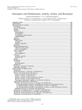
![CONCLUSIONS .........................................................................................................................................................169
REFERENCES ............................................................................................................................................................170
INTRODUCTION
Antiseptics and disinfectants are used extensively in hospi-
tals and other health care settings for a variety of topical and
hard-surface applications. In particular, they are an essential
part of infection control practices and aid in the prevention of
nosocomial infections (277, 454). Mounting concerns over the
potential for microbial contamination and infection risks in the
food and general consumer markets have also led to increased
use of antiseptics and disinfectants by the general public. A
wide variety of active chemical agents (or “biocides”) are
found in these products, many of which have been used for
hundreds of years for antisepsis, disinfection, and preservation
(39). Despite this, less is known about the mode of action of
these active agents than about antibiotics. In general, biocides
have a broader spectrum of activity than antibiotics, and, while
antibiotics tend to have specific intracellular targets, biocides
may have multiple targets. The widespread use of antiseptic
and disinfectant products has prompted some speculation on
the development of microbial resistance, in particular cross-
resistance to antibiotics. This review considers what is known
about the mode of action of, and mechanisms of microbial
resistance to, antiseptics and disinfectants and attempts, wher-
ever possible, to relate current knowledge to the clinical envi-
ronment.
A summary of the various types of biocides used in antisep-
tics and disinfectants, their chemical structures, and their clin-
ical uses is shown in Table 1. It is important to note that many
of these biocides may be used singly or in combination in a
variety of products which vary considerably in activity against
microorganisms. Antimicrobial activity can be influenced by
many factors such as formulation effects, presence of an or-
ganic load, synergy, temperature, dilution, and test method.
These issues are beyond the scope of this review and are
discussed elsewhere (123, 425, 444, 446, 451).
DEFINITIONS
“Biocide” is a general term describing a chemical agent,
usually broad spectrum, that inactivates microorganisms. Be-
cause biocides range in antimicrobial activity, other terms may
be more specific, including “-static,” referring to agents which
inhibit growth (e.g., bacteriostatic, fungistatic, and sporistatic)
and “-cidal,” referring to agents which kill the target organism
(e.g., sporicidal, virucidal, and bactericidal). For the purpose of
this review, antibiotics are defined as naturally occurring or
synthetic organic substances which inhibit or destroy selective
bacteria or other microorganisms, generally at low concentra-
tions; antiseptics are biocides or products that destroy or in-
hibit the growth of microorganisms in or on living tissue (e.g.
health care personnel handwashes and surgical scrubs); and
disinfectants are similar but generally are products or biocides
that are used on inanimate objects or surfaces. Disinfectants
can be sporostatic but are not necessarily sporicidal.
Sterilization refers to a physical or chemical process that
completely destroys or removes all microbial life, including
spores. Preservation is the prevention of multiplication of mi-
croorganisms in formulated products, including pharmaceuti-
cals and foods. A number of biocides are also used for cleaning
purposes; cleaning in these cases refers to the physical removal
of foreign material from a surface (40).
MECHANISMS OF ACTION
Introduction
Considerable progress has been made in understanding the
mechanisms of the antibacterial action of antiseptics and dis-
infectants (215, 428, 437). By contrast, studies on their modes
of action against fungi (426, 436), viruses (298, 307), and pro-
tozoa (163) have been rather sparse. Furthermore, little is
known about the means whereby these agents inactivate prions
(503).
Whatever the type of microbial cell (or entity), it is probable
that there is a common sequence of events. This can be envis-
aged as interaction of the antiseptic or disinfectant with the cell
surface followed by penetration into the cell and action at the
target site(s). The nature and composition of the surface vary
from one cell type (or entity) to another but can also alter as
a result of changes in the environment (57, 59). Interaction at
the cell surface can produce a significant effect on viability (e.g.
with glutaraldehyde) (374, 421), but most antimicrobial agents
appear to be active intracellularly (428, 451). The outermost
layers of microbial cells can thus have a significant effect on
their susceptibility (or insusceptibility) to antiseptics and dis-
infectants; it is disappointing how little is known about the
passage of these antimicrobial agents into different types of
microorganisms. Potentiation of activity of most biocides may
be achieved by the use of various additives, as shown in later
parts of this review.
In this section, the mechanisms of antimicrobial action of a
range of chemical agents that are used as antiseptics or disin-
fectants or both are discussed. Different types of microorgan-
isms are considered, and similarities or differences in the na-
ture of the effect are emphasized. The mechanisms of action
are summarized in Table 2.
General Methodology
A battery of techniques are available for studying the mech-
anisms of action of antiseptics and disinfectants on microor-
ganisms, especially bacteria (448). These include examination
of uptake (215, 428, 459), lysis and leakage of intracellular
constituents (122), perturbation of cell homeostasis (266,
445), effects on model membranes (170), inhibition of en-
zymes, electron transport, and oxidative phosphorylation (162,
272), interaction with macromolecules (448, 523), effects on
macromolecular biosynthetic processes (133), and microscopic
examination of biocide-exposed cells (35). Additional and use-
ful information can be obtained by calculating concentration
exponents (n values [219, 489]) and relating these to mem-
brane activity (219). Many of these procedures are valuable for
detecting and evaluating antiseptics or disinfectants used in
combination (146, 147, 202, 210).
Similar techniques have been used to study the activity of
antiseptics and disinfectants against fungi, in particular yeasts.
Additionally, studies on cell wall porosity (117–119) may pro-
vide useful information about intracellular entry of disinfec-
tants and antiseptics (204–208).
Mechanisms of antiprotozoal action have not been widely
investigated. One reason for this is the difficulty in cultur-
ing some protozoa (e.g., Cryptosporidium) under laboratory
conditions. However, the different life stages (trophozoites
and cysts) do provide a fascinating example of the problem
148 MCDONNELL AND RUSSELL CLIN. MICROBIOL. REV.](data:image/gif;base64,R0lGODlhAQABAIAAAAAAAP///yH5BAEAAAAALAAAAAABAAEAAAIBRAA7)