1. Done by
Dr. Rafid Remthan AL-Temimi.
Clinical Radiology
CABM ,DMRD,MBCHB,
2. The nose consists of the external nose and the nasal cavity, both of which are divided by a septum into right and left halves.
The external nose has two elliptical orifices
called the nostrils, which are separated from
each other by the nasal septum. The lateral
margin, the ala nasi, is rounded and mobile. The
framework of the external nose is made up above
by the nasal bones, the frontal processes of the
maxillae, and the nasal part of the frontal bone.
Below, the framework is formed of plates of
hyaline cartilage.
Dr, Rafid Remthan Al-Temimi, Clinical Radiology, CAMB 2
The Nose
External Nose:
3. Dr, Rafid Remthan Al-Temimi, Clinical Radiology, CAMB 3
Nerve Supply of the External Nose
The external nose is supplied by the infratrochlear and external nasal branches of the ophthalmic nerve (CN V), and the infraorbital branch of the
maxillary nerve (CN V).
Blood Supply and Venous Drainage of the External Nose
The skin of the external nose is supplied by branches of the ophthalmic and the maxillary arteries. The skin of the ala and the lower part of the
septum are supplied by branches from the facial artery. Venous blood from the external nose drains mostly into the facial vein via the angular and
lateral nasal veins.
4. Dr, Rafid Remthan Al-Temimi, Clinical Radiology, CAMB 4
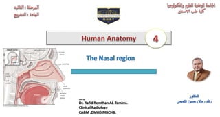
FIGURE : A. Lateral wall of the right nasal cavity. B. Lateral wall of the right
nasal cavity; the superior, middle, and inferior conchae have been partially
removed to show openings of the paranasal sinuses and the nasolacrimal duct into
the meati.
FIGURE : External nose and nasal septum. A. Lateral view of bony and
cartilaginous skeleton of external nose. B. Anterior view of bony and
cartilaginous skeleton of external nose. C. Bony and cartilaginous
skeleton of nasal septum.
5. Dr, Rafid Remthan Al-Temimi, Clinical Radiology, CAMB 5
The nasal cavity extends from the nostrils in front to the posterior nasal apertures or choanae behind, where the nose opens into
the nasopharynx. The nasal vestibule is the area of the nasal cavity lying just inside the nostril. The nasal cavity is divided into
right and left halves by the nasal septum. The septum is made up of the septal cartilage, the vertical plate of the ethmoid, and
the vomer.
Nasal Cavity
6. Dr, Rafid Remthan Al-Temimi, Clinical Radiology, CAMB 6
Mucous Membrane of the Nasal Cavity
The vestibule is lined with modified skin and has coarse hairs.
The area above the superior concha is lined with olfactory
mucous membrane and contains nerve endings sensitive to the
reception of smell. The lower part of the nasal cavity is lined
with respiratory mucous membrane. A large plexus of veins in
the submucous connective tissue is present in the respiratory
region.
The presence of warm blood in the venous plexuses serves to
heat up the inspired air as it enters the respiratory system. The
presence of mucus on the surfaces of the conchae traps foreign
particles and organisms in the inspired air, which are then
swallowed and destroyed by gastric acid.
7. Dr, Rafid Remthan Al-Temimi, Clinical Radiology, CAMB 7
Nerve Supply of the Nasal Cavity
The olfactory nerves from the olfactory mucous membrane ascend through the cribriform plate of the ethmoid bone to the
olfactory bulbs.The nerves of ordinary sensation are branches of the ophthalmic division (V1) and the maxillary division (V2) of
the trigeminal nerve.
8. Dr, Rafid Remthan Al-Temimi, Clinical Radiology, CAMB 8
FIGURE: A. Lateral wall of nasal cavity showing sensory
innervation of mucous membrane. B.
Nasal septum showing sensory innervation of mucous membrane.
9. Dr, Rafid Remthan Al-Temimi, Clinical Radiology, CAMB 9
Blood Supply to the Nasal Cavity
The arterial supply to the nasal cavity is from branches of the maxillary, the ophthalmic, and the facial arteries. The most important branch
is the sphenopalatine artery. The sphenopalatine artery anastomoses with the septal branch of the superior labial branch of the facial artery in
the region of the vestibule. The submucousvenous plexus is drained by veins that accompany the arteries.
The arterial supply of the medial and lateral walls of the nasal cavity can be summarized as follow (Fig. 4):
1. Anterior ethmoidal artery (a branch from the ophthalmic artery).
2. Posterior ethmoidal artery (a branch from the ophthalmic artery).
3. Sphenopalatine artery (a branch from the maxillary artery).
4. Greater palatine artery (a branch from the maxillary artery).
5. Septal branch of the superior labial artery (a branch from the facial artery).
The anterior part of the nasal septum is the site (Kiesselbach area) of an
anastomotic arterial plexus involving all five arteries supplying the septum. The
external nose receives blood from first and fifth arteries listed plus nasal branches
of the infra-orbital artery and the lateral nasal branches of the facial artery.
10. Dr, Rafid Remthan Al-Temimi, Clinical Radiology, CAMB 10
Venous Drainage of the Nasal Cavity
A rich submucosal venous plexus, deep to the nasal mucosa, provides venous drainage of the nose via the sphenopalatine, facial,
and ophthalmic veins. The plexus is an important part of the body’s thermoregulatory system, exchanging heat and warming air
before it enters the lungs.
FIGURE 4: A. Lateral wall of nasal cavity showing the arterial supply of the mucous membrane. B.
Nasal septum showing the arterial supply of the mucous membrane.
12. Dr, Rafid Remthan Al-Temimi, Clinical Radiology, CAMB 12
Lymph Drainage of the Nasal Cavity
The lymph vessels draining the vestibule end in the submandibular nodes. The remainder of the nasal cavity is drained by
vessels that pass to the upper deep cervical nodes.
13. Dr, Rafid Remthan Al-Temimi, Clinical Radiology, CAMB 13
The paranasal sinuses are cavities found in the interior of the maxilla,
frontal, sphenoid,and ethmoid bones.
They are lined with mucoperiosteum and filled with air; they
communicate with the nasal cavity through relatively small apertures.
The maxillary and sphenoidal sinuses are present in a rudimentary form
at birth; they enlarge appreciably after the eighth year and become fully
formed in adolescence.
Drainage of Mucus and Functions of Paranasal Sinuses
The mucus produced by the mucous membrane is moved into the
nose by ciliary action of the columnar cells. Drainage of the mucus
is also achieved by the siphon action created during the blowing
of the nose. The function of the sinuses is to act as resonators to
the voice; they also reduce the weight of the skull. When the
apertures of the sinuses are blocked or they become filled with
fluid, the quality of the voice is markedly changed.
The Paranasal Sinuses
14. Dr, Rafid Remthan Al-Temimi, Clinical Radiology, CAMB 14
Maxillary Sinus
The maxillary sinus is pyramidal in shape and located within the
body of the maxilla behind the skin of the cheek. The roof is formed
by the floor of the orbit, and the floor is related to the roots of the
premolars and molar teeth. The maxillary sinus opens into the middle
meatus of the nose through the hiatus semilunaris .
Frontal Sinuses
The two frontal sinuses are contained within the frontal bone. They
are separated from each other by a bony septum. Each sinus is
roughly triangular, extending upward above the medial end of the
eyebrow and backward into the medialpart of the roof of the orbit.
Each frontal sinus opens into the middle meatus of the nosethrough
the infundibulum .
15. Dr, Rafid Remthan Al-Temimi, Clinical Radiology, CAMB 15
Sphenoidal Sinuses
The two sphenoidal sinuses lie within the body of the sphenoid
bone.Each sinus opens into the sphenoethmoidal recess above the
superior concha.
Ethmoid Sinuses
The ethmoidal sinuses are anterior, middle, and posterior and they
are contained within the ethmoid bone, between the nose and the
orbit.They are separated from the latter by a thin plate of bone so
that infection can readily spread from the sinuses into the orbit.
The anterior sinuses open into the infundibulum; the middle
sinuses open into the middle meatus, on or above the bulla
ethmoidalis; and the posterior sinuses open into the superior
meatus.
16. Dr, Rafid Remthan Al-Temimi, Clinical Radiology, CAMB 16
FIGURE: A. The position of the paranasal sinuses in relation to
the face. B. Coronal section through the nasal cavity showing the
ethmoidal and the maxillary sinuses.
17. Dr, Rafid Remthan Al-Temimi, Clinical Radiology, CAMB 17
Foreign bodies in the nose are common in children. It should be remembered that the nasal septum is rarely
situated in the midline. A severely deviated septum may interfere with drainage of the nose and the paranasal
sinuses.
Trauma to the Nose
Fractures involving the nasal bones are common. Blows directed from the front may cause one or both nasal
bones to be displaced downward and inward. Lateral fracturesalso occur; the nasal septum is usually involved.
Nose Bleeding
Epistaxis, or bleeding from the nose, is a frequent condition. The most common cause is nose picking. The
bleeding may be arterial or venous, and most episodes occur on the anteroinferior portion of the septum.
Clinical Notes
18. Dr, Rafid Remthan Al-Temimi, Clinical Radiology, CAMB 18
Infection of the Nasal Cavity
Infection of the nasal cavity can spread in a variety of directions. The paranasal sinuses are especially prone to infection.
Organisms may spread via the nasal part of the pharynx and the auditory tube to the middle ear. It is possible for organisms
to ascend to the meninges of the anterior cranial fossa, along the sheaths of the olfactory nerves through the cribriform plate,
and produce meningitis.
Sinusitis and the Examination of the Paranasal Sinuses
Infection of the paranasal sinuses is a common complication of nasal infections. Rarely, the cause of maxillary sinusitis is
extension from an apical dental abscess. The frontal, ethmoidal, and maxillary sinuses can be palpated clinically for areas of
tenderness.
The frontal sinus can be examined by pressing the finger upward beneath the medial end of the superior orbital margin.
Here, the floor of the frontal sinus is closest to the surface.
The ethmoidal sinuses can be palpated by pressing the finger medially againstthe medial wall of the orbit.
The maxillary sinus can be examined for tenderness by pressing the finger against the anterior wall of the maxilla below
the inferior orbital margin; pressure over the infraorbital nerve may reveal increased sensitivity.
19. Dr, Rafid Remthan Al-Temimi, Clinical Radiology, CAMB 19
Thank you
References
1. Snell RS: Clinical anatomy by regions. Lippincott Williams & Wilkins, 2011.
2. Keith LM: Clinically Oriented Anatomy, 7th edition. Wolters Kluwer, 2014.