Upper respiratory tract nose pharynx dr kandil l4 final copy (2)
•Download as PPTX, PDF•
0 likes•62 views

NOSE PHARYNX

Recommended
More Related Content
What's hot
What's hot (20)
Similar to Upper respiratory tract nose pharynx dr kandil l4 final copy (2)
Similar to Upper respiratory tract nose pharynx dr kandil l4 final copy (2) (20)
More from mostafa soliman
Recently uploaded
Recently uploaded (20)
Upper respiratory tract nose pharynx dr kandil l4 final copy (2)
- 1. Upper respiratory tract nose &pharynx DR MOSTAFA KANDIL MBCHB MS MD PHD NOSE PHARYNX DR KANDIL 25/06/1441 1
- 2. External Nose Internal Nose Musculature of Nose Vascular Supply. Nerve supply. Lymphatic Drainage Paranasal Sinuses 25/06/1441 NOSE PHARYNX DR KANDIL 2
- 3. External nose 25/06/1441 NOSE PHARYNX DR KANDIL 3
- 4. External Nose It is pyramidal in shape with its root up and the base directed downwards Nares – external opening of nose Choanae - open into the nasopharynx 1. Bony Part: Posterior 1/3rd is bony 1. Nasal Bone 2. Frontal Process of Maxilla 3. Nasal Part of Frontal Bone 2. Cartilaginous Part: Anterior 2/3rd is cartilaginous 1. Upper Lateral Cartilage 2. Lower Lateral Cartilage/Alar Cartilage 3. Septal Cartilage 25/06/1441 NOSE PHARYNX DR KANDIL 4
- 5. Nasal Skin •The skin over the nasal bones and upper lateral cartilages is thin and freely mobile while that covering the alar cartilages is thick and adherent, and contains many sebaceous glands. 25/06/1441 NOSE PHARYNX DR KANDIL 5
- 6. Internal Nose Nasal Cavity • Extends from Nostrils externally to Choanae internally • Nasal Septum divides Nasal Cavity into two compartments Nasal Septum 1. Septal Cartilage 2. Perpendicular Plate of Ethmoid Bone 3. Vomer 25/06/1441 NOSE PHARYNX DR KANDIL 6
- 7. Zeeshan Ali M-1036 25/06/1441 NOSE PHARYNX DR KANDIL 7
- 8. Boundaries of Nasal Cavity • Floor 1. Palatine Process of Maxilla 2. Horizontal Process of Palatine Bone 25/06/1441 NOSE PHARYNX DR KANDIL 8
- 9. Roof 1. Nasal Bone 2. Frontal Bone 3. Cribriform Plate of Ethmoid Bone 4. Sloping body of Sphenoid Bone 25/06/1441 NOSE PHARYNX DR KANDIL 9
- 10. Medial Wall 1. Septal Cartilage 2. Perpendicular Plate of Ethmoid Bone 3. Vomer 25/06/1441 NOSE PHARYNX DR KANDIL 10
- 11. Lateral Wall • 3 Projections • Superior, Middle and Inferior Conchae/Vestibule • Space below each Concha is called Meatus • Spenoethmoidal Recess – Above superior concha > Sphenoid Air Sinus Superior Meatus – Below superior concha > Posterior ethmoid sinuses Middle Meatus – Below middle concha I. Bulla Ethmoidalis > formed by Middle ethmoidal air sinuses II.Hiatus semilunaris – Lies below bulla > Maxillary Sinus • Infundibulum – continuous with frontal sinus • Inferior Meatus – Below inferior concha > Nasolacrimal Duct 25/06/1441 NOSE PHARYNX DR KANDIL 11
- 12. Regions of Nasal Cavities •Each nasal cavity consists of three general regions •Nasal vestibule – small dilated space just internal to the naris that is lined by skin and contains hair follicles •Respiratory region –Largest part of the nasal cavity –Rich neurovascular supply –Lined by respiratory epithelium composed mainly of ciliated and mucous cells •Olfactory region –small, is at the apex of each nasal cavity –Lined by olfactory epithelium which contains the olfactory receptors 25/06/1441 NOSE PHARYNX DR KANDIL 12
- 13. Nerve Supply of Nose • Three cranial nerves – Olfaction - the olfactory nerve [I] – General sensation - the trigeminal nerve [V], • Anterior - ophthalmic nerve [V 1 ] • Posterior - maxillary nerve [V 2 ] – Glands - parasympathetic fibers in the facial nerve [VII] (greater petrosal nerve), • Sympathetic fibers 25/06/1441 NOSE PHARYNX DR KANDIL 13
- 14. 25/06/1441 NOSE PHARYNX DR KANDIL 14
- 15. • (c) Autonomic nerves • Parasympathetic nerve fibres supply the nasal glands and control nasal secretion. They come from greater superficial petrosal nerve, travel in the nerve of pterygoid canal (vidian nerve) and reach the sphenopalatine ganglion where they relay before reaching the nasal cavity. They also supply the blood vessels of nose and cause vasodilation. • Sympathetic nerve fibres come from upper two thoracic segments of spinal cord, pass through superior cervical ganglion, travel in deep petrosal nerve and join the parasympathetic fibres of greater petrosal nerve to form the nerve of pterygoid canal (vidian nerve). They reach the nasal cavity without relay in the sphenopalatine ganglion. Their stimulation causes vasoconstriction. 25/06/1441 NOSE PHARYNX DR KANDIL 15
- 16. 25/06/1441 NOSE PHARYNX DR KANDIL 16
- 17. Lymphatic Drainage •Lymphatics from the external nose and anterior part of nasal cavity drain into submandibular lymph nodes •The rest of nasal cavity drain into upper jugular nodes either directly or through the retropharyngeal nodes. •Lymphatics of the upper part of nasal cavity communicate with subarachnoid space along the olfactory nerves. 25/06/1441 NOSE PHARYNX DR KANDIL 17
- 18. Paranasal sinuses • four paired paranasal sinuses • The paranasal air sinuses are lined with respiratory epithelium 1- Maxillary sinuses, the largest of the paranasal sinuses, are under the eyes, in the maxillary bones (open in the back of the semilunar hiatus of the nose). 2- Frontal sinuses, superior to the eyes, in the frontal bone, which forms the hard part of the forehead. 3- Ethmoidal sinuses, which are formed from several discrete air cells within the ethmoid bone between the nose and the eyes. 4- Sphenoidal sinuses, in the sphenoid bone.25/06/1441 NOSE PHARYNX DR KANDIL 18
- 19. Functions of paranasal sinuses • The presence of these sinuses lightens the skull • They add resonance to speech • They humidify the air entering to respiratory system. 25/06/1441 NOSE PHARYNX DR KANDIL 19
- 20. NOSE PHARYNX DR KANDIL 20 Arteries that supply the nasal cavity include vessels that originate from both the internal and external carotid arteries : • vessels that originate from branches of the external carotid artery include the sphenopalatine, greater palatine, superior labial, and lateral nasal arteries; • vessels that originate from branches of the internal carotid artery are the anterior and posterior ethmoidal arteries. Arterial supply of the nasal cavities. A. Lateral wall of the right nasal cavity. B. Septum (medial wall of right nasal cavity). Arterial supply of the nasal cavities Nosebleed A nosebleed, or epistaxis, is a common occurrence and often involves the richly vascularized region of the vestibule and the anteroinferior aspect of the nasal septum (Kiesselbach’s area). Commonly, nosebleeds occur because of trauma involving the septal branch of the superior labial artery from the facial artery. 25/06/1441
- 21. NOSE PHARYNX DR KANDIL 21 Paranasal sinuses. A. Anterior view. B. Posteroanterior skull radiograph. C. Paramedian view of right nasal cavity. D. Lateral skull radiograph. Paranasal sinuses (Cont’d) • The frontal sinuses are innervated by branches of the supra-orbital nerve from the ophthalmic nerve [V1]. Their blood supply is from branches of the anterior ethmoidal arteries. • The ethmoidal cells, which are divided into anterior, middle, and posterior ethmoidal cells, are innervated by: the anterior and posterior ethmoidal branches of the nasociliary nerve from the ophthalmic nerve [V1]; and the maxillary nerve [V2] via orbital branches from the pterygopalatine ganglion. The ethmoidal cells receive their blood supply through branches of the anterior and posterior ethmoidal arteries. 25/06/1441
- 22. NOSE PHARYNX DR KANDIL 22 • The maxillary sinuses are innervated by infra-orbital and alveolar branches of the maxillary nerve [V2], and receive their blood through branches from the infra- orbital and superior alveolar branches of the maxillary arteries. • The sphenoidal sinuses are innervated by the posterior ethmoidal branch of the ophthalmic nerve [V1]; and the maxillary nerve [V2] via orbital branches from the pterygopalatine ganglion. The sphenoidal sinuses are supplied by branches of the pharyngeal arteries from the maxillary arteries. Paranasal sinuses (Cont’d) 25/06/1441
- 23. NOSE PHARYNX DR KANDIL 23 Drainage of the paranasal sinuses:- • Sphenoethmoidal recess: Sphenoidal air sinus • Superior meatus: Posterior ethmoidal air sinus • Middle meatus : Bulla ethmoidalis: Middle ethmoidal air sinus Hiatus semilunaris: Frontal air sinus Maxillary air sinus Anterior ethmoidal • Inferior meatus: Nasolacrimal duct 25/06/1441
- 24. Pharynx 25/06/1441 NOSE PHARYNX DR KANDIL 24
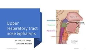
- 25. • Is a wide muscular tube • 12 cm in length • Located posterior to the nasal and oral cavities • Extends inferiorly, posterior to the larynx • Extends from the cranial base to the inferior border of the cricoid cartilage (anteriorly) and inferior border of C6 (posteriorly) [Extends from the base of the skull to the level of the C6 vertebra] where it is continuous with the oesophagus • Widest opposite the hyoid bone and narrowest at the junction where it joins the oesophagus25/06/1441 NOSE PHARYNX DR KANDIL 25
- 26. Pharyngeal wall The wall of the Pharynx consists of five layers: 1.Mucous membrane 2.Submucosa 3.Pharyngobasilar fascia 4.Pharyngeal muscles (3 constrictors) a- Stylopharyngeus b- Salpingopharyngeus C- Palatopharyngeus 5.Buccopharyngeal fascia 25/06/1441 NOSE PHARYNX DR KANDIL 26
- 27. 25/06/1441 NOSE PHARYNX DR KANDIL 27
- 28. • Respiratory function • Roof and Posterior wall: Continuous surface that lies inferior to the body of the sphenoid bone and the basilar part of the occipital bone • Pharyngeal tonsils: Found in the mucous membrane of the roof and the posterior wall of the nasopharynx25/06/1441 NOSE PHARYNX DR KANDIL 28
- 29. 25/06/1441 NOSE PHARYNX DR KANDIL 29
- 30. • Digestive function • Helps in the process of deglutition • Borders Superiorly: Soft Palate Inferiorly: Base of the Tongue Laterally: Palatoglossal and Palatopharyngeal arches and palatine tonsils 25/06/1441 NOSE PHARYNX DR KANDIL 30
- 31. • Palatine tonsils – Collections of lymphoid tissue on either side of the Oropharynx between the arches • Tonsillar bed – Superior constrictor of the pharynx and the pharyngobasilar fascia form the tonsillar bed 25/06/1441 NOSE PHARYNX DR KANDIL 31
- 32. ARTERIAL SUPPLY OF THE TONSIL 1- Tonsillar A. From facial A. 2- Lingual A. 3- Ascending palatine A. 4- Maxillary artery. Acute follicular tonsillitis 25/06/1441 NOSE PHARYNX DR KANDIL 32
- 33. • Extends from the superior border of the epiglottis and the pharyngoepiglottic folds to the inferior border of the cricoid cartilage Borders • Posteriorly: related to the bodies of the C4- C6 vertebrae. • Posterior and lateral walls: Middle and Inferior constrictor muscles 25/06/1441 NOSE PHARYNX DR KANDIL 33
- 34. • Palatopharyngeus and Stylopharyngeus muscles form the walls • Piriform recess: Small depression of the laryngopharyngeal cavity on either side of the laryngeal inlet separated from the laryngeal inlet by the aryepiglottic fold 25/06/1441 NOSE PHARYNX DR KANDIL 34
- 35. 2 layers of voluntary muscle: • External circular layer • Internal Longitudinal layer 25/06/1441 NOSE PHARYNX DR KANDIL 35
- 36. External circular layer • Constrictor muscles Primarily responsible for constricting the pharynx during swallowing Both types are innervated by the vagus nerve, except for the stylopharyngeus, which is innervated by the glossopharyngeal nerve. 25/06/1441 NOSE PHARYNX DR KANDIL 36
- 37. • Internal Longitudinal layer • Elevate/shorten and widen the pharynx during swallowing and speaking • Palatopharyngeus • Stylopharyngeus • Salpingopharyngeus Palatopharyngeus and salpingopharyngeus are innervated by the pharyngeal branch of CNX and the pharyngeal plexus • Stylopharyngeus is innervated by CN IX 25/06/1441 NOSE PHARYNX DR KANDIL 37
- 38. A- Sensory: Each of the three sections of the pharynx have a different innervation: 1- The nasopharynx is innervated by the maxillary nerve (CN V2). 2- The oropharynx by the glossopharyngeal nerve (CN IX). 3- The laryngopharynx by the vagus nerve (CN X). B- Motor: All the muscles of the pharynx are innervated by the vagus nerve (CN X), except for the stylopharyngeus, which is innervated by the glossopharyngeal nerve (CN IX). 25/06/1441 NOSE PHARYNX DR KANDIL 38
- 39. Blood Supply of the pharynx: 1- Arterial supply is via branches of the external carotid artery: • Ascending pharyngeal artery • Ascending palatine artery • Tonsillar branches of the facial artery • Branches of the maxillary and lingual arteries • Pharyngeal branches of the inferior thyroid artery 2- Venous drainage is achieved by the pharyngeal venous plexus, which drains into the internal jugular vein. 25/06/1441 NOSE PHARYNX DR KANDIL 39
- 40. 25/06/1441 NOSE PHARYNX DR KANDIL 40
- 41. Nasopharyngeal tonsil • Adenoids is the hypertrophied mass of lymphoid tissue situated at the junction of roof & post. wall of nasopharynx. • The mass of lymphoid tissue is termed as Adenoids only when it is hypertrophied. • It usually undergoes atrophy by puberty (13- 14 yrs) 25/06/1441 NOSE PHARYNX DR KANDIL 41
- 42. Waldeyer’sRing • Waldeyer's tonsillar ring includes 1. Adenoid tonsil 2. Two tubal tonsils 3. Two palatine tonsils 4. Lingual tonsil. 25/06/1441 NOSE PHARYNX DR KANDIL 42
- 43. Thank you 25/06/1441 NOSE PHARYNX DR KANDIL 43