Vapor growth of binary and ternary phosphorusbased semiconductors into TiO2 n...
REU final poster
1. Raman Time-Resolved Ion Exchange Studies in Natrolite
Rachel Hentz and Aaron Celestian, Geography and Geology
ABSTRACT:
The mechanics of ion exchange and ion mobility within zeolitic materials and
aqueous solutions are not well understood due to the rate of reaction and the
difficulty in probing samples in situ. Knowing the reaction process and
understanding the behaviors of ion mechanics in the solid state can we tailor
materials for specified functions. In this study, we conducted time resolved ion
exchange using Raman spectroscopy, on natrolite with a focus on understanding
the crystallographic and chemical transformations. Natrolite successfully
sequestered ions through its elliptical channels and has previously exhibited high
selectivity for large ion radius cations. Our studies had shown that there is a two
step exchange process: 1) softening of the 8 member rings as K exchanges directly
into the Na site, and 2) after an unmeasured amount of K had exchanged, the 4
member ring columns rapidly distort to open the 8 member rings as K migrates to
one side (see figure below).
Background Methods/Experiment Results and Discussion
DISCUSSION:
Using a combined set of experimental techniques, our measurements
suggest that there are two steps during ion exchange. First, there is a
‘softening’ of the 8MR and 4MR as the polyhedra distort to accommodate
the ingoing K cation. Second, after a maximal strain, the 8MR rapidly
(>1min.) open to allow K to migrate to the walls of the 8MR. Future work
will be focused on detailed structural transitions and on increasing the
effectiveness of ion exchanges to use in the industry field for waste water
filtration.
METHODS:
A single crystal of natrolite was mounted to 0.0125” diameter polyimide
tubing that had been cut to a diagonal point. Larger 0.0625” diameter
polyimide tubing was cut to create a window for viewing the crystal inside
and to allow the solution to escape. The smaller tubing was then inserted
into larger 0.0625” diameter polyimide tubing and glued in position so that
the crystal appeared flat in the window.
The data acquisition routine was set with the following experimental
parameters: laser wavelength, 780nm; laser power at sample, 24mW;
aperture, 50µm slit; grating, 830 line/mm; estimated spot size, 1.6µm;
allowed range, 1871 to 23cm-1
; min range limit, 50cm-1
; max range limit
1868cm-1
; objective, 50x 0.5 N.A.; collect exposure time 10sec; sample
exposures, 3. A dry Raman spectrograph was taken first and followed by a
deionized “wet” graph sampling. Then a macro program was written for 400
loops of the Raman data acquisition routine, automatic smoothing each
graph, and saving each 30 sec. complete scan. The program was started with
the starting of the Masterflex Console Drive, set at .6 (effectively 1 mL/min.),
that connected the ion solution to the crystal. Natrolite crystals were
exchanged with 0.1M solutions of KCl (CsCl and LiCl were also examined but
not presented here) under constant advection for 4 hours at room
temperature. A new crystal was used for each experiment and the finished
crystal was taken to the single crystal x-ray diffractometer for structural
analysis.
AKNOWLEDGEMENTS: This work was supported by a grant through the NSF-REU program
and the Advanced Materials Institute at WKU.
ABOVE: Upon ion exchange the natrolite channel locks in the ion into its nanoporous
crystalline framework. This distorts the channel and traps the ion into the structure, thus
effectively sequestering the ion from solution.
PICTURE SERIES BELOW: This series displays our method setup in the lab. (1) Natrolite
crystal before ion exchange. (2) Natrolite crystal after potassium ion exchange. (3) Single
crystal mount on polyimide tubing. (4) Window setup for ion exchange, with the crystal
mounted on the smaller tubing that lies inside the larger tubing. (5) Smaller tubing was
inserted into the larger tubing. (6) Setup in the Raman DXR machine ready for ion exchange.
LEFT IMAGE: This is the
connection we had setup
between the Raman DXR
to the Masterflex Console
Drive and ion solution
bottles.
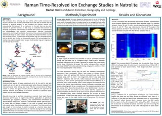
ABOVE: Time resolved data of K exchange into Na-natrolite. Note the two
different exchange steps: (1) a decrease in peak intensity and (2) a shift in
peak position to lower wavenumbers. Peak assignments from Liu et al.
(submitted).
RIGHT: This is the
Iterative Targeted
Transformation Factor
Analysis which helps
determine when
changes are occurring
in the spectra. This
analysis is for the K
exchange into Na-
natrolite and displays
a possible two-step
exchange process.
RESULTS:
For the K exchange with Na-natrolite the Iterative Targeted Transformation
Factor Analysis displays one significant slope decrease (Step 1) in intensity
between frames 10 and 15 and a second slope decrease (Step 2) between
frames 15 and 17. There was a decrease in intensity of the peaks in the
Raman spectrum at 443cm-1 and 535cm-1 at 43 min. (onset of Step 1) and at
second decrease and peak shift after 48 min. (onset of Step 2).
INTRODUCTION:
Particular toxic species of heavy metals such as Cs, Ni, Cu, and Zn, and light
metals such as Li, K, and Na contaminate water when they are released from
industrial waste, mine tailings, and rocks. The low concentrations of these
metals in surface and ground waters make it difficult and expensive for
selective removal, thus offering no economic incentive to reprocess those
metals, such as into a sellable commodity.
Naturally occurring or synthetic nanoporous materials have large channel
structures (on the scale of angstroms) and have applications in petroleum
refinement, gas separation, fertilizers, and environmental remediation to
adsorb/desorb metals from solutions. Time-resolved Raman spectroscopy is
an useful way to observe changes in the shift of energy, which gives
information about the vibrational modes in the system. This information can
give insight to the structure of zeolitic materials as a reaction takes place.
Collecting spectrums and surface maps in real-time during the ion exchange
experiment allows us to monitor chemical and structural properties as cations
diffuse in and out of the crystalline host.
From Liu et al. (submitted to American Mineralogist)
Used with permission.