Basic Civil Engineering first year Notes- Chapter 4 Building.pptx
2.a.lumbricoides and hookworms
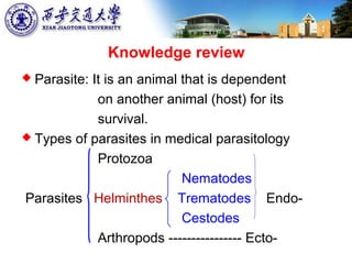
1. Knowledge review
Parasite: It is an animal that is dependent
on another animal (host) for its
survival.
Types of parasites in medical parasitology
Protozoa
Nematodes
Parasites Helminthes Trematodes Endo-
Cestodes
Arthropods ---------------- Ecto-
2. General characteristics of helminths
elongated flat or round worm like parasites
measuring few mm to meters. They are
multicellular and bilaterally symmetrical
They belongs to two phyla
1. Phylum Platyhelminths (flat worms)—
Two classes(important):
− Class: Cestoda (tapeworms)
− Class: Trematoda (flukes)
2. Phylum: Nemathelminths. one class (important):
− Class: Nematoda
6. Introduction to nematodes
Nematodes (worms or roundworms), the
multicellular animals on earth.
The number of nematode species is estimated
at half a million.
Many of them are “free-living” types.
Few are best know in the parasitic realm (E.
vermicularis, A.lumbricoides, Filarial parasites,
T. trichiura and hookworm)
8. 1. shape:
Cylindrical, vermiform, unsegmented and
bilaterally symmetrical.
Adults are dioecious and male worms are smaller
than females.
Characteristics of Nematodes
I
9. Characteristics of Nematodes
II2. Alimentary canal: Complete digestive tract (mouth
opening or buccal cavity / esophagus / intestine /
anus). The host’s gut contents, cells, blood, or
cellular breakdown products are their food.
10. Characteristics of Nematodes
III3. Body wall and Body cavity: pseudocoelom
In transverse section or crosssection
The body wall consists of three layers: cuticle,
hypodermis and an inner layer of muscle
cells. The cuticle is noncellular. The
hypodermis is to secrete the cuticle. The
nematodes move by contraction of muscle.
11. The body wall envelop an inner tube (the
digestive tract) and form the fluid-filled body
cavity (like thoracic cavity or abdominal cavity
of animals), a pseudocoelom without a cellular
lining or peritoneum (like pleural membrane or
abdominal membrane of animals) .
Pseudocoelom lies between these two tubes
and contains the reproductive tract and liquid.
12. Characteristics of Nematodes
IV4. Reproductive
system: tube type
Most male
reproductive
systems are a
single tube type
(testis, vas
deferens and
ejaculatory duct).
The sperms in it.
TESTIS
VAS
DEFERENS
EJACULATORY
DUCT
13. OVARY
OVIDUCT
UTERUS
Most female
reproductive systems
are a pair of tube type.
Two ovaries, each of
which connects to an
oviduct, and a uterus.
The two uteri unite to
a common duct,
vagina, and open to
the outside of the
body by a vulva.
The ova are in it.
14. Characteristics of Nematodes
V
5. Developmental stages of nematodes and life
cycle: six stages; direct or indirect life cycle
Direct life-cycle and soil-transmission (most)
e.g. Ascaris / hookworm / Enterobius
Indirect life-cycle and food or arthropods-
transmission (a few) e.g. Filaria / Trichinella
15. Most of the nematodes inhabitat in the intestine
while some (e.g fillarial worms) reside in various
tissues.
Intestinal (Human) nematodes (Small Intestine;
large intestinne) and Somatic (Human)
nematodes. (Classification based on habitat)
16. Nematodes pass through six developmental
stages: adult, egg and four larval stages (L1–
L4). Each larval stage transforms to the next by
shedding of the cuticle (called as molting).
18. INTRODUCTION
A. lumbricoides is the largest and most
common of the intestinal nematodes.
The name is derived from Askaris means
intestinal worm and Lumbricus means
resembling with common earthworm.
More than 1 billion of the world’s population,
including 86 million Chinese and 4 million
Americans (2013,WHO).
19. INTRODUCTION
A. lumbricoides -----Ascariasis.
Ascariasis is a disease of warm climates and
poor sanitation.
The parasite may also be acquired through
ingestion of egg-contaminated food by the host.
in dry, windy climates, eggs may become
airborne and be inhaled and swallowed.
20. Morphology -- Adult
Similar to earthworm. It has a cylindrical
fusiform body.
Light brown or pink in color, but
eventually white as the sample.
21. The male is slightly smaller, 15 to 31cm long,
than the female with the length of 20 to 35cm.
Male
Female
22. A mouth opening lies at the anterior end with
three lips with finely teeth at the lumen edge.
Electron microscope Biological
microscope
23. The male possesses a curved tail with a pair
of copulatory spicules (conducive to mating,
the sign of identification of species)
MaleSpicules
24. Morphology -- Egg
Eggs are elliptic, yellow. A rough protein
membrane are outside of the egg shells. It
may be desquamated in some eggs.
Fertilized egg and unfertilized egg.
Fertilized
egg Unfertilized
egg
protein
membrane
25. The fertilized egg: broadly oval, a embryo cell
in shell, 60 by 40 µm. A new-moon shaped
space at each end of egg.
New-moon shaped space (crescentic space)
26. The unfertilized egg: narrow (elongated) oval,
about 90 by 44 µm; a mass of disorganized,
highly refractile granules of various sizes in
the shells.
refractile granules
27. Remember (grasp) the feature of eggs
from the following several aspects
Appearance (shape)—(broadly or narrow) oval,
round, Barrel, or planoconvex
Size—small, middle, or large
Colour– yellow, brown, or colorless
Eggshell-- thick or thin
Contents in eggshell—cell (1 or more), larva, or
other material
Special structure—new-moon shaped space,
opercular plug,and so on.
28. Life cycle
Human is
only one host
Development
out of human
(simple)
Development
in human body
(complex)
32. Development of A.l in human body
Infective eggs ingested and hatched
Larvae penetrate intestine wall
Migrate via bloodstream
Liver Heart Lung
Alveoli of lungs
Bronchi Trachea pharynx (Swallowed)
Larvae mature and mate in intestine
Eggs in feces
33. Characteristics of life cycle
Human is the only host (direct life-cycle).
The location of adult: Small intestine.
Infective stage: Infective eggs (Embryonated
eggs containing the L2 larvae).
Mode of transmission: Ingestion of embryonated
eggs from the contaminated soil, food and water.
34. Larvae migration in body: liver, heart, lung
(circulatory system); alveoli, bronchi, trachea,
pharynx (respiratory system); esophagus,
stomach, small intestine (digestive system).
Nutrition: The semidigested food (polypeptide,
polysaccharide, fatty acid) of host.
35. Pathogenesis
Pathogenesis caused by Ascaris infections
is attributed to
The host immune response (Allergic reaction).
Effects of larval migration (Mechanical effects ).
Mechanical effects of the adults.
Nutritional deficiencies due to the presence of
the adult worms (depriving nutrition).
36. Clinical features
Both migrating larvae and adult
worms of A. lumbricoedes can cause
pathological changes and symptoms.
Damage of migrating larvae
Damage of adult worms
38. Damage of adult worms
If the worm burden is small, infections
with worms may be asymptomatic. Clinical
manifestations may result from a heavier
worm load in the intestinal lumen.
Malnutrition and Growth retardation
Allergic reaction
Syndrome of Ascariasis
39. Malnutrition and Growth retardation
In children ( 5), ascariasis may cause severe﹤
malnutrition and growth retardation related to
the worm burden.
Clinical symptoms include anepithymia (loss
of normal appetite, as for food or drink),
nausea, diarrhea, coprostasis (fecal
impaction) , abdominal pains, weight loss,
lassitude (physical or mental weariness).
40. Allergic reaction
The Ascaris allergen is one of the most
potent allergens of parasitic origin.
Allergic manifestations include urticaria, itch,
angioneurotic edema and so on.
41. What is Angioneurotic
Edema?
Angioneurotic Edema: Recurring attacks of
transient edema suddenly appearing in areas
of the skin or mucous membranes and
occasionally of the viscera.
There has been recent exposure to an allergen
42. Syndrome of Ascariasis
Syndrome of ascariasis may be severe.
An adult may migrate to the appendix, bile duct
or pancreatic duct, and cause obstruction and
inflammation of the organs. Biliary ascariasis is
the most common (abdominal pain, gallstone,
gallbladder rupture, peritonitis or liver abscess) .
Large numbers of adults cause mechanical
obstruction of the intestine.
Intestinal penetration or intussusception.
43. About Intussusception
Intussusception occurs when one portion of
the bowel slides into the next, much like the
pieces of a telescope. When this occurs, it
can create a blockage in the bowel, with the
walls of the intestines pressing against one
another. This, in turn leads to swelling,
inflammation, and decreased blood flow to
the part of the intestines involved.
44.
45. Laboratory Diagnosis
Egg detection: Microscopic identification of eggs
collected in stool is the method of choice.
1.The eggs are most easily seen on a direct wet
smear.
2.Concentration techniques by sedimentation method
should be done if direct stool microscopy is
negative.
Adult detection: Detection of adult in feces or
sputum.
46. DIRECT WET SMEAR Procedure
1. Place 1 drop of 0.85% NaCl (normal sodium
chlorine ) on the left side of the slide.
2. Take a small amount of fecal specimen
(about 2 mg , the amount picked up on the
end of an wooden applicator stick when
introduced into the specimen) and thoroughly
emulsify the stool in the saline preparations.
47. 3. Place a 22-mm coverslip on the
suspension.
4. Systematically scan the suspensions with
the 10 x objective. The entire coverslip area
should be examined under low power (total
magnification of x 100).
48. Treatment and prevention
Albendazole / Mebendazole. Albendazole
(400 mg once), mebendazole (100mg twice
daily for 3 days or 500 mg once) is effective.
Sanitary disposal of feces (composting to kill
the eggs).
Pay attention to personal hygiene and eating
habits
50. Introduction to hookworm
Hookworm refers to two organisms,
ancylostoma duodenale (A. duodenale) and
Necator americanus (N. americanus).
Some differences between two adult
hookworms. The egg, larvae and life cycle
are very similar.
Distinct geographic distribution.
51. N. americanus is found in North and South
america, China, India, and Africa.
A. duodenale is seen in the Mediterranean
basin, the Middle East, India, China, and
Japan.
Nearly 900million people are infected by
hookworm (China 400; India 200).
N. americanus infection (835) is more
common than A. duodenale (135) .
52. Morphology – Adult
Adults of both species are pinkish-white. The
females measure about 9 to 12mm. Males are
typical smaller, ranging from 5 to 10mm.
53. The males are equipped with a prominent
posterior copulatory bursa. Two copulatory
spicules.
A.d (spicules free) N.a (spicules fused)
54. The heads of both species are often curved
dorsally.
The end are also curved dorsally in A. d, like
letter “C”. The end are curved ventrally in N. a,
like letter “S”.
55. The mouth capsule of N. Americanus
contains a pair of cutting plates.
The mouth capsule of A. duodenale contains
two pair of teeth.
56. Morphology -- Egg
Ovoid and average 60um ×40um. A thin, clear
and colorless egg-shell. The eggs are in the early
cleavage stage when passed in the stool. They
characteristically have a clear space between the
developing embryo and the thin eggshell.
60. Development of eggs in soilInfectivestage
Filariform
For 1w
in
moist,
warm
soil
Rhabditiform
in 1-2 days
Filariform in 5-7 days
61. Development in human body
Filariforms penetrate skin
Migrate via bloodstream
Heart Lung (No through liver)
Alveoli of lungs
Bronchi Trachea pharynx (Swallowed)
Larvae mature and mate in intestine
Eggs in feces
62. Characteristics of life cycle
Human is the only host (direct life-cycle).
The location of adult: Small intestine.
Infective stage: Filariform (Third stage (L3) larva).
Larvae migration in body: Heart , lung (circulatory
system); alveoli, bronchi, trachea, pharynx
(respiratory system); esophagus, stomach, small
intestine (digestive system).
Nutrition: Blood.
63. Mode of transmission: Through penetration of
skin by the third stage larva.
Though rare, but other routes of transmission of
the larva has been reported through oral, in
utero and trans mammary routes.
64. Pathogenesis
The host immune response (larvae).
Effects of larval migration (larvae).
Mechanical effects of the adults (necrosis of the
intestinal within the adult worm mouth).
Blood loss by direct ingestion of blood by the
worms and continued blood loss from the original
attachment site (the worm secreted anticoagulant
/ stop the blood clotting) and iron deficiency.
65. Clinical features
Both migrating larvae and adult worms of
hookworms can cause pathological changes and
symptoms.
Damage of larval penetration of skin
Damage of migrating larvae
Damage of adult worms
66. Damage of larval penetration of
skin
The larval penetration of skin causes
dermatitis.
A small lesion with an itching and burning
sensations produced at the site of
penetration.
Scratching usually leads to infection by
bacteria.
69. Damage of adult worms
If the worm burden is small, infections with
worms may be asymptomatic.
Clinical manifestations may result from a
heavier worm load in the intestinal lumen.
Gastrointestinal symptoms
Iron deficiency anemia
70. Gastrointestinal
symptoms
Fatigue, nausea, vomiting, abdominal pain,
diarrhea with black to red stools, weakness, and
pallor.
Hookworm,
section of a
worm attached
to the mucosa.
71. Iron deficiency anemia
In chronic infection, the main symptom is iron
deficiency anemia (microcyte, low
hemoglobin ) with pallor, edema of the face
and feet, lassitude, and hemoglobin levels of 5
g/dl(decilite) or less.
There may be cardiomegaly and both mental
and physical retardation.
72. Laboratory Diagnosis
Microscopic identification of eggs collected in
stool is the method of choice. The eggs are
best seen in the direct smear or brine
floatation.
The larvae may be cultivated by fecal culture
in a test tube.
Detection of adult in feces.
73. Brine flotation.
This procedure is easy to perform and it is
used for the recovery of ova. The method is
recommended for the screening of
hookworms in a less than suitable
environment.
74. Reagent
Make a saturated solution of sodium
chloride (table salt suffices) in boiling tap
water. Allow to cool and check the specific
gravity to make sure that it is at least 1:20.
If the specific gravity is too low, reboil the
solution and add more salt. Filter and store
in a cool place.
75. Procedure
STEP 1: Make a 1:1 mixture of feces and the
brine solution in a disposable container.
STEP 2: Transfer to a 20 milliliter round test
tube (about one inch in diameter) and fill
the tube to the brim with salt solution.
76. STEP 3: Place a coverslip on top of the tube
touching the meniscus and allow to stand
undisturbed for 10 to 15 minutes.
STEP 4: With a quick motion, remove the
coverslip and place it on a prepared slide.
STEP 5: Observe microscopically for
parasites.
77. Treatment and prevention
Albendazole / Mebendazole. Antiparasitic drugs
like albendazole (400mg once), mebendazole
(500 mg once) can be given.
Sanitary disposal of feces.
Avoidance of contact with soil by wearing
shoes and gloves.
Modernization of agricultural technique.
78. Questions
What are similarities and differences in the life
cycle between hookworm and Ascaris?
What are the reasons of anemia caused by
hookworm?
What is the most severe damage caused by
hookworm or Ascaris to human body?
What method be respectively used to detect
hookworm or Ascris eggs in feces?
79. Differentiate between:
(a) Acylostoma duodenale and Necator
Americanus
(b) Fertilized and unfertilized egg of Ascaris
(c) Male and female worm of Acylostoma
duodenale
80. Multiple choice questions
Ascaris infects humans by:
(a) Penetration of skin by infective larvae
(b) Ingestion of unembryonated eggs present in
contaminated food and water
(c) Ingestion of embryonated eggs present in
contaminated food and water
(d) Autoinfection