
Recommended
More Related Content
What's hot
What's hot (20)
Similar to A monkey model of auditory scene analysis
Similar to A monkey model of auditory scene analysis (20)
Recently uploaded
Recently uploaded (20)
A monkey model of auditory scene analysis
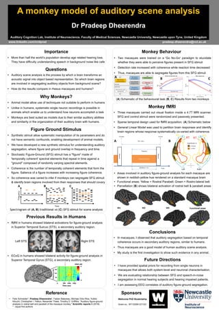
- 1. A monkey model of auditory scene analysis Dr Pradeep Dheerendra Auditory Cognition Lab, Institute of Neuroscience, Faculty of Medical Sciences, Newcastle University, Newcastle upon Tyne, United Kingdom More than half the world’s population develop age related hearing loss. They have difficulty understanding speech in background noise like cafe Importance Auditory scene analysis is the process by which a brain transforms an acoustic signal into object based representation. So which brain regions are involved in segregating auditory objects from background scene? How do the results compare in rhesus macaques and humans? Questions www.linkedin.com/in/dprad pradeep.dheerendra@ncl.ac.uk I have provided spatial priors for recording from single neurons in macaques that allows both system-level and neuronal characterisation. We are evaluating relationship between SFG and speech-in-noise segregation in normal hearing subjects and hearing impaired patients. I am assessing EEG correlates of auditory figure-ground segregation. Future Directions Figure Ground Stimulus Synthetic stimuli allow systematic manipulation of its parameters and do not have semantic confounds, enabling development of animal models. We have developed a new synthetic stimulus for understanding auditory segregation, where figure and ground overlap in frequency and time. Stochastic Figure-Ground (SFG) stimuli has a "figure" made of 'temporally coherent' spectral elements that repeat in time against a "ground" composed of randomly varying spectral elements. Coherence is the number of temporally coherent elements that form the figure. Salience of a figure increases with increasing figure coherence. So coherence was varied to infer if monkeys can segregate SFG stimuli & identify brain regions involved from their responses that should covary Spectrogram of (A, B) traditional vs (C) SFG stimuli for scene analysis Reference • Felix Schneider*, Pradeep Dheerendra*, Fabien Balezeau, Michael Ortiz-Rios, Yukiko Kikuchi, Christopher I. Petkov, Alexander Thiele, Timothy D. Griffiths. "Auditory figure-ground analysis in rostral belt and parabelt of the macaque monkey." Scientific reports 8 (2018). * - equal first authors In macaques, I observed that auditory segregation based on temporal coherence occurs in secondary auditory regions, similar to humans. Thus macaques are a good model of human auditory scene analysis. My study is the first investigation to show such evidence in any animal. Conclusions Animal model allow use of techniques not suitable to perform in humans Unlike in humans, systematic single neuron recordings is possible in animals which enable us to understand how neurons accomplish a task Monkeys are best suited as models due to their similar auditory abilities and similarity in the organization of their auditory brain with humans. Why Monkeys? fMRI in humans showed bilateral activations for figure-ground analysis in Superior Temporal Sulcus (STS), a secondary auditory region. Previous Results in Humans ECoG in humans showed bilateral activity for figure-ground analysis in Superior Temporal Gyrus (STG), a secondary auditory region. Left STS Right STS Sponsors Wellcome PhD Studentship Grant no.: WT102561/Z/13/Z Areas involved in auditory figure-ground analysis for each macaque are shown in reddish-yellow hue rendered on a standard macaque brain Functional areas: Yellow = Rostral Parabelt; Green = Rostro-lateral belt Parcellation (B) shows bilateral activation of rostral belt & parabelt areas Three macaques carried out visual fixation inside a 4.7T MRI scanner. SFG and control stimuli were randomized and passively presented. Sparse temporal design used for fMRI acquisition. (A) Schematic below General Linear Model was used to partition brain responses and identify brain regions whose response systematically co-varied with coherence. Monkey fMRI A B (A) Schematic of the behavioural task (B, C) Results from two monkeys Two macaques were trained on a 'Go No-Go' paradigm to elucidate whether they were able to perceive figures present in SFG stimuli Detection rate increased with coherence while reaction time decreased Thus, macaques are able to segregate figures from this SFG stimuli Monkey Behaviour False alarm rate Hit rate M4 M1 M1 M4 A B C