1. Summary of current research interests
Retinal degenerative diseases are a leading cause of blindness in the
Western world. Photoreceptor replacement therapy provides an outstanding
opportunity to develop novel therapeutic approaches, which may enable
us to replace the photoreceptors lost during degeneration and restore
visual function. We have recently established proof-of-concept for stem cell
therapy in the eye, demonstrating that photoreceptor transplantation is
capable of restoring vision. The focus of my research is the development of
these findings for the provision of cell-based mechanisms for retinal repair
and regeneration.
www.ucl.ac.uk/ioo
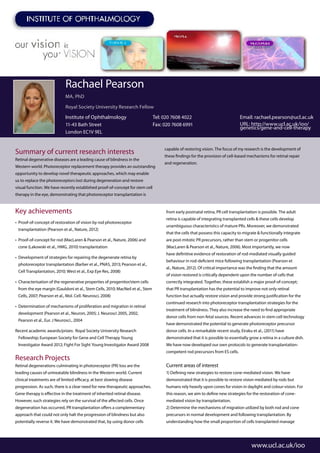
Rachael Pearson
MA, PhD
Royal Society University Research Fellow
Institute of Ophthalmology
11-43 Bath Street
London EC1V 9EL
Tel: 020 7608 4022
Fax: 020 7608 6991
Email: rachael.pearson@ucl.ac.uk
URL: http://www.ucl.ac.uk/ioo/
genetics/gene-and-cell-therapy
Key achievements
• Proof-of-concept of restoration of vision by rod photoreceptor
transplantation (Pearson et al., Nature, 2012)
• Proof-of-concept for rod (MacLaren & Pearson et al., Nature, 2006) and
cone (Lakowski et al., HMG, 2010) transplantation
• Development of strategies for repairing the degenerate retina by
photoreceptor transplantation (Barber et al., PNAS, 2013; Pearson et al.,
Cell Transplantation, 2010; West et al., Exp Eye Res, 2008)
• Characterisation of the regenerative properties of progenitor/stem cells
from the eye margin (Gauldoni et al., Stem Cells, 2010; MacNeil et al., Stem
Cells, 2007; Pearson et al., Mol. Cell. Neurosci, 2008)
• Determination of mechanisms of proliferation and migration in retinal
development (Pearson et al., Neuron, 2005; J. Neurosci 2005, 2002,
Pearson et al., Eur. J Neurosci., 2004
Recent academic awards/prizes: Royal Society University Research
Fellowship; European Society for Gene and Cell Therapy Young
Investigator Award 2012; Fight For Sight Young Investigator Award 2008
Research Projects
Retinal degenerations culminating in photoreceptor (PR) loss are the
leading causes of untreatable blindness in the Western world. Current
clinical treatments are of limited efficacy, at best slowing disease
progression. As such, there is a clear need for new therapeutic approaches.
Gene therapy is effective in the treatment of inherited retinal disease.
However, such strategies rely on the survival of the affected cells. Once
degeneration has occurred, PR transplantation offers a complementary
approach that could not only halt the progression of blindness but also
potentially reverse it. We have demonstrated that, by using donor cells
from early postnatal retina, PR cell transplantation is possible. The adult
retina is capable of integrating transplanted cells & these cells develop
unambiguous characteristics of mature PRs. Moreover, we demonstrated
that the cells that possess this capacity to migrate & functionally integrate
are post-mitotic PR precursors, rather than stem or progenitor cells
(MacLaren & Pearson et al., Nature, 2006). Most importantly, we now
have definitive evidence of restoration of rod-mediated visually guided
behaviour in rod-deficient mice following transplantation (Pearson et
al., Nature, 2012). Of critical importance was the finding that the amount
of vision restored is critically dependent upon the number of cells that
correctly integrated. Together, these establish a major proof-of-concept;
that PR transplantation has the potential to improve not only retinal
function but actually restore vision and provide strong justification for the
continued research into photoreceptor transplantation strategies for the
treatment of blindness. They also increase the need to find appropriate
donor cells from non-fetal sources. Recent advances in stem cell technology
have demonstrated the potential to generate photoreceptor precursor
donor cells. In a remarkable recent study, Eiraku et al., (2011) have
demonstrated that it is possible to essentially grow a retina in a culture dish.
We have now developed our own protocols to generate transplantation-
competent rod precursors from ES cells.
Current areas of interest
1) Defining new strategies to restore cone-mediated vision. We have
demonstrated that it is possible to restore vision mediated by rods but
humans rely heavily upon cones for vision in daylight and colour-vision. For
this reason, we aim to define new strategies for the restoration of cone-
mediated vision by transplantation.
2) Determine the mechanisms of migration utilized by both rod and cone
precursors in normal development and following transplantation. By
understanding how the small proportion of cells transplanted manage
2. to migrate into the recipient retina, we should be able to find ways to
manipulate this migration and drive more cells into the recipient retina.
3) Determine strategies for breaking down barriers within the recipient
retina. We have recently examined transplantation efficiency in a variety
of models of retinal degeneration and found that disease type has a major
impact on outcome (Barber et al., PNAS, 2013). We are working to factors
within the degenerating retina that impede (or enhance) transplanted cell
integration and find ways to manipulate them to improve transplantation
outcome (West et al., 2012; Pearson et al., 2010; West et al., 2008)
4) Determine whether purinergic signalling as an evolutionarily restricted
signalling mechanism in the control of retinal stem cell proliferation.
Unlike lower vertebrates, the mammalian retina lacks the ability to
generate. Understanding the mechanisms behind these differences
is crucial to knowing whether it might be possible to stimulate the
mammalian retina to repair itself. We believe that the presence or absence
of purinergic signaling may be important in determining this capability
(see Pearson et al., Neuron, 2005).
Techniques used in the lab
Multi-photon, confocal and fluorescence microscopy, stem cell culture,
calcium imaging, proliferation assays, viral vector production, molecular
biology, transplantation, RNAi, multielectrode array recordings,
electroretinogram recordings, intrinsic imaging of visual cortex,
behavioural tests of vision.
www.ucl.ac.uk/ioo
Dr. Rachael Pearson
Funding:
The Royal Society
MRC
The Wellcome Trust
BBSRC
RP FIghting Blindness
Collaborators:
Professor Robin Ali
Dr Jane Sowden
Pofessor David Becker
Alumni:
Dr Amanda Barber, Post-Doctoral Research
Associate, University of East Anglia
Legend for Images:
A. When transplanted into the subretinal space
of adult eyes, rod photoreceptor precursor cells
(green) migrate into and integrate within the
photoreceptor layer of the recipient eye.
B. Transplanted cells develop normally and have
all the morphological characteristics of mature
photoreceptors.
C. Rod precursor cells can also integrate into
diseased eyes and replace the proteins missing
in the degenerating retina. In this case, a
transplanted cell (green) has integrated within
the photoreceptor layer of an animal model of
retinal disease. The integrated cell expresses
rhodopsin (red), a photopigment essential for
light detection, which is normally absent in the
diseased eye.
D. The eye itself is a source of stem cells, which
may be a potential source of donor cells for
transplantation. Picture shows a neurospheres,
formed by proliferating stem cells in vitro. The
sphere is stained for the neural progenitor marker
nestin (green). Cell nuclei are stained in red.
E. Illustration used for cover of Nature.
Images: