More Related Content
Similar to nanoscale (20)
nanoscale
- 1. Nanoscale
REVIEW
Cite this: Nanoscale, 2015, 7, 12773
Received 3rd May 2015,
Accepted 26th June 2015
DOI: 10.1039/c5nr02878g
www.rsc.org/nanoscale
Reduction-sensitive polymeric nanocarriers in
cancer therapy: a comprehensive review†
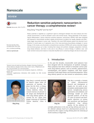
Bing Deng,a
Ping Mab
and Yan Xie*a
Redox potential is regarded as a significant signal to distinguish between the extra-cellular and intra-
cellular environments, as well as between tumor and normal tissues. Taking advantage of this physio-
logical differentiation, various reduction-sensitive polymeric nanocarriers (RSPNs) have been designed
and explored to demonstrate excellent stability during blood circulation but rapidly degrade and effec-
tively trigger drug release in tumor cells. Therefore, this smart RSPN delivery system has attracted much
attention in recent years, as it represents one of the most promising drug delivery strategies in cancer
therapy. In this review, we will provide a comprehensive overview of RSPNs with various reducible linkages
and functional groups up to date, including their design and synthetic strategies, preparation methods,
drug release behavior, and their in vitro and in vivo efficacy in cancer therapy. In addition, dual- and
triple-sensitive nanocarriers based on reducible disulfide bond-containing linkages will also be discussed.
1. Introduction
In the past few decades, innumerable novel polymers have
been designed with the development of synthetic strategies.
These polymers have been utilized to formulate various poly-
meric drug delivery systems, such as micelles, polymersomes,
nanohydrogels, and polymer complexes, with their unique
nanometer scale-range feature and structural properties. These
drug delivery systems are also termed as nanocarriers, which
Bing Deng
Bing Deng is currently pursuing
his Master’s degree under the
supervision of Associate Pro-
fessor Yan Xie, Ph.D., at Shang-
hai University of Traditional
Chinese Medicine, China. His
research interests are focused on
the design and development of
novel drug delivery systems. He
received his B.S. degree from
Jiangxi University of Traditional
Chinese Medicine, China in
2013. Ping Ma
Ping Ma is currently a Scientist
in Global Pharmaceutical
Research and Development at
Hospira Inc. He works on pre-
formulation and formulation devel-
opment, manufacturing process
optimization and validation, and
technology transfer of sterile
injectable products. Dr Ma has
15+ years of research and devel-
opment experience on various
oral, parenteral, and lipid-based
nanoparticle drug delivery
systems. He has authored 20+
scientific publications in the area of drug delivery and serves on
the editorial advisory board of 4 pharmaceutical journals. He
received his Ph.D. degree in Drug Delivery from the University of
North Carolina at Chapel Hill.
†Electronic supplementary information (ESI) available. See DOI: 10.1039/
c5nr02878g
a
Research Center for Health and Nutrition, Shanghai University of Traditional
Chinese Medicine, Shanghai 201203, China. E-mail: rosexie_1996@hotmail.com;
Fax: +86 (21)51322407; Tel: +86 (21)51322440
b
Global Pharmaceutical Research and Development, Hospira Inc., McPherson,
Kansas, USA
This journal is © The Royal Society of Chemistry 2015 Nanoscale, 2015, 7, 12773–12795 | 12773
- 2. provide a promising and feasible technology platform in
cancer therapy because of their ability to encapsulate, attach,
and adsorb anti-cancer small molecules as well as biothera-
peutics.1,2
Additionally, these drug-loaded nanocarriers
demonstrate some significant advantages, such as prolonged
circulation time, passive targeting to tumor tissue via the
enhanced permeability and retention effect, reduced side
effects, and improved bioavailability.3–5
For a long time, the
most common drug-loaded polymeric nanocarriers were based
on hydrolytically biodegradable aliphatic polyesters and poly-
carbonates.6,7
Unfortunately, this is far from optimal due to
their slow drug release at tumor sites although they are stable
during blood circulation, which significantly compromises
their anti-cancer efficacy both in vitro and in vivo.8,9
To this end, tremendous effort has been directed to
maximize the therapeutic index of anti-cancer drugs, such as
bio-responsive polymeric nanocarriers,10–14
which are not only
sensitive to intra-cellular stimuli (pH, redox potential, lyso-
somal enzymes, etc.) but also simultaneously release drugs
rapidly and efficiently in cancer cells. Among these bio-respon-
sive polymeric nanocarriers, reduction-sensitive polymeric
nanocarriers (RSPNs) have drawn particular attention for intra-
cellular anti-cancer drug delivery in recent years.13,15,16
Com-
pared to other bio-responsive polymeric nanocarriers, RSPNs
demonstrate higher stability against hydrolytic degradation in
circulation, as well as a faster response to intra-cellular redu-
cing conditions, thus triggering drug release in the cytosol and
the cell nucleus, where many anti-cancer drugs exert their
therapeutic effects. The high reducing potential in cells is
mainly attributed to glutathione (GSH), an abundant biological
reducing agent.17
It has been reported that the intra-cellular
concentration of GSH is approximately 2–10 mM, particularly
in certain organelles such as the cytosol, mitochondria, and
the cell nucleus, while GSH levels are a thousand-fold lower
(approximately 2–20 μM) in normal body fluids (blood and the
extra-cellular matrix).18,19
Moreover, the GSH concentration in
tumor tissues is four times higher than in normal tissues
(approximately 4 μM g−1
vs. 1 μM g−1
).20
It should be noted
that the endo/lysosome also possesses a high reducing poten-
tial which is modulated by gamma interferon-induced lyso-
somal thiol reductase (GILT) in the copresence of L-cysteine.21,22
This significant difference in reducing properties between the
extra- and intra-cellular environments, as well as between
tumor and normal tissues, provides RSPNs a unique advan-
tage, given that they are stable in the extra-cellular environ-
ment but rapidly and efficiently release drug in the intra-
cellular environment due to their hydrolytically biodegradable
properties, which is the premise of RSPN design and also
suggests that it is possible for RSPNs to deliver drugs to tumor
cells. Therefore, the high reducing potential of the intra-cellu-
lar environment has been recognized as a stimulus for the dis-
assembly of RSPNs to rapidly and efficiently achieve drug
release in cancer cells.
Generally, RSPNs are synthesized from reducible polymers
by incorporating a reduction cleavable linkage or introducing
a reduction-sensitive functional group into their structures. In
this comprehensive review, we present recent progress on
RSPNs for the intra-cellular delivery of anti-cancer drugs and
bio-therapeutics. The design and synthesis of reduction-sensi-
tive polymers with more than 15 different reducible linkages
and functional groups (Table 1), such as endogenous disulfide
linkages (cystamine, 3,3′-dithiodipropionic acid, 2,2′-dithio-
diethanol, and cystamine bisacrylamide), synthesized disulfide
linkages (based on lipoic acid (LA), cysteine, pyridyl disulfide
group), diselenium linkages, platinum(IV), and trimethyl-
locked benzoquinone (TMBQ), as well as their in vitro and
in vivo drug release behavior and anti-tumor efficacy in cancer
therapy are discussed. In addition, dual- and triple-stimuli-
responsive drug delivery systems containing reduction-sensi-
tive linkages, such as pH/reduction, temperature/reduction,
and pH/temperature/reduction-sensitive nanocarriers, are also
discussed. The current review will be beneficial for the design
and development of novel RSPN systems for better cancer
therapy in the future.
2. Nanocarriers with disulfide bond
linkages
The disulfide bond (SS) is an extremely valuable functional
group that exists in a variety of chemical and biological
agents and can be elegantly applied in the synthesis of
RSPNs.12,23–26
In general, there are two synthetic methods to
incorporate the disulfide bond into drug carriers at different
positions (Fig. 1). One is the direct introduction of endogen-
ous disulfide bond-containing linkages, including disulfide
bond-containing cross-linkers,27–29
some disulfide bond-con-
taining living or controlled polymerization initiators,30,31
and
disulfide bond-containing and olefin-based monomers.32
The
Yan Xie
Yan Xie is Associate Professor at
the Research Center for Health
and Nutrition, Shanghai Univer-
sity of Traditional Chinese Medi-
cine (SUTCM). Her research
focuses on the development of
various nano-delivery systems for
poorly water-soluble traditional
Chinese medicine compounds.
She has authored 50+ scientific
publications in the area of drug
delivery of Traditional Chinese
Medicine (TCM). Dr Xie has
received numerous awards,
including Shanghai Science and Technology Advancement Prize,
Shanghai Medical Technology Award, and Honorary Title of
Shanghai Rising-Star of Science and Technology. She has obtained
her Ph.D. degree in Pharmaceutical Sciences with specialization in
TCM from SUTCM and a Master’s degree in Pharmaceutics from
China Pharmaceutical University.
Review Nanoscale
12774 | Nanoscale, 2015, 7, 12773–12795 This journal is © The Royal Society of Chemistry 2015
- 3. Table 1 Summary of reducible linkages and functional groups used in the preparation of RSPNs
Types of reducible
linkages and
functional groups Chemicals Chemical structures
Disulfide bond
containing linkages
Cystamine
3,3′-Dithiodipropionic acid
2,2′-Dithiodiethanol
Cystamine bisacrylamide
3,3′-Dithiobis(sulfosuccinimidyl
propionate)
L-Cystine N-carboxyanhydride
N-Succinimidyl 3-(2-pyridyldithio)
propionate
3-[(2-Aminoethyl)dithiol]
propionic acid
Thiol containing or
generated groups
Lipoic acid
Cysteine
Pyridyldithio residue
Diselenide bond
containing linkages
Di-(1-hydroxylundecyl) diselenide
2-(2-(2-Hydroxyethoxy)ethoxy)-
ethyl diselenide
3,3′-Diselanediyldipropanoic acid
Nanoscale Review
This journal is © The Royal Society of Chemistry 2015 Nanoscale, 2015, 7, 12773–12795 | 12775
- 4. other method is the introduction of thiol groups into block
polymers such that a thiol–disulfide exchange reaction can
occur to transform thiol groups into disulfides.33
These strat-
egies play an important role in the design of different struc-
tural polymers containing disulfide, which could be further
utilized to prepare various RSPNs for anti-cancer drug delivery.
2.1. Endogenous disulfide bond-linked RSPNs
Thus far, numerous polymeric nanocarriers based on
endogenous disulfide bond linkages have been prepared to
incorporate different types of anti-cancer drugs and biothera-
peutics. These endogenous disulfide linkers usually exist in
the main chain, at the side chain, or as the cross-linker of the
polymers (Table 2). Cystamine, 3,3′-dithiodipropionic acid,
2,2′-dithiodiethanol, and cystamine bisacrylamide have been
the most useful small linkages for the synthesis of disulfide-
containing polymers.13
2.1.1. Cystamine-linked nanocarriers. Cystamine is a di-
sulfide bond containing linkage with a short alkyl spacer in
the middle and terminating in a primary amine, and can
easily react with N-carboxyanhydride (NCA) or carboxylic acid
groups on polymers. Consequently, reduction-sensitive poly-
mers containing cystamine at different positions can be
achieved. These polymers primarily form three types of RSPNs:
shell-sheddable micelles, core disassemblable micelles, and
crosslinked micelles. These cystamine-containing micelles are
stable in the blood circulation but will disassemble or collapse
in the intra-cellular reductive environment or under tumor
cell-relevant GSH levels, and subsequently, the encapsulated
drug cargos are rapidly released.
Shell-sheddable micelles have been engineered from
various amphiphilic block polymers, including linear,34–36,38
graft,52,53
star-shaped,54
and prodrug39
polymers, in which
cystamine exists between the hydrophobic and hydrophilic seg-
ments. The cleavage of cystamine in a reducing environment
leads to the shedding of hydrophilic shells from micelles and
subsequently results in rapid drug release. For example, Park
et al. developed reduction-sensitive shell-sheddable micelles
based on poly(ethylene glycol)-SS-poly(γ-benzyl-L-glutamate)
(PEG-SS-PBLG) linear polymers.34
PEG-SS-PBLG was syn-
thesized by ring opening polymerization (ROP) of benzyl
glutamate-NCA using PEG-cystamine as a macroinitiator
(Scheme 1). The solvent casting method was utilized to
prepare camptothecin (CPT)-loaded micelles, and the micelles
released CPT completely within 20 h in the presence of 10 mM
GSH in vitro, whereas only 40% of CPT was released in the absence
of GSH. The in vitro cytotoxicity results also showed that CPT-
loaded micelles exhibited higher toxicity to squamous carci-
noma cancer cells (SCC7) than micelles without the disulfide
Table 1 (Contd.)
Types of reducible
linkages and
functional groups Chemicals Chemical structures
Other reducible
functional groups
Platinum(IV)
Trimethyl-locked benzoquinone
Fig. 1 The position of the disulfide bond in polymers.
Review Nanoscale
12776 | Nanoscale, 2015, 7, 12773–12795 This journal is © The Royal Society of Chemistry 2015
- 5. bond. Similarly, the cystamine-linked hybrid poly(ethylene
glycol) methyl ether-SS-poly(D-leucine-L-leucine) (mPEG-SS-Pleu)
and mPEG-SS-poly(ε-benzyloxycarbonyl-L-lysine) (mPEG-SS-PzLL)
amphiphilic linear polymers were designed for the preparation
of shell-sheddable micelles.35,36
These micelles underwent a
fast shell-shedding process and simultaneously accelerated
doxorubicin (DOX) release in the presence of 10 mM GSH or
10 mM dithiothreitol (DTT). Zhou et al. synthesized a hyal-
uronic acid-g-SS-deoxycholic acid (HA-g-SS-DOCA) graft conjugate
via a 1-ethyl-3 (3-dimethylaminopropyl) carbodiimide (EDC)-
mediated coupling reaction.53
In aqueous media, HA-g-
SS-DOCA conjugates self-assembled into nanosized micelles,
which rapidly disassembled in response to 20 mM GSH. By
using paclitaxel (PTX) as a model drug molecule, the obtained
PTX-loaded micelles were taken up by human breast adeno-
carcinoma cells (MDA-MB-231) via HA receptor-mediated
endocytosis and exhibited significantly higher antitumor
activity (IC50 = 25.6 ng mL−1
) compared to the insensitive PTX-
loaded HA-DOCA micelles (IC50 = 56.6 ng mL−1
) as well as
Taxol (IC50 = 51.7 ng mL−1
) after 72 h of incubation. Moreover,
in vivo imaging analysis in tumor-bearing mice also indicated
that near infrared reflection (NIR) dye-loaded HA-g-SS-DOCA
micelles exhibited nearly 2.5-fold higher fluorescence intensity
at tumor sites than insensitive micelles at 24 h following
injection. Cystamine-containing star-shaped cationic polymers
based on cyclodextrin (CD) and ethanolamine-functionalized
poly(glycidyl methacrylate) (PGEA) were also successfully syn-
thesized via atom transfer radical polymerization (ATRP).54
The cationic shell-sheddable micelles possessed good DNA
condensation ability due to the plentiful secondary amine and
hydroxyl groups in their structure, low cytotoxicity, and
efficient gene delivery attributable to the PGEA arms that
could be cleaved from the micelles under reducing conditions.
Similarly, Li et al. designed a prodrug polymer by reacting
Table 2 Summary of RSPNs based on endogenous disulfide bond containing polymers
Position of disulfide
bond Polymers
Disulfide bond containing
linkage
Model drug
or gene Ref.
In the main chain PEG-SS-PBLG Cystamine CPT 34
mPEG-SS-Pleu DOX 35
mPEG-SS-PzLL DOX 36
PzLL-SS-PEG-SS-PzLL DOX 37
DEX-SS-PBLG MTX 38
CPT-SS-PEG-SS-CPT CPT 39
PLA-SS-P(OEOMA) 2,2′-Dithiodiethanol — 40
PLA-SS-PMPC NR 41
SS(PLA-b-P(OEOMA)) DOX 42
P(OEOMA)-SS-P(GMA-TEPA) DNA 43
mPEG-SS-CPT CPT 44
OEI-SS-TetK 3,3′-Dithiodipropionic acid DNA 45
mPEG-SS-C16 DOX 46
H40-star-PLA-SS-PEP DOX 47
In the main chain SS-PAA-g-PEG Cystamine bisacrylamide MTX 48
SS-PCA-g-PCL PTX 49
SS-PAA-g-Chol siRNA 50
mPEG-g-SS-PAA-CPT CPT 51
At the side chain CS-g-SS-Chol Cystamine Quercetin 52
HA-g-SS-DOCA PTX 53
CD-g-SS-PGEA DNA 54
DEX-g-SS-DPDPE DNA 55
P(Asp-Az)x-g-SS-PEI gene 56
P(OEOMA-co-FAMA) 2,2′-Dithiodiethanol DNA 57
P(BSSMA-g-PDMAEMA)
PEG-b-P(MAOHD-g-SS-Py) Py 58
Chi-g-SS-PCL 3,3′-Dithiodipropionic acid DOX 59
PHEA-g-SS-C16 DOX 60
As the cross-linker PEG-b-PLG/SS-b-(PLA)2 Cystamine DOX 28
PEG-b-PAEG-b-PAAs/SS DOX 61
PEG-P(HEMA-co-AC/SS) CC 62
PEO-b-P(α-N3CL)/SS-b-PCL 2,2′-Dithiodiethanol DOX 63
P(PEGMEMA-b-PMAU/SS) — 64
mPEG-CPT-SS-P(TA-OCA)/SS CPT 65
SS/PEI-g-mPEG 3,3′-Dithiodipropionic acid MTX 66
SS/DEX-PBLG DOX 67
SS/PEI siRNA 68
SS/PMA Cystamine bisacrylamide OR 69
PA-b-PEI/SS DNA 70
SS/PEI-RGD DNA 71
SS/PEI-PAAs DNA 72
Nanoscale Review
This journal is © The Royal Society of Chemistry 2015 Nanoscale, 2015, 7, 12773–12795 | 12777
- 6. H2N-SS-PEG-SS-NH2 with succinate-activated CPT, in which the
prodrug spontaneously arranged into shell-sheddable micelles
with an average size of approximately 226 nm in aqueous
medium.39
Interestingly, the release rate of CPT from
CPT-SS-PEG-SS-CPT micelles demonstrated apparent biphasic
kinetics when treated with 10 mM DTT in that CPT was
released at a rapid rate of 4% h−1
within the first 10 h, while
the rate was only 0.4% h−1
during the following 240 h.
Reduction-sensitive micelles with a disassemblable core are
prepared from amphiphilic polymers bearing cystamine
embedded in the main chain. For example, DOX-loaded dis-
assemblable micelles were prepared with the amphiphilic poly
(amide amine)-g-poly(ethylene glycol) (SS/PAA-g-PEG) polymer
containing multiple cystamines throughout the main chain.73
The micelles released approximately 95% DOX within 10 h in
the presence of 1 mM DTT. Cell experiments showed that
DOX-loaded SS/PAA-g-PEG micelles effectively transported
DOX into the nuclei of both HeLa and HepG2 cells, and the
IC50 of micelles was approximately 1.73 times of that of the DOX
solution control, and 2.35 times lower than that of common
non-sensitive micelles, which indicated that the SS/PAA-g-PEG
micelles had potent cytotoxicity against cancer cells.
In recent years, reduction-sensitive crosslinked nanocarriers
have been designed to elegantly resolve the dilemma between
extra-cellular stability and intra-cellular drug release or
address the difficulty between transfection efficiency and cyto-
toxicity when delivering anti-tumor drugs or biotherapeutics.
Cystamine is a homo-bifunctional crosslinker that can be
utilized to prepare reduction-sensitive crosslinked nanocarriers
via carbodiimide coupling chemistry, Michael addition reac-
tions, and click chemistry. Jing et al. prepared interlayer cross-
linked (ICL) micelles based on Y-shaped amphiphilic block
polymers mPEG-b-poly(L-glutamate)-b-(polylactide)2 (mPEG-b-PLG-b-
(PLA)2) using cystamine as a crosslinker via carbodiimide
coupling chemistry (Scheme 2). The in vitro release studies
showed that crosslinked micelles released DOX upon reduction
stimulus in that the ICL micelles released approximately
45% and 52% DOX within 14 h in the presence of 10 mM and
15 mM DTT, respectively, whereas less than 30% DOX was
released in the absence of DTT. Moreover, the in vivo pharmaco-
kinetic results demonstrated that the mean residence time
(1.49 ± 0.18 h) of crosslinked micelles was longer than that of
non-crosslinked micelles (0.76 ± 0.079 h) and free DOX (0.62 ±
0.096 h), indicating the improved in vivo stability of cross-
linked micelles compared to non-crosslinked micelles and free
DOX.28
Similarly, reduction-sensitive core crosslinked (CCL)
micelles self-assembled from an mPEG-b-polycaprolactone-b-
poly(2-(2-oxo-1,3,2-dioxaphospholoyloxy)ethyl methacrylate)
(mPEG-b-PCL-b-PPEMA) triblock polymer via the Michael
addition reaction between cystamine and carbon–carbon
double bond of PPEMA segments. Compared to the uncross-
linked micelles, the CCL structure improved micellar stability
and achieved higher cytotoxic activity against HeLa cells.74
Cystamine-crosslinked low-molecular-weight polyethylenimine
(SS/PEI800) derivatives were synthesized via a click reaction
between alkyne-modified cystamine and azide-functionalized
PEI800.29
The gene transfection activity of SS/PEI800 polyplexes
was 5–10 times higher than that of polyplexes prepared using
25 kDa PEI, and the IC50 of SS/PEI800 polymers was 8–10 times
higher than that of 25 kDa PEI. This indicated that the
SS/PEI800 polymers enhanced gene transfection with lower
cytotoxicity.
2.1.2. 3,3′-Dithiodipropionic acid-linked nanocarriers.
Similar to cystamine, 3,3′-dithiodipropionic acid also contains
a disulfide bond in its chemical structure, but both sides end
with carboxylic acid groups. A covalent bond could be easily
formed by reacting the carboxylic acid groups with hydroxy or
amino groups. As of now, several 3,3′-dithiodipropionic acid-
containing shell-sheddable and crosslinked micelles for deli-
vering anti-cancer drugs or genes have been developed.
Polymers based on 3,3′-dithiodipropionic acid are usually
synthesized by carbodiimide chemistry, click chemistry, and
ATRP. These polymers can further form shell-sheddable
micelles. For instance, Huang et al. exploited 3,3′-dithiodi-
propionic acid to synthesize mPEG-SS-hexadecyl (mPEG-SS-C16)
block polymers via two dicyclohexylcarbodiimide (DCC)-
mediated coupling reactions.46
mPEG-SS-C16 micelles with an
average size of 137 nm were prepared using a dialysis method.
Treated wih 10 mM DTT, micelle size increased from 137 nm
to approximately 300 nm within 10 min. Moreover, cell experi-
ments showed that DOX-loaded mPEG-SS-C16 micelles efficien-
tly delivered DOX to the cell nuclei, displaying higher
Scheme 1 Synthesis of the PEG-SS-PLGB copolymer via disulfide bond
conjugation. Reproduced with permission from ref. 34. Copyright 2011
American Chemical Society.
Review Nanoscale
12778 | Nanoscale, 2015, 7, 12773–12795 This journal is © The Royal Society of Chemistry 2015
- 7. Scheme 2 Synthetic route of the Y-shaped block copolymer mPEG-b-PLG-b-(PLA)2 and the preparation of drug-loaded, middle-shell-crosslinked micelles and their reduction response. Repro-
duced with permission from ref. 28. Copyright 2011 Royal of Society of Chemistry.
NanoscaleReview
Thisjournalis©TheRoyalSocietyofChemistry2015Nanoscale,2015,7,12773–12795|12779
- 8. cytotoxicity against HeLa cells compared to reduction-insensi-
tive micelles. In another study, reduction-sensitive amphi-
philic chitosan (Chi)-g-SS-PCL graft polymers were formulated
by conjugating PCL onto the backbone of Chi with 3,3′-dithiodi-
propionic acid via a DCC-mediated coupling reaction.59
The
reduction-sensitive behavior of Chi-g-SS-PCL micelles was
investigated using DTT as a reductive reagent, and DOX
release was accelerated when the micelles were treated with
10 mM DTT. Similar to Chi-g-SS-PCL, another graft polymer,
poly{α,β-[N-(2-hydroxyethyl)-L-aspartamide]}-g-SS-C16 (PHEA-g-
SS-C16), was synthesized based on C16, PHEA, and 3,3′-dithiodi-
propionic acid.60
Cell experiments demonstrated that DOX
efficiently entered the nuclei and exhibited higher cell cytotoxi-
city compared to its reduction-insensitive counterpart, which
was due to the fact that PHEA-g-SS-C16 micelles achieved rapid
release of DOX in reducible cancer cells. Yan et al. synthesized
an amphiphilic hyperbranched multiarm polymer with 3,3′-
dithiodipropionic acid between the hydrophobic polyester core
and hydrophilic polyphosphate arms through a carbodiimide
coupling reaction.47
Benefiting from the amphiphilic struc-
ture, this hyperbranched polymer was able to self-assemble
into shell-sheddable micelles in aqueous solution. The hydro-
philic shell of the micelles could be detached under 10 mM
DTT and consequently resulted in rapid DOX release in gluta-
thione monoester (GSH-OEt)-pretreated HeLa cells, which sig-
nificantly enhanced cell proliferation inhibition.
Differing from the DCC-mediated coupling reaction, Oh
et al. reported a reduction-sensitive polymer that was syn-
thesized by the ATRP of 3,3′-dithiodipropionic acid functiona-
lized methacrylate in the presence of poly(ethylene oxide)
monomethyl ether (mPEO)-functionalized bromoisobutyrate.75
The micelles disassembled when treated with intra-cellular
GSH because the hydrophobic/hydrophilic balance was broken
upon the cleavage of pendant 3,3′-dithiodipropionic acid. This
micellar destabilization also led to enhanced release of en-
capsulated DOX. In another study, a reduction-sensitive cat-
ionic polymer was synthesized via click chemistry with 1-azido-
3-aminopropane functionalized 3,3′-dithiodipropionic, amide-
triazole moieties, and secondary amine groups.76
This polymer
efficiently condensed DNA into nanoparticles but also released
DNA rapidly under reducing conditions due to the cleavage of
3,3′-dithiodipropionic acid.
In addition to shell-sheddable micelles, shell-crosslinked
nanocarrier (SCL) micelles based on 3,3′-dithiodipropionic
acid-crosslinked amphiphilic PBLG-b-dextran/SS (PBLG-b-DEX/
SS) were recently designed. Briefly, the PBLG-b-DEX/SS
polymer was synthesized via click reaction of α-azido PBLG
and α-alkyne DEX, and then, the DEX segment of PBLG-b-DEX
was linked by 3,3′-dithiodipropionic acid via an esterification
reaction to form the SCL micelles in phosphate buffered saline
(PBS). Micelles after crosslinking showed good stability in PBS
compared to non-crosslinked micelles, and the release kinetics
of DOX-loaded SCL micelles exhibited accelerated DOX release
under intra-cellular mimicking reductive conditions. Further-
more, the micelles demonstrated higher cellular proliferation
inhibition against GSH-OEt-pretreated HeLa and HepG2 cells
than non-pretreated cells.67
Similarly, the Zhang group
reported that 3,3′-dithiodipropionic acid-crosslinked PEG-SS-
poly(L-lysine)-b-Pleu (PEG-SS-PLL-b-Pleu) SCL micelles exhibi-
ted much faster CPT release compared to irreversible SCL
micelles in 10 mM DTT.27
In addition, CPT-loaded reversible
SCL micelles demonstrated 2-fold stronger fluorescence and
higher cytotoxicity in GSH-OEt-pretreated HeLa cells than non-
pretreated cells.
2.1.3. 2,2′-Dithiodiethanol-linked nanocarriers. Currently,
various types of reduction-sensitive polymers based on the
2,2′-dithiodiethanol linkage have been exploited, such as
brush block polymers,57
graft block polymers,44,58
linear block
polymers,42,77
and others. For example, Zhang et al. syn-
thesized a prodrug polymer based on mPEG and CPT by utiliz-
ing 2,2′-dithiodiethanol as a linkage through the radical
addition–fragmentation chain transfer (RAFT) reaction and
esterification reaction.44
This prodrug polymer self-assembled
into micelles with an average size of 122 nm in PBS. Interest-
ingly, another anti-cancer drug (DOX) was introduced into the
prodrug micelles, and thus, this system simultaneously deli-
vered two anticancer drugs. More importantly, both CPT and
DOX were released from the micelles under reducing con-
ditions, leading to a synergistic cytotoxicity toward human
tongue squamous cell carcinoma (TCA8113) cells and rat
adrenal pheochromocytoma (PC12) cells.
Reduction-sensitive micelles were prepared from amphi-
philic hyperbranched homopolyphosphates (HPHSEP), which
were fabricated by self-condensing the ROP of 2-[(2-hydroxy-
ethyl)-disulfanyl]ethoxy-2-oxo-1,3,2-dioxaphospholane.78
Con-
focal microscopy observations showed that the micelles
quickly and efficiently transported DOX into the nuclei of
HeLa cells when pretreated with 10 mM GSH-OEt, and exhibi-
ted enhanced cell inhibition against GSH-OEt-pretreated HeLa
cells but reduced inhibition toward buthionine sulfoximine
(BSO)-pretreated cells. This suggested that the degradation of
GSH-responsive 2,2′-dithiodiethanol accelerated intra-cellular
DOX release from the micelles.
Reduction-sensitive CCL CPT prodrug micelles were also
prepared via the coprecipitation of 2,2′-dithiodiethanol-con-
taining CPT-poly(5-[4-(prop-2-yn-1-yloxy)benzyl]-1,3-dioxolane-
2,4-dione) (CPT-poly(tyrosine(alkynyl)-OCA)) conjugates and
mPEG-b-poly(tyrosine(alkynyl)-OCA), followed by crosslinking
the micellar core via an azide–alkyne click reaction. The size of
the reduction-sensitive CPT-loaded CCL micelles exhibited a
negligible change from 57.4 nm to 59.3 nm after incubation in
PBS for 8 days, demonstrating the excellent stability of CCL
micelles under physiological conditions, while they underwent
rapid dissociation under reducing conditions, resulting in a
burst release of CPT and enhanced cytotoxicity against human
breast cancer cells.65
2.1.4. Cystamine bisacrylamide-linked nanocarriers. Cyst-
amine bisacrylamide is a derivative of cystamine that is
obtained by reacting primary amines with acryloyl chloride via
an amidation reaction. As the olefin group exists at both ends,
cystamine bisacrylamide was employed as a reducible linker to
connect primary amines of PEI or PAAs through a Michael
Review Nanoscale
12780 | Nanoscale, 2015, 7, 12773–12795 This journal is © The Royal Society of Chemistry 2015
- 9. addition to synthesize various structure types of bioreducible
cationic polymers with multiple disulfide bonds (SS/PEI, SS/
PAAs). Most of the cationic polymers were used for delivering
biotherapeutics (siRNA, DNA, and gene),50,70,71,79–81
and these
positively charged polyplexes were stable under neutral con-
ditions but were rapidly destabilized in a reducing environ-
ment, consequently leading to increased transfection
efficiency with relatively low toxicity.
It should be noted that cystamine bisacrylamide has also
been applied for the delivery of anti-cancer drugs. For
example, reduction-sensitive starch nanoparticles with di-
sulfide crosslinkers were prepared by the free radical reaction
of hydroxy groups of starch to ethylene groups of cystamine
bisacrylamide via the reversed-phase microemulsion method.
The drug loading of nanoparticles (0.302 g g−1
) crosslinked by
cystamine bisacrylamide was higher than that of non-disulfide
crosslinked nanoparticles (0.275 g g−1
). Furthermore, the
in vitro drug release results demonstrated that the crosslinked
starch nanoparticles exhibited accelerated drug release behav-
ior in the presence of 20 mM DTT.82
In another study, an
amphiphilic polymer poly(N,N′-bis(acryloyl)cystamine-ethanol-
amine)-g-PCL (PCA-g-PCL) with multiple disulfide bonds in the
backbone was synthesized by reacting cystamine bisacrylamide
with ethanolamine via a Michael addition reaction to obtain
cationic polymer PCA, and then PCL was grafted onto PCA by
ROP. Under normal circumstances, these micelles were able to
maintain stability and hold 70% PTX in the core, while under
40 mM DTT, all loaded PTX was quickly released from micelles
within 9 h due to the reduction triggered breakage property of
the micelles.49
Amphiphilic mPEG-b-SS/PAA-b-mPEG triblock
polymers based on cystamine bisacrylamide, phenylethyl-
amine, and mPEG were synthesized in a simple one-step
process under mild conditions through a Michael addition
reaction.83
Reduction-sensitive micelles were prepared by dia-
lyzing against distilled water, and rapid aggregation of micelles
was observed in 10 mM DTT, in which micelle size increased
from 70 nm to 560 nm in 0.5 h. Notably, methotrexate (MTX)-
incorporated micelles effectively inhibited the growth of
several cancer cell lines due to the rapid intra-cellular MTX
release.
2.1.5. Other disulfide bond-linked nanocarriers. Other
than cystamine, 3,3′-dithiodipropionic acid, 2,2′-dithiodietha-
nol, and cystamine bisacrylamide, there are a few disulfide-
containing linkages that have been less explored for the design
of RSPNs. These linkages are classified into two categories:
homo- and hetero-bifunctional linkages. Homo-bifunctional
linkages terminated with the same functional groups are
ideally suitable for the connection of similar polymers, while
hetero-bifunctional linkages are generally chosen when two
different polymers need to be connected selectively. These lin-
kages are also utilized in the fabrication of disulfide-linked
RSPNs for desired stability in the blood circulation and intra-
cellular reduction-responsive drug release.
Lee et al. synthesized the PEG-b-PLL-b-poly(L-phenylalanine)
(PEG113-PLL19-PPhe24) polymer via the one-pot two-step ROP
and a subsequent deprotection process. SCL micelles were
formed by crosslinking the PLL of PEG113-PLL19-PPhe24 using
the homo-bifunctional linkage of 3,3′-dithiobis(sulfosuccin-
imidyl propionate) as a disulfide linker, and the PPhe inner cores
were used for the loading of docetaxel (DTX).84
DTX release
was facilitated by the reductive cleavage of disulfide bonds. At
33 days after injection, DTX-loaded SCL micelles exhibited
enhanced anti-tumor activity over non-crosslinked micelles by
44.4% in terms of tumor volume regression. Another CCL
micelle was prepared through one-step ROP of L-phenyl-
alanine-NCA and L-cystine-NCA (a homo-bifunctional disulfide
bond linker) with mono-amino terminated PEG-NH2 as the
macroinitiator.85
The drug release behavior of these micelles
could be adjusted by various concentrations of GSH, and
enhanced intra-cellular drug release occurred in GSH-pre-
treated HeLa cells. Furthermore, the micelles were biocompati-
ble based on an in vitro cytotoxicity experiment.
Although crosslinked micelles can be directly obtained by
incorporating homo-bifunctional disulfide linkages, this
approach lacks selectivity. If a conjugation between two
different polymers is needed, the application of hetero-bifunc-
tional disulfide linkages should be considered. For example,
Yang et al. utilized N-succinimidyl-3-(2-pyridyldithio) propio-
nate as a hetero-bifunctional disulfide linkage to conjugate
PTX onto fluorescent mesoporous silica nanoparticles (FMSN).
Next, PTX prodrug modules conjugated with FMSN were acti-
vated to their cytotoxic form inside tumor cells upon internal-
ization owing to the breakage of the disulfide bond. The intra-
cellular drug delivery results indicated that the prodrug exhibi-
ted much higher cellular proliferation inhibition against
10 mM GSH-OEt-pretreated cells (IC50 = 5.0 µg mL−1
) than
untreated cells (IC50 = 49.6 µg mL−1
).86
Graft-reducible N-(2-
hydroxypropyl)methacrylamide (HPMA)-co-oligolysine poly-
mers based on HPMA, methacrylamido-functionalized oligo-L-
lysine peptide monomers, and 3-[(2-aminoethyl)dithiol] pro-
pionic acid linker were polymerized via RAFT polymerization.87
This cationic polymer was used for nucleic acid delivery. The
stability studies showed that reducible polymers were more
stable than non-reducible polymers under saline and serum-
containing conditions, and the cytotoxicity results demon-
strated that polymers were not cytotoxic at the tested charge
ratios.
2.2. Synthesized disulfide bond-linked RSPNs
Aside from endogenous disulfide linkages, disulfide-contain-
ing polymers can also be fabricated from thiolated polymers
by oxidation reactions or thiol–disulfide exchange reactions.
Oxidation of thiol to disulfide can be easily accomplished
with oxygen or with a mild oxidizing reagent such as dimethyl
sulfoxide (DMSO).88
Typical thiol-generated reagents or
thiol-containing reagents, such as LA, cysteine, and the pyridyl
disulfide group, have been frequently utilized to prepare
thiol modified polymers, which can be used to fabricate
various disulfide-containing CCL nanocarriers,89,90
SCL
nanocarriers,91,92
ICL nanocarriers,93,94
and shell-sheddable
nanocarriers.95–97
Nanoscale Review
This journal is © The Royal Society of Chemistry 2015 Nanoscale, 2015, 7, 12773–12795 | 12781
- 10. 2.2.1. Disulfide-linked nanocarriers synthesized using
LA. LA is a natural and biocompatible compound containing a
lipoyl ring, which can generate dihydrolipoyl groups (the
reduced form of lipoyl groups) under reducing conditions via
ROP; then, core crosslinking occurs with the formation of new
linear disulfide bonds between different dihydrolipoyl groups
under a mild oxidizing environment. Given this, several di-
sulfide core crosslinking conjugates have been synthesized by
LA to obtain reducible nanocarriers that are extremely stable
under physiological conditions but robustly release drugs in a
reducing environment.89,98,99
For example, Chen et al. pre-
pared reduction-sensitive disulfide CCL micelles based on an
amphiphilic graft starch-g-PEG/LA polymer. The polymer was
conveniently synthesized by grafting PEG-COOH and LA onto a
starch backbone with an esterification reaction. The starch and
LA here acted as the hydrophobic inner core, and PEG acted as
the hydrophilic outer shell. Under catalytic amounts of DTT,
lipoyl rings of LA in the micelles were broken into dihydroli-
poyl groups and then were oxidated under air atmosphere to
form disulfide bond-crosslinked micelles.99
The crosslinked
micelles were stable under normal physiological conditions
but degraded under reducing conditions. Moreover, higher cel-
lular proliferation inhibitory activity was achieved against
GSH-OEt-pretreated HeLa and HepG2 cells compared to non-
pretreated and BSO-pretreated cells. Similarly, another
reduction-sensitive amphiphilic block polymer of PEG and
PCL containing two lipoyl functional groups at their interface
was synthesized by Zhong et al.100
A thiol–disulfide exchange
reaction was used for the development of ICL micelles. The
crosslinked micelles demonstrated markedly enhanced stabi-
lity against a diluted physiological salt concentration. In the
presence of 10 mM DTT, micelles were subjected to rapid de-
crosslinking and released DOX.
It must be noted that introducing LA into the polymers
using postpolymerization modification is difficult to control
and calculate the degree of substitution of LA. Utilizing con-
trolled RAFT polymerization of functionalized LA is one
method to address this problem. For instance, PEG-b-poly(N-2-
hydroxypropyl methacrylamide)–LA (PEG-b-PHPMA–LA) conju-
gates with known and tunable LA content were obtained by
two reactions: RAFT polymerization of HPMA using
PEG-CPADN (CPADN: 4-cyanopentanoic acid dithionaphtha-
lenoate) as a macro-RAFT agent to synthesize PEG-b-PHPMA,
followed by grafting of LA onto the main chain of PEG-b-
PHPMA via lipoylation.90
The substitution degree of LA was
determined to be 71. The preparation of CCL micelles was
carried out in PBS (pH 7.4, 10 mM) by introducing 10 mol%
DTT relative to the lipoyl units in the PEG-b-PHPMA–LA conju-
gates (Scheme 3). These CCL micelles exhibited several com-
bined advantages, including excellent biocompatibility,
superior drug loading, and high extra-cellular stability.
Additionally, DOX-loaded micelles demonstrated pronounced
antitumor effects following 48 h of incubation in HeLa and
HepG2 cells with IC50 values of 6.7 µg mL−1
and 12.8 µg mL−1
,
respectively.
2.2.2. Disulfide-linked nanocarriers synthesized using
cysteine. Cysteine is an aliphatic amino acid containing a
thiol group that is commonly used to synthesize thiol-contain-
Scheme 3 Synthetic route of PEG-b-PHPMA–LA conjugates (A) and the illustration of robust reversibly core-cross-linked PEG-b-PHPMA–LA
micelles for the efficient loading and reduction-triggered release of DOX (B). Reproduced with permission from ref. 90. Copyright 2012 American
Chemical Society.
Review Nanoscale
12782 | Nanoscale, 2015, 7, 12773–12795 This journal is © The Royal Society of Chemistry 2015
- 11. ing oligomeric peptides and polypeptides, which can also be
oxidated into disulfide, and subsequently construct disulfide-
containing nanocarriers. Compared to the direct incorporation
of endogenous disulfide linkages for crosslinking, the utiliz-
ation of cysteine crosslinking provides some advantages as
follows: (1) no additional crosslinkers are required; (2) the
purification process is straightforward; (3) the polymer concen-
tration and the molar ratio of cysteine to polymer could be pre-
cisely adjusted during synthesis; (4) thiol groups are inherently
present in cysteine; (5) the amount of cysteine can be easily
quantitatively determined by a thiol detection reagent, such as
Ellman’s reagent; and (6) the crosslinking conditions are a
mild oxidizing environment such that micelle aggregation can
be avoided. Given the above advantages, Zhang and co-workers
designed cysteine incorporated linear bioreducible polypeptide
nanocarriers for gene delivery, in which cysteine was intro-
duced at the two sides of the peptides to obtain thiol-ending
short peptides, followed by crosslinking via an oxidative
polymerization reaction to obtain various ratios of disulfide-
linked polypeptides under 30% DMSO. The gene-loaded vector
demonstrated an excellent ability to bind and condense DNA
and exhibited higher transfection efficiency with lower cyto-
toxicity compared to 25 kDa PEI.101
Recently, two disulfide-
linked highly branched polypeptides based on cysteine-termi-
nated three-armed peptides were reported. These polypeptides
were able to bind and compact DNA strongly and tightly, thus
reducing DNA loss in the circulation and enhancing its cellular
uptake. Furthermore, the transfection efficiency mediated by
the two polypeptides was nearly 100-fold higher than that of
linear PLL (4–15 kDa).102
In addition, ICL micelles were also prepared for controlled
drug release by crosslinking cysteine-containing micelles. PEG-
b-poly(L-cysteine)-b-PPhe (PEG-PCys-PPhe) triblock polymers
were prepared by the sequential ROP of L-cysteine-NCA and
L-Phe-NCA. The polymers self-assembled into disulfide-cross-
linked biodegradable micelles via the oxidation of thiol groups
in the middle of PCys segments. The in vitro drug release
showed that approximately 60% DOX was released in the pres-
ence of 10 mM GSH in 24 h, whereas only approximately 15%
DOX was released in PBS (without GSH) at the same time. The
results suggested that the crosslinked micelles were helpful to
reduce drug loss in the extra-cellular environment and acceler-
ated drug release under intra-cellular GSH.93
Ahn et al.
exploited a short peptide comprising three cysteines (Cys(trt))
to conjugate PEG and PCL and thereby obtained the PEG-Cys-
(trt)-PCL block polymer, which was further used to prepare
reduction-sensitive crosslinked micelles via oxygen reaction in
the air.103
The polymeric micelles after crosslinking remained
stable under diluted conditions as well as in the bloodstream
and quickly destabilized to release encapsulated DOX after the
addition of 1 mM DTT. Lam et al. designed and synthesized
thiolated linear-dendritic polymers by introducing cysteine into
the dendritic oligo-lysine backbone of telodendrimers com-
prised of PEG and a dendritic cluster of cholic acids.104
These
telodendrimers self-assembled into micelles in PBS, and di-
sulfide CCL micelles were then prepared by the oxidization of
thiol groups. The resulting CCL micelles were able to load PTX
with a superior loading capacity up to 35.5%, and the release
of PTX from the CCL micelles was significantly slower than
that from non-crosslinked micelles but was facilitated by
increasing the concentration of GSH. Moreover, the CCL
micelles were found to preferentially accumulate at the tumor
site in nude mice, and the disulfide-crosslinked micellar for-
mulation of PTX was more efficacious than both free PTX and
the non-crosslinked formulation at equivalent doses of PTX in
an ovarian cancer xenograft mouse model.
2.2.3. Disulfide-linked nanocarriers synthesized by pyridyl-
dithio residues. Apart from LA and cysteine, another impor-
tant strategy to fabricate disulfide-containing polymers is to
form new disulfide bonds via the preparation of pyridyldithio
residue-modified polymers and the thiol–disulfide exchange
reaction of these residues with other thiol-containing poly-
mers. The pyridyl disulfide group is excellently suited for the
formation of disulfide-containing polymers as it exhibits a
highly efficient thiol–disulfide exchange reaction with sulf-
hydryl groups under mild reaction conditions. Moreover, the
pyridine-2-thione (a byproduct of this pyridyl disulfide group)
is a good leaving group possessing a distinctive absorption
band in the visible region, which allows for the quantitative
monitoring of the reaction progress. Taking advantage of this
strategy, Zhong et al. synthesized a disulfide-linked diblock
polymer DEX-SS-PCL, which was prepared through two steps
of a thiol–disulfide exchange reaction: DEX orthopyridyl di-
sulfide (DEX-SS-Py) was obtained by the thiol–disulfide
exchange reaction between cysteamine-modified DEX and
2,2′-dithiodipyridine, and DEX-SS-PCL was then prepared via
another thiol–disulfide exchange reaction between DEX-SS-Py
and mercapto PCL (PCL-SH) (Scheme 4). The DEX-SS-PCL
polymer formed reduction-sensitive shell-sheddable micelles
with an average size of 60 nm by the solvent exchange
method, and large aggregates with a size of over 1000 nm
were rapidly observed in response to 10 mM DTT. A cell viabi-
lity assay showed that approximately 20% and 70% cells
were observed alive when cells were treated with drug-loaded
DEX-SS-PCL micelles and reduction-insensitive DEX-PCL
micelles for 2 days, respectively, which indicated that the
anti-tumor efficacy of the drug in DEX-SS-PCL micelles was
markedly enhanced compared to that of DEX-PCL micelles.105
Similarly, another disulfide-containing polymer was prepared
by reacting (2-(pyridyldithio)-ethylamine hydrochloride)-
modified HA with mercapto dodecanethiol (DDT) under
ambient conditions via a thiol–disulfide exchange reac-
tion.106
DOX-loaded HA-SS-DDT micelles were prepared by
the water-in-oil emulsion method, and the micelles triggered
release of DOX in the presence of 10 mM GSH. Furthermore,
the in vitro cellular uptake results showed that a weak DOX
fluorescence signal was observed when the cells were
treated with reduction-insensitive micelles, but a strong DOX
signal was detected in the intra-cellular compartments of
cells after treatment with HA-SS-DDT micelles, which was
probably due to the rapid release of DOX from HA-SS-DDT
micelles.
Nanoscale Review
This journal is © The Royal Society of Chemistry 2015 Nanoscale, 2015, 7, 12773–12795 | 12783
- 12. Reduction-sensitive grafted polymers based on pyridyl di-
sulfide functionalized cyclic carbonate (PDSC) were also deve-
loped in recent years. Zhong et al. first reported the synthesis
of the grafted polymer PCL-g-SS-PEG by ROP of ε-caprolactone
and PDSC followed by reacting with thiolated PEG via a thiol–
disulfide exchange reaction.107
DOX was loaded into PCL-g-
SS-PEG micelles with a decent drug loading of 10.1%. Notably,
in vitro release studies revealed that approximately 82.1% of
DOX was released in 12 h in a reductive environment, whereas
only approximately 17.5% DOX was released in 24 h under
non-reductive conditions. Following the same protocol, they
synthesized another similar grafted polymer PCL-g-SS-lacto-
bionic acid (PCL-g-SS-LBA) based on ε-caprolactone, PDSC, and
thiolated LBA. The PCL-g-SS-LBA micelles with an average size
of approximately 80 nm were obtained by the solvent exchange
method, and the micelles remained stable under physiological
conditions (37 °C, pH 7.4), although they were prone to rapid
shell shedding and aggregation in the presence of 10 mM
DTT. MTT assays showed that DOX-loaded micelles exhibited
high anti-tumor activities toward HepG2 cells, which was com-
parable to free DOX, and approximately 18-fold higher than
their reduction-insensitive counterparts, while blank PCL-g-
SS-LBA micelles were nontoxic up to a tested concentration of
1.0 mg mL−1
.95
3. Nanocarriers with the diselenium
linkage
Selenium and sulfur belong to the same group in the periodic
table, and thus, they exhibit many similar chemical pro-
perties. Since the radius of the selenium atom is larger
than that of sulfur, and the electro-negativity of selenium
is weaker than that of sulfur, the bond energy of SeSe
(172 kJ mol−1
) is lower than that of SS (240 kJ mol−1
), which
makes it possible to synthesize reduction-sensitive polymers
to fabricate nanocarriers for controlling drug release in
cancer therapy.108
To date, only a few examples have been shown to synthesize
diselenium-containing main chain polymers by stepwise
polymerization. A diselenide-containing amphiphilic linear tri-
block amphiphilic polymer PEG-polyurethane/SeSe-PEG
(PEG-PU/SeSe-PEG) with good solubility was developed by
Zhang’s group. In order to do so, the diselenide group was
first introduced into a diol with long alkyl chains, which pos-
sessed the desirable solubility in organic solvent. The dialkyl
diselenide-containing polyurethane was then synthesized via
stepwise polymerization of toluene diisocyanate and finally ter-
minated by PEG.109
The polymer self-assembled in an aqueous
environment to form micelles with a 76 nm average diameter
due to its amphiphilic property, and the micelles exhibited a
unique disassembling behavior with both reduction and oxi-
dation stimuli. The in vitro drug release results indicated that
the micelles were very sensitive even under 0.01 mg mL−1
GSH
in that the encapsulated Rhodamine B was completely released
in almost 5 h, while the micelles were stable without redox
stimuli. Similarly, Yan et al. designed a hyperbranched polydi-
selenide (HPSe) (Scheme 5) consisting of hydrophobic disele-
nide groups and hydrophilic phosphate segments. The HPSe
was synthesized via the polymerization of diol-modified disele-
nide with phosphorus oxychloride under an alkalescent
environment.110
HPSe self-assembled into unique multi-core/
shell spherical micelles in water with an average size of 50 nm
and a critical micelle concentration of 5 µg mL−1
. The IC50
values of HPSe micelles in all the tested cancer cell lines were
in the range of 1–2.5 µg mL−1
with an incubation time of 72 h.
Moreover, DOX was able to be encapsulated into HPSe micelles
for combination therapy.
Scheme 4 Synthetic pathway of the Dex-SS-PCL copolymer (A), and the micelles of the Dex-SS-PCL block copolymer for efficient triggered intra-
cellular release of DOX (B). Reproduced with permission from ref. 105. Copyright 2010 American Chemical Society.
Review Nanoscale
12784 | Nanoscale, 2015, 7, 12773–12795 This journal is © The Royal Society of Chemistry 2015
- 13. Apart from the linear and hyperbranched diselenide-con-
taining polymers, a crosslinked reduction-sensitive oligoethyl-
enimine (OEI) cationic polymer, OEI800-SeSex, was also
synthesized using diselenide as the linkage.111
First, OEI800-
SeSex was started by preparing diselenide bonds containing
the linker 3,3′-diselanediyldipropanoic acid (DSeDPA). Second,
the carboxyl group at the terminal end of DSeDPA was reacted
with the primary amine group of OEI800 by a carbodiimide-
mediated coupling reaction, resulting in the diselenide bonds
crosslinking OEI800-SeSex. The cytotoxicity assay results
demonstrated that the reducible OEI800-SeSex had much lower
cytotoxicity and higher transfection activity compared to the
nondegradable PEI25k control, and confocal laser scanning
microscopy experiments directly demonstrated that OEI800-
SeSex was cleaved in a cellular reductive environment.
Indeed, it should be noted that diselenide-containing nano-
carriers have been far less studied compared to the disulfide-
containing nanocarriers because of the inefficient synthetic
methods, resulting in their poor solubility. To address this
challenge, Zhu et al. recently prepared a new RAFT mediator
based on diselenocarbonyl compounds that can form a com-
paratively universal selenium-containing RAFT agent after the
optimization of substitution groups,112
and this was a promising
candidate for the fabrication of well-defined diselenide-con-
taining polymers with predetermined molecular weights,
narrow molecular weight distributions, and excellent yields.
4. Nanocarriers with other
reduction-sensitive functional groups
Recently, some novel reduction-sensitive functional groups
were also used to construct RSPNs for controlled anti-cancer
drug release, but they have been less explored compared to the
disulfide and diselenide bonds. cis-Diamminedichloroplati-
num(II) (cisplatin) is a first-line anti-cancer drug with severe
toxic side effects to normal cells and is usually conjugated
onto polymers to alleviate these side effects as well as to
Scheme 5 Detailed synthetic route of HPSe. Reproduced with permission from ref. 110. Copyright 2012 Elsevier.
Nanoscale Review
This journal is © The Royal Society of Chemistry 2015 Nanoscale, 2015, 7, 12773–12795 | 12785
- 14. enhance its anti-cancer effects. Among the explored platinum
compounds, octahedrally coordinated platinum(IV) com-
pounds are considered the promising prodrugs of platinum(II)
that overcome the toxicity to normal cells caused by cispla-
tin.113,114
This is because they can be reduced to the cytotoxic
substance platinum(II) by intra-cellular reduction bio-
molecules, such as GSH and L-cysteine.115,116
Owing to this,
the reduction-sensitive platinum(IV)-coordinate polymers
based on diamminedichlorodihydroxy-platinum(IV) (DHP) or
diacid diamminedichlorodisuccinato-platinum(IV) (DSP) co-
monomers were synthesized using a condensation polymeriz-
ation reaction (Scheme 6).117
These polymerized polymers not
only assembled into micelles in aqueous solutions with high
and fixed platinum contents but also degraded and released
cisplatin under intra-cellular reducing conditions. These
polymer micelles demonstrated increased cytotoxicity to
various tumor cell lines compared to the corresponding plati-
num(IV) monomer. More importantly, a platinum biodistribu-
tion study in nude mice bearing human ovarian cancer
SKOV-3 tumors showed that the administration of poly-
(diamminedichlorodisuccinatoplatinum(IV)-ethylenediamine)
P(DSP-EDA) resulted in a significantly higher platinum con-
centration in tumor tissue than free DSP (6.6 mg g−1
vs. 2.4 mg
g−1
, p < 0.05) at 12 h after administration. This is the first
example of using reduction-sensitive anti-cancer drugs to
block drug carriers, which not only represents a novel strategy
for cisplatin conjugation but also provides a new idea for the
design of RSPNs.
Another reduction-sensitive functional group was based on
TMBQ. TMBQ and corresponding hydrocoumarin were
assessed, ranging from their transformation mechanisms and
kinetics to their applications in prodrug design, solid-phase
synthesis, probes, and biological switches.118
Even if
reduction-triggered liposomes that can electrochemically
release chemicals have been designed using TMBQ redox sen-
sitive chemistry,119
little attention has been paid to its utiliz-
ation in reduction-sensitive polymer fabrication. Jo’s group
applied TMBQ reduction-responsive chemistry to design the
first polymeric drug carriers.120
In order to do so, a reduction-
sensitive polymer was synthesized from a monomer containing
TMBQ. Initially, benzoquinone carboxylic acid (β,β,2,4,5-penta-
methyl-3,6-dioxo-1,4-cyclohexadiene-1-propanoic acid) was
activated by N-hydroxysuccinimide, and the activated com-
pound was coupled with serinol (2-amino-1,3-propanediol) to
yield a reduction-sensitive diol monomer. Afterwards, a
reduction-sensitive polymer was obtained by polymerizing the
serinol monomer. Polymeric nanoparticles were prepared from
this polymer by an emulsion method, and under the reduction
of sodium dithionite, TMBQ was shed from the polymer back-
bone, resulting in disassembly of the nanoparticles. In vitro
drug release experiments showed that the nanoparticles
released 52% of drugs within 3 h in the presence of a reducing
agent, while only 13% of drugs were released over 12 h without
the reducing agent. Furthermore, in vitro cytotoxicity studies
revealed that the blank nanoparticles were nontoxic even at
the concentration of 1 mM and PTX released from the PTX-
Scheme 6 Synthesis of Pt(IV)-conjugated polymers. Reproduced with permission from ref. 117. Copyright 2011 Elsevier.
Review Nanoscale
12786 | Nanoscale, 2015, 7, 12773–12795 This journal is © The Royal Society of Chemistry 2015
- 15. loaded nanoparticles efficiently suppressed the growth of both
human breast tumor T47D and MDA-MB-231 cells.121
5. Dual- and triple-stimuli-sensitive
nanocarriers based on reducible
linkages
As described above, RSPNs have already demonstrated
improved drug release behavior, rapid responses, and
enhanced anti-tumor activity to some extent. In an effort to
further fine-tune drug release and augment the therapeutic
efficacy of drugs, dual- and triple-stimuli-responsive polymeric
nanocarriers based on reduction-sensitive linkages were
aggressively pursued and designed (Table 3). These nano-
carriers are not only conducive to sophisticated and intricate
drug delivery processes but also facilitate precisely controlled
drug release at tumor sites, thus leading to superior anti-
cancer potency.
5.1. Reduction and pH dual-sensitive nanocarriers
Redox potential and pH are the two most attractive stimuli
because both exist naturally in all cancer cells.148
Therefore,
they have been well-recognized as ideal and universal signals
for triggering the prompt destabilization of micelle structures
inside cancer cells. Polymeric micelles that are responsive to
both intra-cellular reduction and pH stimuli have been
designed to achieve rapid drug release in recent years. For
example, Xing et al. reported a reduction and pH-sensitive
amphiphilic graft polymer with poly(amino ester)s (PAE) as the
main chain and PEG as side chains.139
With disulfide bonds
in each repeat unit of PAE, the novel polymer was synthesized
via Michael addition polymerization from 2,2′-dithiodiethanol
diacrylate, 4,4′-trimethylene dipiperidine, and mPEG-NH2. The
tertiary amines of PAE and the repeated disulfide in the main
chain conferred the micelles with dual sensitivity. These
micelles were able to release the loaded DOX under mildly
acidic (pH 6.5) or reductive conditions (5 mM DTT), and faster
drug release was observed with both stimuli. MTT assays
showed that DOX-loaded micelles exhibited higher cytotoxicity
against HepG2 tumor cells than free DOX at drug concen-
trations higher than 10 µg mL−1
.
In another study, reduction and pH dual-sensitive ICL
nanocarriers were prepared by self-assembly of mPEG-b-poly-
(L-aspartic acid/mercaptoethylamine)-b-poly(L-aspartic acid/
2-(diisopropylamino)ethylamine) (mPEG-PAsp(MEA)-PAsp(DIP))
triblock polymers at pH 10, and followed by interlayer cross-
linking upon disulfide formation. These crosslinked micelles
were stable, and drug leakage was avoided at pH 7.4. Impor-
tantly, the release of DOX was accelerated at pH 5.0 and was
even faster in the presence of 10 mM DTT at pH 7.4. Notably,
the fastest drug release was observed both at pH 5.0 and in
10 mM DTT. In vivo studies in nude mice bearing the Bel-7402
xenograft showed that DOX-loaded dual-sensitive micelles sup-
pressed tumor growth to a volume of 45 mm3
, which was
Table 3 Summary of dual- and triple-stimuli responsive nanocarriers based on reduction sensitive linkages
Stimuli Nanocarriers Drugs Ref.
Reduction/pH HSA/PLL colloidal spheres DOX 122
hPG-DOX prodrug nanogels DOX 123
DOX/PEG-SS-HA nanoparticles DOX 124
PEG-PLV nanoparticles DOX 125
FA-PEG-PLV nanoparticles DOX 126
TCM-Chi nanoparticles MTX 127
mPEG-SS-P(BLA-APILA) micelles DOX 128
P(BAC-AMPD)-g-PEG-g-Chol micelles DOX 129
DEX-g-BM/CD-SS host–guest nanogels DOX 130
PCL-PDEA/mPEG-SS-PCL mixed micelles Curcumin 131
PEG-P(LL-CCA/LA) micelles DOX 132
PCL-PA/SS-BPEG micelles DOX 133
FA-P(HPMAm-MAA) nanohydrogels DOX 134
PCL-b-P(OEGMA-MAEBA) micelles CPT 135
Reduction/pH PEG-SS-PTMBPEC micelles DOX 136
SS/PMAA nanohydrogels DOX 137
mPEG-b-P(LG-CELG) micelles DOX 138
PAE-PEG micelles DOX 139
Reduction/thermo HMSN@P(MEO2MA-SS-OEGMA) DOX 140
PEU nanoparticles DOX 141
P(PEG-MEMA-Boc-Cyst-MMAm)-b-PEG micelles PTX 142
Reduction/photo PEO-SS-PS-ONB-PDMAEMA micelles Dye 143
PEI-SS-NBN micelles DOX 144
PEO-b-P(SS-ONB)-b-PEO micelles — 145
Reduction/ultrasound PEG-PU(SS)-PEG micelles Py 146
Reduction/pH/thermo P(PEG-MEMA-Boc-Cyst-MMAm-VI)-PEG micelles PTX 147
PNIPAAM-SS-P(THP-HEMA) micelles NR 31
Nanoscale Review
This journal is © The Royal Society of Chemistry 2015 Nanoscale, 2015, 7, 12773–12795 | 12787
- 16. smaller than DOX-loaded PEG-PCL micelles (55 mm3
) and free
DOX (850 mm3
), demonstrating that the DOX-loaded dual-sen-
sitive micelles resulted in a synergistic effect.149
Fulton et al.
prepared dual-sensitive CCL polymeric nanoparticles that were
crosslinked through pH-sensitive imine bonds and reduction-
sensitive disulfide bonds. RAFT polymerization was utilized to
prepare acrylamide-based linear polymers grafting pyridyl di-
sulfide appendages, aldehyde, or amine functional groups.150
These polymer chains were inter-molecularly crosslinked
through imine bond formation at pH 8.0, and then, nano-
particles were formed with disulfide bond crosslinking. Either
lowering the pH to 5.5 or adding a reducing agent caused
partial de-crosslinking of the nanoparticles but did not destroy
the nanostructure, whereas nanoparticles completely dis-
assembled under both conditions. This indicated that the
unique crosslinking method efficiently promoted nanoparticles
to target the tumor microenvironment.
5.2. Reduction and temperature dual-sensitive nanocarriers
Reduction and temperature dual-sensitive nanocarriers are
generally prepared by introducing thiol groups and poly(N-iso-
propylacrylamide) (PNIPAAM) into one polymer, and then con-
verting the free thiols to form intermolecular disulfides. For
example, Akiyoshi et al. reported a dual-responsive nanogel
that was prepared from thiol-terminated PNIPAAM-grafted
pullulan by increasing the solution temperature from 25 to
50 °C followed by oxidative crosslinking of thiol ends.151
The
size of crosslinked nanogels was 86.7 nm at 25 °C but
decreased to 54.5 nm when the nanogel solution was heated to
40 °C. Interestingly, when treating the crosslinked nanogels
with the reducing agent for 24 h, the size of the nanogels
increased to 95 nm. Both of the above results demonstrated
that nanogels were simultaneously sensitive to temperature
and reduction stimuli.
Another method to fabricate reduction and temperature
dual-sensitive nanocarriers is by crosslinking temperature-
sensitive polymers using disulfide-containing crosslinkers.
The response of nanocarriers to reduction and temperature
stimuli was achieved by increasing the temperature of the
PEO-poly(acrylic acid)-PNIPAAM (PEO-PAA-PNIPAAM) triblock
polymer aqueous solution to above its lower critical solution
temperature and subsequently crosslinking with cystamine via
a carbodiimide-mediated coupling reaction.152,153
These cross-
linked nanocarriers maintained their structural integrity after
dilution and even exhibited remarkable stability in organic
solvent, high salt conditions, and temperature change in PBS,
but they were rapidly disassembled in response to 10 mM
DTT. Next, fluorescein isothiocyanate-labeled cytochrome
C (FITC-CC) was loaded into nanocarriers with high protein
loading efficiencies. In vivo experiments showed that FITC-CC-
loaded dual-sensitive nanoparticles not only efficiently deli-
vered and released FITC-CC in the cytosol of MCF-7 cells after
12 h incubation but also induced markedly enhanced apopto-
sis of MCF-7 cells compared to free CC and reduction-insensi-
tive crosslinked counterparts.153
5.3. Other dual-sensitive nanocarriers
Apart from endogenous pH and temperature stimuli, exogen-
ous stimuli such as photo and ultrasound stimuli are also uti-
lized to combine with reduction stimulus to design dual-
sensitive nanocarriers. For instance, photo and reduction
dual-responsive crosslinked micelles have been developed for
efficient tumor-targeted drug delivery by Luo’s group.154
Cou-
marin and a disulfide bond were introduced precisely into a
linear-dendritic polymer via peptide chemistry. The results
demonstrated that coumarin responded to photo-irradiation
reversibly to crosslink and de-crosslink nanocarriers under
ultraviolet (UV) light at different wavelengths, and the nano-
carriers disassembled under tumor cell-relevant GSH concen-
trations. In vivo and ex vivo small animal images revealed that
the crosslinked micelles exhibited preferred tumor accumu-
lation and prolonged tumor residence of the encapsulated
drugs compared to the non-crosslinked micelles due to
increased stability (Fig. 2).
Disulfide bonds are weak with low bond energy and can be
easily cleaved under reducing conditions as well as under
high-intensity focused ultrasound (HIFU) irradiation.146
This
property of disulfide bonds is utilized for the preparation of
dual-sensitive nanocarriers with fast and controlled release be-
havior under the combined stimulation of HIFU and
reduction. For example, Xia et al. synthesized a series of triple
block polymers with different amounts of disulfide bonds in
the main chain by a condensation reaction of 2,2′-dithiodie-
thanol, dicyclohexylmethane-4,4′-diisocyanate, and PEG. The
polymer assembled into micelles in aqueous solution, but dis-
rupted release of the encapsulated drug in the presence of the
reducing agent of DTT. Notably, the release rate of the
entrapped drug was significantly enhanced under the com-
bined treatment of HIFU irradiation and DTT and was remo-
tely controlled by adjusting the HIFU power.146
5.4. Triple-sensitive nanocarriers
Polymers that are responsive to temperature, pH, and redox
can be synthesized by simultaneously linking temperature-sen-
sitive polymers and pH-sensitive polymers via disulfide bonds.
For example, triple-sensitive block polymers were designed as
follows: the polymers consisted of an acid-sensitive tetrahydro-
pyran (TPH)-protected 2-hydroxyethyl methacrylate (HEMA) as
the hydrophobic block and a temperature-sensitive PNIPAM as
the hydrophilic part that were linked by an intervening di-
sulfide bond.31
In aqueous solution, this amphiphilic block
polymer self-assembled into micellar assemblies that were
responsive not only to single redox potential, pH, and tempera-
ture but also to the simultaneous presence of multiple stimuli
(Scheme 7). The results showed that the release kinetics of
encapsulated Nile Red (NR) were relatively slow or incomplete
when treated with pH or reduction stimulus alone, while its
release was significantly accelerated and complete with the
combination of these stimuli.
Li et al. prepared another triple-sensitive nanogel with a
temperature-sensitive shell and a disulfide-crosslinked pH-sen-
Review Nanoscale
12788 | Nanoscale, 2015, 7, 12773–12795 This journal is © The Royal Society of Chemistry 2015
- 17. sitive core. The polymers were synthesized by miniemulsion
radical polymerization of monomethyl oligo(ethylene glycol)
acrylate (OEGA) and 2-(5,5-dimethyl-1,3-dioxan-2-yloxy)ethyl
acrylate (DMDEA) containing an acid-labile ortho ester bond
and bis(2-acryloyloxyethyl)disulfide.155
These nanogels exhibi-
ted thermo- and pH-sensitive properties and shrank at a
higher temperature or quickly swelled in mildly acidic media
due to the hydrolysis of the ortho ester groups. Notably, the
nanogels were rapidly disassembled in 20 mM DTT at pH 7.4
due to the cleavage of disulfide crosslinkers. In vitro drug
release was accelerated by either decreasing the solution pH or
adding 20 mM DTT in nanogels.
6. Conclusions and future
perspectives
In this review, we have discussed a variety of RSPNs with more
than 15 reduction-sensitive linkages and functional groups, as
well as their synthetic methods, to date. The diversity of these
reducible polymers with different architectures demonstrates
great flexibility in the preparation of RSPNs in cancer therapy.
Compared to reduction-insensitive polymeric nanocarriers, the
advantages of RSPNs have been confirmed, such as good stabi-
lity under physiological conditions, rapid response to a redu-
Fig. 2 Illustration of the cross-linking and de-cross-linking processes of the coumarin-containing photosensitive phase-segregated micelle nano-
carriers. Reproduced with permission from ref. 154. Copyright 2014 American Chemical Society.
Scheme 7 Schematic illustration of the amphiphilic block copolymer that responds to three stimuli: pH, temperature, and redox. Reproduced with
permission from ref. 31. Copyright 2009 American Chemical Society.
Nanoscale Review
This journal is © The Royal Society of Chemistry 2015 Nanoscale, 2015, 7, 12773–12795 | 12789
- 18. cing environment, triggering anti-cancer therapeutics for loca-
lized release in the cytosol and the cell nucleus, and excellent
anti-tumor efficacy. In addition, dual and triple stimuli-respon-
sive nanocarriers containing disulfide bonds have also been
developed for even more precisely controlled anti-tumor drug
delivery, which allows for the construction of multiple func-
tions and thereby demonstrates the potential to overcome
different barriers in cancer therapy. Therefore, RSPN has
attracted more and more attention over the past several years,
and remarkable progress has been made in the design and
development of various RSPNs. Ideally, RSPNs should have the
following features: (1) minimal toxicity; (2) high drug loading
efficiency by a convenient and safe method; (3) good stability
and minimal drug loss in systemic circulation; (4) precise
tumor tissue targeting and enhanced cancer cell uptake; (5)
rapid drug release into cancer cells; (6) the capacity to scale up
with good reproducibility. The first disulfide-containing anti-
body conjugate to gain marketing approval from the US FDA is
gemtuzumab ozogamicin (Mylotarg®), which consists of a cali-
cheamicin compound, a CD33 antibody, and a disulfide bond-
containing linker system.156
It is used for the treatment of
acute myelocytic leukemia. However, gemtuzumab ozogamicin
was withdrawn from the market in 2010 due to toxicity con-
cerns and low clinical benefits.157
However, it should be noted
that recent studies suggest that gemtuzumab ozogamicin may
provide clinical benefits with modified dosing regimens.158
Although RSPNs hold great promise for drug delivery in
cancer therapy, there are still a number of challenges. First, for
most reduction-sensitive polymers, the complexity of their
architectural design and difficulties in the scale-up of their
synthesis will inevitably hinder the development of sophisti-
cated RSPNs. Thus, more attention should be paid to polymer
design by a much simpler and easily scaled-up synthetic tech-
nology. Second, the crosslinking reagents and catalysts are
commonly utilized in RSPN preparation, thereby an extensive
purification process is necessary to remove those excessive
crosslinking reagents and catalysts, as well as the possible
intermediates or byproducts. However, sometimes it is very
difficult to completely remove them, which probably leads to
the potential toxicity of RSPNs and thus limits their clinical
application. Therefore, simple, clean, and biocompatible cross-
linking agents, or even non-crosslinking agents159
are pre-
ferred in the design of RSPNs. Third, the intra-cellular and
systemic fates of RSPNs remain unclear, including the mech-
anism, accurate action site, and rate of reduction reaction. Bio-
physical studies of RSPNs should be performed to better
understand their degradation and drug release behaviors
inside tumor cells. Fourth, although in vitro proofs of concept
have been reported for most RSPNs, only a few comprehensive
in vivo experiments have been performed.53,84,104,117,160
There-
fore, more systemic in vivo studies should be investigated,
such as in vivo stability, in vivo biodistribution, and in vivo
anti-tumor efficacy. Fifth, RSPNs mostly rely on passive tumor-
targeting, which leads to compromised therapeutic efficacy
due to inefficient tumor cell uptake. With regard to this,
tumor targeting ligands (such as antibodies, peptides, apta-
mers, etc.) should be tethered on the RSPNs to enhance their
cellular uptake through receptor-mediated endocytosis. Last
but not least, many of the reported RSPNs still could not fulfil
the basic requirement of biodegradability for drug delivery
systems, and thus, more effort should be focused on incorpo-
rating reducible linkages into degradable nanocarriers based
on lipids, polypeptides, and natural polymers. Nevertheless,
we are convinced that RSPNs will be widely applied and
thereby advance to address different barriers in cancer
therapy.
List of abbreviations
AC Acryloyl carbonate
APILA N-(3-Aminopropyl)imidazole-L-aspartamide
ATRP Atom transfer radical polymerization
BLA Benzyl-L-aspartate
BM Benzimidazole
Boc-Cyst-MMAm tert-Butyloxycarbonylaminoethyldithioethyl
BPEG Highly pH-sensitive benzoic-imine linkage
BSO Buthionine sulfoximine
BSSMA 2-(2-(2-(2-Bromo-2-methylpropanoyloxy)-
ethyl)disulfanyl)ethyl methacrylate
C16 Hexadecyl
CC Cytochrome C
CCL Core crosslinked
CD Cyclodextrin
Chi Chitosan
Chol Cholesterol
CPADN 4-Cyanopentanoic acid dithionaphthalenoate
CPT Camptothecin
CS Chondroitin sulfate
Cys(trt) Short peptide comprising three cysteines
DCC Dicyclohexylcarbodiimide
DDT Mercapto dodecanethiol
DEX Dextran
DHP Diamminedichlorodihydroxy-platinum(IV)
DIP 2-(Diisopropylamino)ethylamine
DMDEA 2-(5,5-Dimethyl-1,3-dioxan-2-yloxy)ethyl
acrylate
DMSO Dimethyl sulfoxide
DOCA Deoxycholic acid
DOX Doxorubicin
DPDPE Poly(2-(dimethylamino)ethyl methacrylate)-
poly(ethylene glycol)ethyl ether methacrylate
DSeDPA 3,3′-Diselanediyldipropanoic acid
DSP Diacid diamminedichlorodisuccinato-
platinum(IV)
DTT Dithiothreitol
DTX Docetaxel
EDA Ethylenediamine
EDC 1-Ethyl-3(3-dimethylaminopropyl)carbodiimide
FA Folic acid
FAMA Poly(methacrylate-co-folic acid methacrylate)
FITC Fluorescein isothiocyanate
Review Nanoscale
12790 | Nanoscale, 2015, 7, 12773–12795 This journal is © The Royal Society of Chemistry 2015
- 19. FMSN Fluorescent mesoporous silica nanoparticles
g Gram
GILT Gamma interferon-induced lysosomal thiol
reductase
GSH Glutathione
GSH-OEt Glutathione monoester
h Hour
H40 Boltorn H40
HA Hyaluronic acid
HEMA 2-Hydroxyethyl methacrylate
HIFU High intensity focused ultrasound
HMSN Hollow mesoporous silica nanoparticles
hPG Hyperbranched polyglycerol
HPHSEP Hyperbranched homopolyphosphates
HPMA N-2-Hydroxypropyl methacrylamide
HPSe Hyperbranched polydiselenide
IC50 Half maximal inhibitory concentration
ICL Interlayer crosslinked
LA Lipoic acid
LBA Lactobionic acid
MAEBA p-(Methacryloxyethoxy)benzaldehyde
MEA Mercaptoethylamine
MEMA Methyl methacrylate
MEO2MA 2-(2-Methoxyethoxy)ethyl methacrylate
methacrylamide
mg Milligram
min Minute
mL Milliliter
mM Millimole
mm3
Cubic millimeter
mPEG Poly(ethylene glycol)methyl ether
mPEO Poly(ethylene oxide)monomethyl ether
MTX Methotrexate
NBN 1-(4,5-Dimethoxy-2-nitrophenyl)ethyl
(2,5-dioxopyrrolidin-1-yl) carbonate
NCA N-Carboxyanhydride
ng Nanogram
NIR Near infrared reflection
nm Nanometer
NR Nile Red
OEGA Monomethyl oligo(ethylene glycol)acrylate
OEGMA Oligo(ethylene glycol)monomethyl ether
methacrylate
OEI Oligoethylenimine
ONB o-Nitrobenzyl
OR Oil red
P(Asp-Az) Poly(aspartic acid-azide)
P(BAC-AMPD) Poly(N,N-cystaminebis(acrylamide)-
4-(aminomethyl)piperidine)
P(GMA-TEPA) Poly(glycidyl methacrylate-
tetraethylenepentamine)
P(HPMAm-
MAA)
Poly(N-(2-hydroxypropyl)methacrylamide-co-
methacrylic acid)
P(LG-CELG) Poly(L-glutamic acid-co-γ-2-chloroethyl-
L-glutamate)
P(LL-CCA/LA) Poly(L-lysine-cis-1,2-cyclohexanedicarboxylic
acid/lipoic acid)
P(MAOHD) Poly(2-(methacryloyl)oxyethyl-2′-hydroxy-
ethyl disulfide)
P(OEOMA) Poly(oligo(ethylene glycol)monomethyl
ether methacrylate)
P(TA-OCA) Poly(5-[4-(prop-2-yn-1-yloxy)benzyl]-
1,3-dioxolane-2,4-dione)
P(α-N3CL) Poly(α-azido-ε-caprolactone)
PA Pluronic-diacrylate
PAA Poly(acrylic acid)
PAAs Poly(amide amine)
PAE Poly(amino ester)
PAEG Poly(2-acryloxyethyl-galactose)
PAsp Poly(L-aspartic acid)
PBLG Poly(γ-benzyl-L-glutamate)
PBS Phosphate buffered saline
PCA Poly(N,N′-bis(acryloyl)cystamine-ethanolamine)
PCL Polycaprolactone
PCys Poly(L-cysteine)
PDEA Poly(2-(diethylamino)ethyl methacrylate)
PDMAEMA Poly[2-(dimethylamino)ethylmethacrylate]
PDSC Pyridyl disulfide functionalized cyclic carbonate
PEG Poly(ethylene glycol)
PEGMEMA Poly(polyethylene glycol methyl ether
methacrylate)
PEI Polyethylenimine
PEO Poly(ethylene oxide)
PEP Hydrophilic polyphosphate arms
PEU Poly(ether urethane)
PGEA Poly(glycidyl methacrylate)
PHEA Poly{α,β-[N-(2-hydroxyethyl)-L-aspartamide]}
PHPMA Poly(N-2-hydroxypropyl methacrylamide)
PLA Polylactide
Pleu Poly(D-leucine-L-leucine)
PLG Poly (L-glutamate)
PLL Poly(L-lysine)
PLV Polymeric lipid vesicles
PMA Polyacrylamide
PMAA Poly(methacrylic acid)
PMAU Poly(5′-O-methacryloyluridine)
PMPC Poly(2-methacryloyloxyethyl
phosphorylcholine)
PNIPAAM Poly(N-isopropylacrylamide)
PPEMA Poly(2-(2-oxo-1,3,2-dioxaphospholoyloxy)-
ethyl methacrylate)
PPhe Poly(L-phenylalanine)
PS Polystyrene
PTMBPEC Poly(2,4,6-trimethoxybenzylidene-
pentaerythritol carbonate)
PTX Paclitaxel
PU Polyurethane
PU/SeSe Diselenide containing polyurethane
Py Pyrenyl
PzLL Poly(ε-benzyloxycarbonyl-L-lysine)
Nanoscale Review
This journal is © The Royal Society of Chemistry 2015 Nanoscale, 2015, 7, 12773–12795 | 12791
- 20. RAFT Radical addition–fragmentation chain transfer
Ref Reference
RGD L-Arginine-glycine-aspartic acid
ROP Ring opening polymerization
RSPN Reduction sensitive polymeric nanocarrier
SCL Core crosslinked
SeSe Diselenide bond
SS Disulfide bond
TCM Thiolated carboxymethyl
TetK Disulfide containing tetralysine
THP Tetrahydropyran
TMBQ Trimethyl-locked benzoquinone
µg Microgram
µM Micromole
UV Ultraviolet
VI Vinylimidazole
vs. Versus
Acknowledgements
This work was supported by the National Science Foundation
of China (81303304), the Innovation Program of the Shanghai
Municipal Education Commission (14YZ057), the Specialized
Research Fund for the Doctoral Program of Higher Education
(20133107120006), the First-Class Subjects of Chinese Materia
Medica (ZYX-NSFC-013), and the Shanghai Talent Develop-
ment Fund (201369).
References
1 Y. Bae and K. Kataoka, Adv. Drug Delivery Rev., 2009, 61,
768–784.
2 D. Peer, J. M. Karp, S. Hong, O. C. FaroKHzad, R. Margalit
and R. Langer, Nat. Nanotechnol., 2007, 2, 751–760.
3 V. Torchilin, Adv. Drug Delivery Rev., 2011, 63, 131–135.
4 J. Hrkach, D. Von Hoff, M. M. Ali, E. Andrianova, J. Auer,
T. Campbell, D. De Witt, M. Figa, M. Figueiredo,
A. Horhota, S. Low, K. McDonnell, E. Peeke, B. Retnarajan,
A. Sabnis, E. Schnipper, J. J. Song, Y. H. Song, J. Summa,
D. Tompsett, G. Troiano, T. V. Hoven, J. Wright,
P. LoRusso, P. W. Kantoff, N. H. Bander, C. Sweeney,
O. C. Farokhzad, R. Langer and S. Zale, Sci. Transl. Med.,
2012, 4, 128–139.
5 J. Gong, M. W. Chen, Y. Zheng, S. P. Wang and Y. T. Wang,
J. Controlled Release, 2012, 159, 312–323.
6 X. Shuai, H. Ai, N. Nasongkla, S. Kim and J. Gao, J. Con-
trolled Release, 2004, 98, 415–426.
7 G. Y. Liu, C. J. Chen and J. Ji, Soft Matter, 2012, 8, 8811–8821.
8 W. M. Saltzman and L. K. Fung, Adv. Drug Delivery Rev.,
1997, 26, 209–230.
9 G. Gaucher, R. H. Marchessault and J. C. Leroux, J. Con-
trolled Release, 2010, 143, 2–12.
10 M. Motornov, Y. Roiter, I. Tokarev and S. Minko, Prog.
Polym. Sci., 2010, 35, 174–211.
11 W. Gao, J. M. Chan and O. C. Farokhzad, Mol. Pharm.,
2010, 7, 1913–1920.
12 H. Wei, R. X. Zhuo and X. Z. Zhang, Prog. Polym. Sci.,
2013, 38, 503–535.
13 F. Meng, W. E. Hennink and Z. Zhong, Biomaterials, 2009,
30, 2180–2198.
14 P. Meers, Adv. Drug Delivery Rev., 2001, 53, 265–272.
15 R. Cheng, F. Feng, F. H. Meng, C. Deng, J. Feijen and
Z. Y. Zhong, J. Controlled Release, 2011, 152, 2–12.
16 R. L. McCarley, Annu. Rev. Anal. Chem., 2012, 5, 391–
411.
17 G. Y. Wu, Y. Z. Fang, S. Yang, J. R. Lupton and
N. D. Turner, J. Nutr., 2004, 134, 489–492.
18 G. K. Balendiran, R. Dabur and D. Fraser, Cell Biochem.
Funct., 2004, 22, 343–352.
19 F. Q. Schafer and G. R. Buettner, Free Radicals Biol. Med.,
2001, 30, 1191–1212.
20 P. Kuppusamy, H. Q. Li, G. Ilangovan, A. J. Cardounel,
J. L. Zweier, K. Yamada, M. C. Krishna and J. B. Mitchell,
Cancer Res., 2002, 62, 307–312.
21 R. L. Lackman, A. M. Jamieson, J. M. Griffith, H. Geuze
and P. Cresswell, Traffic, 2007, 8, 1179–1189.
22 T. Kurz, J. W. Eaton and U. T. Brunk, Antioxid. Redox
Signaling, 2010, 13, 511–523.
23 M. H. Lee, Z. Yang, C. W. Lim, Y. H. Lee, S. Dongbang,
C. Kang and J. S. Kim, Chem. Rev., 2013, 113, 5071–5109.
24 L. Brulisauer, M. A. Gauthier and J. C. Leroux, J. Con-
trollled Release, 2014, 195, 147–154.
25 Y. Shao, W. Huang, C. Shi, S. T. Atkinson and J. Luo, Ther.
Delivery, 2012, 3, 1409–1427.
26 R. Lehner, X. Wang, M. Wolf and P. Hunziker, J. Controlled
Release, 2012, 161, 307–316.
27 K. Wang, Y. Liu, W. J. Yi, C. Li, Y. Y. Li, R. X. Zhuo and
X. Z. Zhang, Soft Matter, 2013, 9, 692–699.
28 J. Yue, R. Wang, S. Liu, S. Wu, Z. Xie, Y. Huang and
X. Jing, Soft Matter, 2012, 8, 7426–7435.
29 J. Liu, X. Jiang, L. Xu, X. Wang, W. E. Hennink and
R. Zhuo, Bioconjugate Chem., 2010, 21, 1827–1835.
30 J. Y. Liu, W. Huang, Y. Pang, P. Huang, X. Y. Zhu,
Y. F. Zhou and D. Y. Yan, Angew. Chem., Int. Ed., 2011, 50,
9162–9166.
31 A. Klaikherd, C. Nagamani and S. Thayumanavan, J. Am.
Chem. Soc., 2009, 131, 4830–4838.
32 W. F. Dong, A. Kishimura, Y. Anraku, S. Chuanoi and
K. Kataoka, J. Am. Chem. Soc., 2009, 131, 3804–3805.
33 Z. Ge and S. Liu, Chem. Soc. Rev., 2013, 42, 7289–7325.
34 T. Thambi, H. Y. Yoon, K. Kim, I. C. Kwon, C. K. Yoo and
J. H. Park, Bioconjugate Chem., 2011, 22, 1924–1931.
35 T. B. Ren, W. J. Xia, H. Q. Dong and Y. Y. Li, Polymer,
2011, 52, 3580–3586.
36 J. Ding, J. Chen, D. Li, C. Xiao, J. Zhang, C. He, X. Zhuang
and X. Chen, J. Mater. Chem. B, 2013, 1, 69–81.
37 T. Ren, W. Wu, M. Jia, H. Dong, Y. Li and Z. Ou, ACS Appl.
Mater. Interfaces, 2013, 5, 10721–10730.
38 H. K. Yang and L. M. Zhang, Mater. Sci. Eng., C, 2014, 41,
36–41.
Review Nanoscale
12792 | Nanoscale, 2015, 7, 12773–12795 This journal is © The Royal Society of Chemistry 2015
- 21. 39 X. Q. Li, H. Y. Wen, H. Q. Dong, W. M. Xue, G. M. Pauletti,
X. J. Cai, W. J. Xia, D. Shi and Y. Y. Li, Chem. Commun.,
2011, 47, 8647–8649.
40 B. Khorsand Sourkohi, A. Cunningham, Q. Zhang and
J. K. Oh, Biomacromolecules, 2011, 12, 3819–3825.
41 Y. Wang, H. Wang, G. Liu, X. Liu, Q. Jin and J. Ji, Macro-
mol. Biosci., 2013, 13, 1084–1091.
42 A. Cunningham and J. K. Oh, Macromol. Rapid Commun.,
2013, 34, 163–168.
43 H. Wei, J. G. Schellinger, D. S. Chu and S. H. Pun, J. Am.
Chem. Soc., 2012, 134, 16554–16557.
44 Z. Xu, D. Wang, S. Xu, X. Liu, X. Zhang and H. Zhang,
Chem. – Asian J., 2014, 9, 199–205.
45 H. Urakami, J. Hentschel, K. Seetho, H. Zeng, K. Chawla
and Z. Guan, Biomacromolecules, 2013, 14, 3682–3688.
46 C. Cui, Y. N. Xue, M. Wu, Y. Zhang, P. Yu, L. Liu,
R. X. Zhuo and S. W. Huang, Biomaterials, 2013, 34, 3858–
3869.
47 J. Liu, Y. Pang, W. Huang, X. Huang, L. Meng, X. Zhu,
Y. Zhou and D. Yan, Biomacromolecules, 2011, 12, 1567–
1577.
48 Y. Huang, J. Liu, Y. Cui, H. Li, Y. Sun, Y. Fan and
X. Zhang, Biomed. Res. Int., 2014, 2014, 904634.
49 H. Huang, X. Zhang, J. Yu, J. Zeng, P. R. Chang, H. Xu and
J. Huang, Colloids Surf., B, 2013, 110, 59–65.
50 C. J. Chen, J. C. Wang, E. Y. Zhao, L. Y. Gao, Q. Feng,
X. Y. Liu, Z. X. Zhao, X. F. Ma, W. J. Hou, L. R. Zhang,
W. L. Lu and Q. Zhang, Biomaterials, 2013, 34, 5303–5316.
51 H. Fan, J. Huang, Y. Li, J. Yu and J. Chen, Polymer, 2010,
51, 5107–5114.
52 C. Yu, C. Gao, S. Lü, C. Chen, Y. Huang and M. Liu, Chem.
Eng. J., 2013, 228, 290–299.
53 J. Li, M. Huo, J. Wang, J. Zhou, J. M. Mohammad,
Y. Zhang, Q. Zhu, A. Y. Waddad and Q. Zhang, Biomater-
ials, 2012, 33, 2310–2320.
54 Y. Hu, Y. Zhu, W. T. Yang and F. J. Xu, ACS Appl. Mater.
Interfaces, 2013, 5, 703–712.
55 Z. H. Wang, Y. Zhu, M. Y. Chai, W. T. Yang and F. J. Xu,
Biomaterials, 2012, 33, 1873–1883.
56 G. Zhang, J. Liu, Q. Yang, R. Zhuo and X. Jiang, Bioconju-
gate Chem., 2012, 23, 1290–1299.
57 Y. Li, T. Liu, G. Zhang, Z. Ge and S. Liu, Rapid Commun.
Mass Spectrom., 2014, 35, 466–473.
58 L. Yuan, J. Liu, J. Wen and H. Zhao, Langmuir, 2012, 28,
11232–11240.
59 C. Moyuan, J. Haixia, Y. Weijuan, L. Peng, W. Liqun and
J. Hongliang, J. Appl. Polym. Sci., 2012, 123, 3137–3144.
60 C. Cui, Y. N. Xue, M. Wu, Y. Zhang, P. Yu, L. Liu,
R. X. Zhuo and S. W. Huang, Macromol. Biosci., 2013, 13,
1036–1047.
61 Y. Wang, X. Zhang, P. Yu and C. Li, Int. J. Pharm., 2013,
441, 170–180.
62 W. Chen, M. Zheng, F. Meng, R. Cheng, C. Deng, J. Feijen
and Z. Zhong, Biomacromolecules, 2013, 14, 1214–1222.
63 S. Cajot, D. Schol, F. Danhier, V. Preat, M. C. Gillet De
Pauw and C. Jerome, Macromol. Biosci., 2013, 13, 1661–1670.
64 L. Zhang, W. Liu, L. Lin, D. Chen and M. H. Stenzel, Bio-
macromolecules, 2008, 9, 3321–3331.
65 H. Wang, L. Tang, C. Tu, Z. Song, Q. Yin, L. Yin, Z. Zhang
and J. Cheng, Biomacromolecules, 2013, 14, 3706–3712.
66 S. S. Abolmaali, A. Tamaddon, G. Yousefi, K. Javidnia and
R. Dinarvand, Int. J. Nanomed., 2014, 9, 2833–2848.
67 A. Zhang, Z. Zhang, F. Shi, C. Xiao, J. Ding, X. Zhuang,
C. He, L. Chen and X. Chen, Macromol. Biosci., 2013, 13,
1249–1258.
68 W. Xia and C. Lin, Ther. Delivery, 2012, 3, 439–442.
69 N. Grant, H. Wu and H. Zhang, Ind. Eng. Chem. Res., 2014,
53, 246–252.
70 L. Zhang, Z. Chen and Y. Li, Int. J. Nanomed., 2013, 8,
3689–3701.
71 H. Y. Wang, Y. X. Sun, J. Z. Deng, J. Yang, R. X. Zhuo and
X. Z. Zhang, Int. J. Pharm., 2012, 438, 191–201.
72 J. Z. Deng, Y. X. Sun, H. Y. Wang, C. Li, F. W. Huang,
S. X. Cheng, R. X. Zhuo and X. Z. Zhang, Acta Biomater.,
2011, 7, 2200–2208.
73 Y. Sun, X. Yan, T. Yuan, J. Liang, Y. Fan, Z. Gu and
X. Zhang, Biomaterials, 2010, 31, 7124–7131.
74 Y. Lv, B. Yang, Y. M. Li, Y. Wu, F. He and R. X. Zhuo,
Colloids Surf., B, 2014, 122, 223–230.
75 B. Khorsand, G. Lapointe, C. Brett and J. K. Oh, Biomacro-
molecules, 2013, 14, 2103–2111.
76 Y. Gao, L. Chen, Z. Zhang, Y. Chen and Y. Li, Biomaterials,
2011, 32, 1738–1747.
77 X. Zhang, K. Liu, Y. Huang, J. Xu, J. Li, X. Ma and S. Li,
Bioconjugate Chem., 2014, 25, 1689–1696.
78 J. Liu, Y. Pang, W. Huang, Z. Zhu, X. Zhu, Y. Zhou and
D. Yan, Biomacromolecules, 2011, 12, 2407–2415.
79 M. Piest and J. F. Engbersen, J. Controlled Release, 2011,
155, 331–340.
80 X. Zeng, Y. X. Sun, W. Qu, X. Z. Zhang and R. X. Zhuo, Bio-
materials, 2010, 31, 4771–4780.
81 M. Zhang, Y. N. Xue, M. Liu, R. X. Zhuo and S. W. Huang,
Nanoscale Res. Lett., 2010, 5, 1804–1811.
82 J. Yang, Y. Huang, C. Gao, M. Liu and X. Zhang, Colloids
Surf., B, 2014, 115, 368–376.
83 Y. Sun, Y. Huang, S. Bian, J. Liang, Y. Fan and X. Zhang,
Colloids Surf., B, 2013, 112, 197–203.
84 A. N. Koo, K. H. Min, H. J. Lee, S. U. Lee, K. Kim,
I. C. Kwon, S. H. Cho, S. Y. Jeong and S. C. Lee, Bio-
materials, 2012, 33, 1489–1499.
85 T. Xing, B. Lai, X. Ye and L. Yan, Macromol. Biosci., 2011,
11, 962–969.
86 L. Yuan, W. Chen, J. Hu, J. Z. Zhang and D. Yang, Lang-
muir, 2013, 29, 734–743.
87 J. Shi, R. N. Johnson, J. G. Schellinger, P. M. Carlson and
S. H. Pun, Int. J. Pharm., 2012, 427, 113–122.
88 S. Bauhuber, C. Hozsa, M. Breunig and A. Gopferich, Adv.
Mater., 2009, 21, 3286–3306.
89 S. McRae Page, M. Martorella, S. Parelkar, I. Kosif and
T. Emrick, Mol. Pharm., 2013, 10, 2684–2692.
90 R. Wei, L. Cheng, M. Zheng, R. Cheng, F. Meng, C. Deng
and Z. Zhong, Biomacromolecules, 2012, 13, 2429–2438.
Nanoscale Review
This journal is © The Royal Society of Chemistry 2015 Nanoscale, 2015, 7, 12773–12795 | 12793
- 22. 91 X. Ai, J. Sun, L. Zhong, C. Wu, H. Niu, T. Xu, H. Lian,
X. Han, G. Ren, W. Ding, J. Wang, X. Pu and Z. He, Macro-
mol. Biosci., 2014, 14, 1415–1428.
92 L. Jia, D. Cui, J. Bignon, A. Di Cicco, J. Wdzieczak-Bakala,
J. Liu and M. H. Li, Biomacromolecules, 2014, 15, 2206–
2217.
93 K. Wang, G. F. Luo, Y. Liu, C. Li, S. X. Cheng, R. X. Zhuo
and X. Z. Zhang, Polym. Chem., 2012, 3, 1084–1090.
94 Y. C. Wang, Y. Li, T. M. Sun, M. H. Xiong, J. Wu,
Y. Y. Yang and J. Wang, Rapid Commun. Mass Spectrom.,
2010, 31, 1201–1206.
95 W. Chen, Y. Zou, F. Meng, R. Cheng, C. Deng, J. Feijen
and Z. Zhong, Biomacromolecules, 2014, 15, 900–907.
96 E. Ranucci, M. A. Suardi, R. Annunziata, P. Ferruti,
F. Chiellini and C. Bartoli, Biomacromolecules, 2008, 9,
2693–2704.
97 J. H. Ryu, R. Roy, J. Ventura and S. Thayumanavan, Lang-
muir, 2010, 26, 7086–7092.
98 M. Zheng, Y. Zhong, F. Meng, R. Peng and Z. Zhong, Mol.
Pharm., 2011, 8, 2434–2443.
99 A. P. Zhang, Z. Zhang, F. H. Shi, J. X. Ding, C. S. Xiao,
X. L. Zhuang, C. L. He, L. Chen and X. S. Chen, Soft
Matter, 2013, 9, 2224–2233.
100 Y. Xu, F. Meng, R. Cheng and Z. Zhong, Macromol. Biosci.,
2009, 9, 1254–1261.
101 S. Chen, K. Han, J. Yang, Q. Lei, R. X. Zhuo and
X. Z. Zhang, Pharm. Res., 2013, 30, 1968–1978.
102 Q. Lei, Y. X. Sun, S. Chen, S. Y. Qin, H. Z. Jia, R. X. Zhuo
and X. Z. Zhang, Macromol. Biosci., 2014, 14, 546–556.
103 J. E. Kim, E.-J. Cha and C.-H. Ahn, Macromol. Chem. Phys.,
2010, 211, 956–961.
104 Y. Li, K. Xiao, J. Luo, W. Xiao, J. S. Lee, A. M. Gonik,
J. Kato, T. A. Dong and K. S. Lam, Biomaterials, 2011, 32,
6633–6645.
105 H. Sun, B. Guo, X. Li, R. Cheng, F. Meng, H. Liu and
Z. Zhong, Biomacromolecules, 2010, 11, 848–854.
106 J. M. Shin, S. R. Hwang, R. Heo, G. Saravanakumar and
J. H. Park, Polym. Degrad. Stab., 2014, 109, 398–404.
107 W. Chen, Y. Zou, J. Jia, F. Meng, R. Cheng, C. Deng,
J. Feijen and Z. Zhong, Macromolecules, 2013, 46, 699–
707.
108 M. Huo, J. Yuan, L. Tao and Y. Wei, Polym. Chem., 2014, 5,
1519–1528.
109 N. Ma, Y. Li, H. Xu, Z. Wang and X. Zhang, J. Am. Chem.
Soc., 2010, 132, 442–443.
110 J. Liu, Y. Pang, J. Chen, P. Huang, W. Huang, X. Zhu and
D. Yan, Biomaterials, 2012, 33, 7765–7774.
111 G. Cheng, Y. He, L. Xie, Y. Nie, B. He, Z. Zhang and Z. Gu,
Int. J. Nanomed., 2012, 7, 3991–4006.
112 J. D. Zeng, J. Zhu, X. Q. Pan, Z. B. Zhang, N. C. Zhou,
Z. P. Cheng, W. Zhang and X. L. Zhu, Polym. Chem., 2013,
4, 3453–3457.
113 A. M. Montana and C. Batalla, Curr. Med. Chem., 2009, 16,
2235–2260.
114 W. H. Ang, S. Pilet, R. Scopelliti, F. Bussy, L. Juillerat-Jean-
neret and P. J. Dyson, J. Med. Chem., 2005, 48, 8060–8069.
115 A. Nemirovski, Y. Kasherman, Y. Tzaraf and D. Gibson,
J. Med. Chem., 2007, 50, 5554–5556.
116 M. D. Hall and T. W. Hambley, Coord. Chem. Rev., 2002,
232, 49–67.
117 J. Yang, W. Liu, M. Sui, J. Tang and Y. Shen, Biomaterials,
2011, 32, 9136–9143.
118 M. N. Levine and R. T. Raines, Chem. Sci., 2012, 3, 2412–
2420.
119 W. Ong, Y. Yang, A. C. Cruciano and R. L. McCarley, J. Am.
Chem. Soc., 2008, 130, 14739–14744.
120 H. Cho, J. Bae, V. K. Garripelli, J. M. Anderson, H. W. Jun
and S. Jo, Chem. Commun., 2012, 48, 6043–6045.
121 J. Bae, M. A. Nael, L. Z. Jiang, P. T. Hwang, F. Mahdi,
H. W. Jun, W. M. Elshamy, Y. D. Zhou, S. N. Murthy,
R. J. Doerksen and S. Jo, J. Appl. Polym. Sci., 2014, 131,
40461.
122 F. Zhao, G. Shen, C. Chen, R. Xing, Q. Zou, G. Ma and
X. Yan, Chemistry, 2014, 20, 6880–6887.
123 X. Zhang, K. Achazi, D. Steinhilber, F. Kratz, J. Dernedde
and R. Haag, J. Controlled Release, 2014, 174, 209–216.
124 M. Xu, J. Qian, A. Suo, H. Wang, X. Yong, X. Liu and
R. Liu, Carbohydr. Polym., 2013, 98, 181–188.
125 S. Wang, S. Zhang, J. Liu, Z. Liu, L. Su, H. Wang and
J. Chang, ACS Appl. Mater. Interfaces, 2014, 6, 10706–
10713.
126 S. Wang, H. Wang, Z. Liu, L. Wang, X. Wang, L. Su and
J. Chang, Nanoscale, 2014, 6, 7635–7642.
127 C. Gao, T. Liu, Y. Dang, Z. Yu, X. Zhang, G. He, H. Zheng,
Y. Yin and X. Kong, Carbohydr. Polym., 2014, 111, 964–
970.
128 Y. Chu, H. Yu, Y. Ma, Y. Zhang, W. Chen, G. Zhang,
H. Wei, X. Zhang, R. Zhuo and X. Jiang, J. Polym. Sci., Part
A: Polym. Chem., 2014, 52, 1771–1780.
129 W. Cheng, J. N. Kumar, Y. Zhang and Y. Liu, Macromol.
Biosci., 2014, 14, 347–358.
130 X. Chen, L. Chen, X. Yao, Z. Zhang, C. He and J. Zhang,
Chem. Commun., 2014, 50, 3789–3791.
131 M. Cai, K. Zhu, Y. Qiu, X. Liu, Y. Chen and X. Luo, Colloids
Surf., B, 2014, 116, 424–431.
132 L. Wu, Y. Zou, C. Deng, R. Cheng, F. Meng and Z. Zhong,
Biomaterials, 2013, 34, 5262–5272.
133 N. Song, M. Ding, Z. Pan, J. Li, L. Zhou, H. Tan and Q. Fu,
Biomacromolecules, 2013, 14, 4407–4419.
134 Y. J. Pan, D. Li, S. Jin, C. Wei, K. Y. Wu, J. Guo and
C. C. Wang, Polym. Chem., 2013, 4, 3545–3553.
135 X. Hu, H. Li, S. Luo, T. Liu, Y. Jiang and S. Liu, Polym.
Chem., 2013, 4, 695–706.
136 W. Chen, P. Zhong, F. H. Meng, R. Cheng, C. Deng,
J. Feijen and Z. Y. Zhong, J. Controlled Release, 2013, 169,
171–179.
137 Y. J. Pan, Y. Y. Chen, D. R. Wang, C. Wei, J. Guo, D. R. Lu,
C. C. Chu and C. C. Wang, Biomaterials, 2012, 33, 6570–
6579.
138 J. Ding, C. Xiao, L. Yan, Z. Tang, X. Zhuang, X. Chen
and X. Jing, J. Controlled Release, 2011, 152(Suppl. 1),
e11–e13.
Review Nanoscale
12794 | Nanoscale, 2015, 7, 12773–12795 This journal is © The Royal Society of Chemistry 2015
- 23. 139 J. Chen, X. Qiu, J. Ouyang, J. Kong, W. Zhong and
M. M. Xing, Biomacromolecules, 2011, 12, 3601–3611.
140 Y. Jiao, Y. Sun, B. Chang, D. Lu and W. Yang, Chemistry,
2013, 19, 15410–15420.
141 Y. Y. Wang, G. L. Wu, X. M. Li, J. T. Chen, Y. N. Wang and
J. B. A. Ma, J. Mater. Chem., 2012, 22, 25217–25226.
142 X. Jiang, L. Li, J. Liu, W. E. Hennink and R. Zhuo, Macro-
mol. Biosci., 2012, 12, 703–711.
143 J. Xuan, D. Han, H. Xia and Y. Zhao, Langmuir, 2014, 30,
410–417.
144 Q. Huang, T. Liu, C. Bao, Q. Lin, M. Ma and L. Zhu,
J. Mater. Chem. B, 2014, 2, 3333–3339.
145 D. Han, X. Tong and Y. Zhao, Langmuir, 2012, 28, 2327–
2331.
146 R. Tong, H. Xia and X. Lu, J. Mater. Chem. B, 2013, 1, 886–
894.
147 X. Huang, X. Jiang, Q. Yang, Y. Chu, G. Zhang, B. Yang
and R. Zhuo, J. Mater. Chem. B, 2013, 1, 1860–1868.
148 N. Rapoport, Prog. Polym. Sci., 2007, 32, 962–990.
149 J. Dai, S. Lin, D. Cheng, S. Zou and X. Shuai, Angew.
Chem., Int. Ed., 2011, 50, 9404–9408.
150 A. W. Jackson and D. A. Fulton, Macromolecules, 2012, 45,
2699–2708.
151 N. Morinloto, X. P. Qiu, F. M. Winnik and K. Akiyoshi,
Macromolecules, 2008, 41, 5985–5987.
152 R. Cheng, F. H. Meng, S. B. Ma, H. F. Xu, H. Y. Liu,
X. B. Jing and Z. Y. Zhong, J. Mater. Chem., 2011, 21,
19013–19020.
153 H. F. Xu, F. H. Meng and Z. Y. Zhong, J. Mater. Chem.,
2009, 19, 4183–4190.
154 Y. Shao, C. Shi, G. Xu, D. Guo and J. Luo, ACS Appl. Mater.
Interfaces, 2014, 6, 10381–10392.
155 Z. Y. Qiao, R. Zhang, F. S. Du, D. H. Liang and Z. C. Li,
J. Controlled Release, 2011, 152, 57–66.
156 N. K. Damle and P. Frost, Curr. Opin. Pharmacol., 2003, 3,
386–390.
157 S. H. Petersdorf, K. J. Kopecky, M. Slovak, C. Willman,
T. Nevill, J. Brandwein, R. A. Larson, H. P. Erba, P. J. Stiff,
R. K. Stuart, R. B. Walter, M. S. Tallman, L. Stenke and
F. R. Appelbaum, Blood, 2013, 121, 4854–4860.
158 J. M. Rowe and B. Lowenberg, Blood, 2013, 121, 4838–4841.
159 Y. C. Wang, Y. Li, T. M. Sun, M. H. Xiong, J. Wu,
Y. Y. Yang and J. Wang, Macromol. Rapid Commun., 2010,
31, 1201–1206.
160 Y. Y. He, G. Cheng, L. Xie, Y. Nie, B. He and Z. W. Gu, Bio-
materials, 2010, 31, 7124–7131.
Nanoscale Review
This journal is © The Royal Society of Chemistry 2015 Nanoscale, 2015, 7, 12773–12795 | 12795