3. INTRODUCTION
Trauma involving the central
nervous system can be life
threatening. Even if not life-
threatening, brain and spinal cord
injury may result in major physical
and psychological dysfunction
and can alter the patient’s life
completely.
5. INCIDENCE
Groups at highest risk for
traumatic brain injury are
persons age 15 to 24 years
and males, who suffer
traumatic brain injury at a rate
almost twice that of females.
9. CLOSED INJURY
It refers to the injury in which
there is no break in the tissue
(scalp and skull) that separates
the intra cranial cavity from
external environment.
Eg: sub dural hematoma.
10. OPEN INJURY
It occurs when an object
penetrates the skull,
enters the brain and
damages the soft brain
tissues.
11. DIFFUSE INJURY
It occurs when a blow is
received that does not result
in fracture but causes the
brain to move enough to
shear or tear some of the
veins going from the cortical
surface of the brain to the
skull.
12. COUNTER COUP
Injury caused due to
rebound of the cranial
contents may result in an
area of injury opposite to
the point of impact.
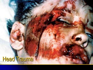
15. Scalp injuries
It is considered as minor head injury
Abrasion (brush wound)
Laceration- it is an external
head trauma which is
relatively small wound and
can bleed significantly.
32. INTRA CEREBRAL
HEMORRHAGE
IT IS BLEEDING INTO THE
SUBSTANCE OF THE BRAIN.
IT IS COMMONLY SEEN IN
HEAD INJURIES WHEN
FORCE IS EXERTED TO THE
HEAD OVER A SMALL AREA.
Eg: BULLET WOUND.
34. Brain suffers traumatic
injury
Brain swelling or bleeding
increases intra cranial volume
Rigid cranium allows no room for
expansion of contents so ICP
increases
Pressure on blood vessels within
the brain causes blood flow to the
brain to slow
35. ICP continues to raise.
Brian may herniate
Cerebral blood flow
ceases
Cerebral hypoxia and
ischemia occurs
Brain
Death
38. CONT..
Halo Sign: A blood stain
surrounded by a yellowish stain. It
may seen on bed linens or the
head dressings and is highly
suggestive of a CSF leak. Bloody
CSF suggests a brain laceration
or contusion.
39.
40. CLINICAL MANIFESTATIONS
OF BRAIN INJURY
Altered level of consciousness
Confusion
Pupillary abnormalities
Absent gag reflex
Absent corneal reflex
Sudden onset of neurologic
deficit
41. Cont..
Changes in vital signs
Vision and hearing
impairment
Sensory dysfunction
Headache
Vertigo
seizures
42. DIAGNOSIS
PHYSICAL AND NEUROLOGICAL
ASSESSMENT (particularly GCS)
X RAYS
C. T
M.R.I
POSITRON EMISSION
TOMOGRAPHY
LUMBAR PUNCTURE
44. PRE-HOSPITAL
MANAGEMENT
It refers to the immediate
resuscitation ,stabilization
,immobilization of a patient at
the scene of injury and en route
to the hospital,improvements in
airway management,fluid
replacement,and immobilization
of the neck in case of cervical
fracture and safe transport by
land or air
45. Major goals in the care of severely
head injury clients
Prompt recognition and
treatment of hypoxia and acid
base disorders that can
contribute to cerebral edema
Controlling of increased ICP
Stabilization of other
conditions
46. ARRIVAL IN THE
EMERGENCY DEPT:
.Quick history of the injury
.Base line vital signs
.Stabilization and support of vital signs
.Cervical neck assessment
.Triage neurological examination
.Control of seizures if any
.ICP management
.LAB data on blood
47. AFTER STABILIZATION
Additional information for history
Ongoing assessment of neurological
and vital signs
Comprehensive physical examinations
Radiological data-CT scan,X-rays
Tetanus vaccination
Antibiotics
Drainage of bladder and stomach
50. Surgical management
Craniotomy
Burr hole surgery- opening into
cranium to remove fluid and blood
beneath the dura.
Shunt procedure is an alternative
pathway to redirect CSF.
52. Controlling ICP in Severely
Brain-Injured Patients
Elevate the head of bed 30 degrees.
• Maintain the patient’s head and
neck in neutral alignment (no
twisting).
• Initiate measures to prevent the
Valsalva maneuver (eg, stool
softeners).
Maintain normal body temperature.
53. Administer O2 to maintain PaO2 > 90 mm
Hg.
Maintain fluid balance with normal saline
solution.
Avoid noxious stimuli (eg, excessive
suctioning, painful procedures).
Administer sedation to reduce agitation.
Maintain cerebral perfusion pressure > 70
mm Hg.
54. NURSING DIAGNOSES
Altered cerebral tissue perfusion related
to increased ICP
Ineffective breathing pattern rlt increased
ICP or brain stem injury
Altered nutrition less than body
requirement rlt compromised
neurological function and stress of injury.
55. Altered thought process rlt physiology of
injury
Risk for injury rlt altered thought
process
Ineffective family coping related to
unpredictability of out come.
56. Cont…
Ineffective airway clearance related to
coma or bleeding into the air way
altered cerebral tissue perfusion related to
hypotension ICH
impaired physical mobility related to motor
sensory deficits
57. Altered nutrition
altered urinary elimination
high risk for fluid volume deficit
sleep pattern disturbance
high risk for seizures
high risk for infection
altered thought process
altered health maintenance
62. PATIENT EDUCATION
Review with family the signs of increased ICP
Reinforce the ability pf cognitive , language
and physical functioning of the person with
brain injury and lengthy recovery period
Teach family therapeutic use of touch and
music to calm the patient
Provde them information regarding agencies
like BRAIN INJURY ASSOCIATION
66. CONTROL OF INCREASED ICP
Osmotic diuretics:-eg: Manitol
cortico steroids-eg:Dexamethasone
Elevation of head end to 30* angle
Hyper ventilation
Continuous CSF drainage
Prevent increase of ICP
Maintenance of normo-thermia
71. Community and home care
Observe for signs of post contusion
syndrome:- like
Had ache ,lack of concentration
Irritability,dizziness,insomnia
Restlessness,anxiety,
Easy fatigability,
Alcohol intolerance
72. Encourage family members to report
these symptoms
Act as laison in home care services the
pt will need,while keeping in touch with
the patients primary care provider
Provide necessary education to the
family members in
feeding,positioning,range of motion
exercises