
Recommended
More Related Content
Similar to TRAUMA-HYDROCHEPALUS-LAMINECTOMY.ppt
Similar to TRAUMA-HYDROCHEPALUS-LAMINECTOMY.ppt (20)
Recently uploaded
Recently uploaded (20)
TRAUMA-HYDROCHEPALUS-LAMINECTOMY.ppt
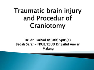
- 1. Traumatic brain injury and Procedur of Craniotomy Dr. dr. Farhad Bal’afif, SpBS(K) Bedah Saraf - FKUB/RSUD Dr Saiful Anwar Malang
- 2. ANATOMY and PHYSIOLOGY OF BRAIN The brain is one of the largest and most complex organs in the human body. It is made up of more than 100 billion nerves that communicate in trillions of connections called synapses. The brain is made up of many specialized areas that work together: The cortex is the outermost layer of brain cells. The basal ganglia are a cluster of structures in the center of the brain
- 4. SKULL The purpose of the bony skull is to protect the brain from injury. All the arteries, veins and nerves exit the base of the skull through holes, called foramina. The big hole in the middle (foramen magnum) is where the spinal cord exits.
- 6. Sutures of the Skull
- 7. Brain
- 8. The brain is composed of three parts: CEREBELLUM CEREBRUM. BRAINSTEM
- 11. MENINGES The brain and spinal cord are covered and protected by three layers of tissue called meninges. From the outermost layer inward they are:The Dura mater, Arachnoid mater, and Piamater.
- 12. Ventricles and Cerebrospinal fluid The brain has hollow fluid-filled cavities called ventricles. Inside the ventricles is a ribbon-like structure called the choroid plexus that makes clear colorless cerebrospinal fluid. CSF flows within and around the brain and spinal cord to help cushion it from injury. This circulating fluid is constantly being absorbed and replenished.
- 13. Nervous system The nervous system is divided into central and peripheral systems. The central nervous system (CNS) is composed of the brain and spinal cord. The peripheral nervous system(PNS) is composed of spinal nerves. That branch from the spinal cord and cranial nerves that branch from the brain.
- 14. Cranial nerves
- 15. Number Name Function I olfactory Smell II optic sight III oculomotor moves eye, pupil IV trochlear moves eye V trigeminal face sensation VI abducens moves eye THE TWELVE CRANIAL NERVES
- 16. VII facial moves face, salivate VIII vestibulocochlear hearing, balance IX glossopharyngeal taste, swallow X vagus heart rate, digestion XI accessory moves head XII hypoglossal moves tongue
- 17. Blood supply Blood is carried to the brain by two paired arteries, the internal carotid arteries and the vertebral arteries. The internal carotid arteries supply most of the cerebrum. The vertebral arteries supply the cerebellum, brainstem, and the underside of the cerebrum
- 19. Head Injury • Scalp injury • Skull injury • Meningeal injury • Traumatic Brain Injury
- 20. Epidemic in Indonesia Major cause of death and permanent disability 70% of all road fatalities 50% of trauma death 10-20% of head injury: death on arrival Degree 10% mild head injury 10% moderate head injury 80% severe head injury
- 21. Severity of primary injury Intracranial complications Hypoxaemia Hypercarbia Hypotension Anaemia Multiple injuries, proportional to Injury Severity Score (ISS) Age
- 22. Prolonged prehospital time Admission to inappropriate hospital Delayed or inappropriate interhospital transfer/retrieval Delay in definitive surgical treatment
- 23. Traumatic Brain Injury Blunt(Closed) Penetrating Explosion Fall GSW Stab Blast Fragment Motor vehicle crashes (MVC)
- 25. 1. Headache 2. Vomiting 3. Papilloedema occur in chronic condition 4. Cushing response ◦ Bradicardia ◦ Hipertension ◦ Alteration of ventilatory pattern
- 26. 5. Herniation • Uncal - ipsilateral dilated pupil - contralateral hemiparesis - ipsilateral hemiparesis (Kernohan’s notch phenomenom) • Central - rostrocaudal sign • Subfalcine • Tonsiller herniation
- 27. Mild – GCS 14-15 Patient typically mildly lethargic, disoriented Moderate – GCS 9-13 Patient typically sleepy or obtunded, able to follow commands with arousal. Confused. Severe – GCS 3-8 Patient comatose, unable to follow command or perform purposeful motor activity. Range of motor activity: localizes, withdraws, decorticate posturing, decerebrate posturing, nil.
- 28. Primary Injury: Function of energy transmitted to brain ◦ Very little can be done by health care providers to influence ◦ Cerebral concussion, contusion and degeneration, Diffuse Axonal Injury (DAI) Secondary Injury: Function of damage to brain from systemic physiology ◦ Systemic Hypotension: Acute and easily treatable Hypoxia: Acute and easily treatable Fever and Electrolyte Imbalances ◦ Seizures ◦ Intracranial Pressure Can Lead to Herniation
- 29. Primary Scalp contusion, abrasion, laceration Skull fracture open, closed (note-compound base of skull fracture without a scalp laceration), linier, depressed Meningeal injury dural tear Brain injury concussion contussion diffuse axonal focal – contusion laceration and penetration
- 30. Secondary Intracranial haemorrhage Cerebral swelling cerebral hypoxia CSF leakage and pneumocephalus methabolic disorders infection epilepsy
- 31. Factors influencing outcome - airway - breathing - control of haemorrhage - prevention and shock treatment - avoidance of factors ↑ ICP • head down position • hypoxia • hypercarbia • vomiting - recognition of serious associated injury - effective communication and transport
- 32. Lateral position for airway control in Px with susp spinal injury The face in turned slightly down words the tongue to fall forwards saliva and vomit will drain out
- 33. The indication: ◦ Airway is inadequate ◦ GCS ≤ 8 ◦ Herniation ◦ Rapid deterioration Should be performed only by a competent medical practitioner
- 34. Early management of severe trauma The management plan is based on: 1. Primary survey 2. Resuscitation 3. Secondary survey 4. Definitive care
- 35. Primary survey ◦ Airway with cervical spine immobilized in neutral position ◦ Breathing pattern and adequacy ◦ Circulation and haemorrhage ◦ Disability, minineurological examination: GCS Pupils Motor deficit ◦ Exposure: completely expose the patient for an adequate examination but protect against hypothermia
- 36. Resuscitation ◦ Airway Ensure patient airway Unconscious patient: intubated if skilled Note: maintain cervical spine immobilization until radiological examination excludes spinal injury ◦ Breathing and oxygenation Ensure adequate ventilation Mechanically ventilate if intubated Give supplemental oxygen initially
- 37. ◦ Circulation support and control haemorrhage Treat shock aggressively to improve tissue perfusion Control external haemorrhage ◦ Assess response to resuscitation using physiological parameters: pulse, blood pressure, skin colour, capilary refill and urine output ◦ Nasogastric tube and urinary catheter unless contraindicated
- 38. ◦ Head injury alone, without scalp injury, does not cause hypotension. If hypotension is present, identify the cause, e.g.: • Hypovolaemic shock, • spinal injury. • Rarely, may be due to medulallary failure. • Blood loss from a scalp or head injury may cause hypotension (hypovolaemic shock) in children
- 39. Secondary survey ◦ Special neurosurgical assessment including Glasgow Coma Score (GCS) and external sign of injury to the head ◦ Record the pulse, blood pressure, respiratory rate and temperature ◦ Systematically examine each region of the body, i.e. head-to-toe examination ◦ Connect to monitors as available ◦ Re-evaluate the GCS ◦ Radiological examination-lateral X-ray spine, chest, pelvis, other areas as indicated, skull X-ray and CT head scan ◦ History
- 40. CNS examination ◦ Glasglow Coma Scale (GCS) ◦ Pupillary responses are they equal or unequal? Were the pupils equal at the time of the incident (report from ambulance officer) and have they the same response now? ◦ Motor pattern hemiparesis, quadriparesis flexion or extension to pain (from supraorbital, trapezius or tendo achilles pressure) ◦ Inspection of the face and scalp ◦ Palpation of the face and scalp and any laceration for a depressed fracture ◦ Palpation of the spine for the tenderness and deformity
- 41. Glasgow Coma Scale (GCS) This scale examines three areas of behaviour: eye opening, response to voice and motor responses. The score can be quantitative with 3 being the lowest score and 15 normal
- 42. Eye opening ◦ Spontaneous E4 ◦ To speech 3 ◦ To pain 2 ◦ Nil 1 Verbal response ◦ Orientated V5 ◦ Confused conversation 4 ◦ Inappropriate words 3 ◦ Incomprehensible sound 2 ◦ Nil 1 Best motor response ◦ Obeys M 6 ◦ Localizes 5 ◦ Withdraws 4 ◦ Abnormal flexion 3 ◦ Extension 2 ◦ Nil 1 Coma Score (E+V+M) = 3-15
- 43. CT head scan guidelines ◦ GCS < 15 after resuscitation ◦ Neurological deterioration, i.e 2 point or more on the GCS, hemiparesis, squint ◦ Drowsiness or confusion (GCS 9-14 persisting>2 h) ◦ Persistent headache, vomiting ◦ Focal neurological signs ◦ Fracture – known or suspected ◦ Penetrating injury – known or suspected ◦ Age – over 50 years of age ◦ Post-operative assessment
- 44. Skull X-ray guidelines In rural area where a CT scan is not available or readily accessible, a plan skull X-ray can provide useful information. The pictures required are AP, lateral, Towne’s view and tangential to the point of impact for demonstrating a depressed fracture
- 45. Indications ◦ Loss of consciousness, amnesia ◦ Persisting headache ◦ Focal neurological signs ◦ Scalp injury ◦ Suspected penetrating injury ◦ CSF or blood from nose or ear ◦ Palpable or visible skull deformity ◦ Difficulty in clinical assessment ◦ Patient with GCS 15, essntially asymptomatic but “at risk” bacause of a defect blow or fall onto a hard surface, etc, especially in a patient over 50 years of age
- 46. Criteria for admission to hospital with head injury: • Confusion or any other decreased level of consciousness • Neurological symtoms or sign – including persistent headache, vomiting • Difficulty in clinical assessment, e.g. alcohol, epilepsy • Other medical condition, e.g. coagulation defects, diabetes mellitus • Skull fracture • Abnormal CT brain scan • Responsible observation not available outside the hospital • Age – patients over 50 years of age • Children under 5 years of age
- 47. Criteria for admission in Minor head injury ◦ A minor head injury is defined as one where the Glasgow Coma Score is 14-15 ◦ Admit and observe the patient if: a). There has been loss of consciousness or a period of post-traumatic amnesia b). The patient remains confused c). The patient is under 5 years of age or over 50 years of age d). Presence or development of focal neurological signs e). Severe headache with or without vomiting
- 48. Discharge of minor head injury patient ◦ Orientated in time and place ◦ No focal neurological signs ◦ No skull fracture ◦ A responsible person is available to continue observation of the patient ◦ Discharge check list – advise to report back to hospital immediately if there is: a) Vomiting b) Complains of severe headache or dizziness c) Becomes restless, drowsy or unconscious d) Had a convulsion or fit
- 49. Intubate & ventilate with GCS<9 the goal: PaO2=100mmHg, PaCO2 35mmHg, O2 sat 96%, hyperventilation (PaCO2< 30mmHg) should be avoided Cerebral perfusion the goal: CPP>70mmHg, MAP>90mmHg hypotension (systolic BP<90mmHg) must be avoided Intravenous fluid electrolites normovolaemic is the goal, avoid dehydration on or ever hydration normal serum electrolyte should be maintaned Head posture: should be elevated to 20-30° Corticosteroid: are not recommended Transfer to CT and / or neurosurgical unit
- 50. Active treatment of intracranial pressure Only be under taken of there is evidence of - rapid neurological deterioration - signs of uncal herniation - ↑ ICP from the ICP monitoring - modality (should be decided by neurosurgeon) • CSF drainage • intravenous mannitol • hyperventilation • barbiturate • mild hypothermia • decompressive craniectomy
- 51. Restlessness and analgesia o Before prescribing analgesia, it is important to determine the cause of restlestness, e.g.: cerebral hypoxia from airway inadequacy or poor ventilation or poor perfusion, raised intracranial pressure, pain, alcohol intoxication or a full bladder. oDrugs other than paracetamol or codeine phosphate require neurosurgical consultation
- 52. Post traumatic epilepsy The risk factors for epilepsy are: intradural haematomas, dural laceration with cortical injury, depressed fractures, a post-traumatic amnesia period of 24 h early post traumatic epilepsy The indication for prophylactic anti-convulsant therapy is controversial. A neurosurgical consultation is indicated both for the cause of the epilepsy and for consideration for anti- convulsant therapy.
- 54. Admit to ICU S/P TBI Cond: critical Vitals q1hr w/ neuro checks (if on Propofol, stop and check q4 hrs) Bedrest, HOB to 30*, loosen c-collar when patient sedated NPO IVF ◦ ½ NS w/ 20 K @ 80-100 cc/hr ◦ If significant brain edema, start 3%NS @ 15/hr, increase up to 50/hr (keep serum Na at 145-155, serum osmol 300-320) Vent ◦ No or low PEEP ◦ Keep PaCO2 at 30-35 (see hyperventilation above) Meds ◦ Propofol drip or Ativan 2-10mg iv q1hr for sedation or ICP>20 for>5’ ◦ MSO4 2-10 mg iv q1hr prn pain or ICP>20 for>5’ ◦ Mannitol 25g iv q4hrs prn ICP>20 for>5’ (hold if serum Na >155 or osmol>320) ◦ Cerebyx 1g iv now (loading dose), then 100mg q8 ◦ Pepcid 20mg iv bid ◦ Ancef 1g iv q8 if scalp wound or ICP monitor Nursing – per ICU routine Labs ◦ CBC, CMP, Dilantin level qAM ◦ Serum Na and osmol q6 if on 3% NaCl or Mannitol Repeat head CT in am (at least 2 CTs per patient, one on arrival and one next day) Call for problems
- 56. Absence of brainstem reflexes ◦ Fixed pupils ◦ Absent corneal reflexes ◦ Absent oculovestibular reflex (cold water calorics) ◦ Absent oculocephalic reflex (not if C-spine not cleared) ◦ Absent gag and cough reflex No response to deep central pain Apnea test (last test to perform!) Vital signs ◦ Core temp > 32.2*C (90*F) ◦ SBP>90 mm Hg No drugs in the system!
- 60. Treatment • Burr hole trephination. A hole is drilled in the skull over the area of the subdural hematoma, and the blood is suctioned out through the hole. • Craniotomy. A larger section of the skull is removed, to allow better access to the subdural hematoma and reduce pressure. • Craniectomy. A section of the skull is removed for an extended period of time, to allow the injured brain to expand and swell without permanent damage
- 61. craniotomy Craniotomy is a cut that opens the cranium.During this surgical procedure, bone flap, is removed to access the brain underneath.Craniotomies are often named for the bone being removed. Some common craniotomies include frontotemporal, parietal, temporal, and suboccipital.A craniotomy is cut with a special saw called a craniotome.
- 63. STEPS OF PROCEDURE There are 6 main steps craniotomy.. Step 1: prepare the patient Step 2: make a skin incision. Step 3: perform a craniotomy, open the skull Step 4: exposure the brain
- 64. Step 5: correct the problem Step 6: close the craniotomy
- 66. TREPANASI INSTRUMENT Handvat mess no. 3 (Scalp blade and handle) : 2 buah Handvat mess no. 7 (Scalp blade and handle) : 1 buah Nald voeder (Needle holder) : 2 buah Gunting metzembaum (Metzemboum scissor) : 1 buah Gunting benang (Surgical scissor ) : 1 buah Gunting mayo/kasar (Surgical scissor straight) : 1 buah Gunting duramater : 1 buah Pincet anatomis (Tissue forceps) : 2 buah Pinset chirurgis (Dissecting forceps) : 2 buah Pinset bebek (Adson forceps) : 2 buah Disinfeksi klem (washing and dressing forcep) : 1 bauh Duk klem (towel klem) : 5 buah Klem pean cantik (nissen) : 1 buah Bipolar / monopolar couter : 1 set Cannule suction : 2 buah Langenbeck retractor : 2 buah Sharp retractor : 2 buah Raney scalp hemostatic clips : 15 buah Raney aplikator removing forceps : 2 buah Dissector : 1 buah Elevator : 1 buah Raspatories : 1 buah Handle gigli saw : 2 buah Conductor gigli saw : 1 buah Brain spatula : 4 buah Leyla retractor : 2 buah
- 67. CRANIOPLASTY INSTRUMENT Handvat mess no. 3 (Scalp blade and handle) : 2 buah Nald voeder (Needle holder) : 2 buah Gunting metzembaum (Metzemboum scissor) : 1 buah Gunting benang (Surgical scissor ) : 1 buah Gunting mayo/kasar (Surgical scissor straight) : 1 buah Pincet anatomis (Tissue forceps) : 2 buah Pinset chirurgis (Dissecting forceps) : 2 buah Disinfeksi klem (washing and dressing forcep) : 1 bauh Duk klem (towel klem) : 5 buah Klem pean cantik (nissen) : 1 buah Bipolar / monopolar couter : 1 set Cannule suction : 1 buah Langenbeck retractor : 2 buah Sharp retractor : 2 buah Raney scalp hemostatic clips : 15 buah Raney aplikator removing forceps : 2 buah Elevator : 1 buah Raspatories : 1 buah Kikir (bone levers) : 1 buah Spatula : 1 buah Screwdriver : 1 buah
- 68. Quantity Name Description Size 2 Jansen Retractor Blunt 3x3 Blades 4" 2 Weitlaner Retractor Blunt 3x4 Teeth 6-1/2" 1 Scalpel Handle #3 1 Scalpel Handle #4 1 Scalpel Handle #7 4 Solid Bar Handle For Gigli Saw Pack of 2 2 Adson (Ewald) Dressing Forceps Serrated 4-3/4" 2 Adson Tissue Forceps 1x2 Teeth 4-3/4" 12 Backhaus Towel Clamp 5-1/4" 2 Cushing Brain Forceps Delicate Serrated 7" 2 Cushing Brain Forceps Delicate 1x2 Teeth 7" 6 Ruskin Rongeur Straight 7-1/4" 6 Foerster Sponge Forceps Serrated 9-1/2" 18 Halsted Mosquito Forceps Straight 5" 18 Halsted Mosquito Forceps Curved 5" 1 Luer Bone Rongeur Curved 8mm x 10mm Bite 7" 1 Stille-Liston Bone Forceps Curved Double Action 10-1/2" 2 Mayo-Hegar Nh Serrated 7" 1 Gigli Saw Wire 12" 1 Gigli Saw Wire 20" 1 Operating Scissors Straight Sharp/Blunt 6" 1 Mayo-Stille Dissecting Scissors Straight 6-3/4" 1 Mayo-Stille Dissecting Scissors Curved 6-3/4" 1 Metzenbaum Dissecting Scissors Curved 7" 1 Taylor Dural Scissors w/ Probe Tip 5-1/2" 1 Cover for Instrument Tray
- 72. Labs: CBC, lytes, Cr, INR/PTT Crossmatch (at least 2 U PRBC) 2 physician consent Spinal precautions ◦ May need to log roll patient
- 73. Scalp Wounds 1. shave at least 3 cm around the wound 2. gently palpate the laceration with a gloved finger. This may provide information regarding an underlying fracture 3. if a fracture is found unexpectedly, do not remove bone fragments: contact your neurosurgeon at once. 4. Scalp wounds may bleed profusely and cause hypertension. Secure haemostasis by pressure or suturing early 5. if the wound edges are badly torn, excise non – viable scalp and where possible suture the scalp in two layers
- 75. Seldom used by neurosurgeons in CT era Position supine Shave entire head & drape to allow access to frontal, parietal, & temporal areas Burr holes typically on side of localizing neuro findings – ipsilateral to dilated pupil or skull fracture, contralateral to abN motor response ◦ If no hematoma found on suspected side, other side should be explored Initially burr hole placed in temporal region 2.5 cm above zygomatic arch Following dx of either ASDH or EDH, 2 additional burr holes can be appropriately placed in parietal & frontal regions Skin incision should be made in such a manner that if formal craniotomy required, they can be joined to form skin flap
- 76. Scalp Incision ◦ Large question mark incision starting 1 cm in front of tragus at zygomatic arch & curved backward & upward above auricle to reach midline, carried forward to frontal region ◦ Raney clips along skin edges ◦ Bovie incision in superficial temporal fascia & temporalis muscle down to the bone, close to margin of skin opening ◦ Myocutaneous flap reflected inferiorly Rapid Temporal Decompression ◦ In patient who is herniated or is deteriorating, temporal end of incision rapidly opened & burr hole placed ◦ Burr hole is then enlarged to form a limited craniectomy 3 cm in diameter ◦ If hematoma in subdural space, dura is opened in a cruciate manner & underlying hematoma is promptly evacuated – helps to reduce ICP before completion of craniotomy which can then be completed more slowly, w/ better hemostasis
- 77. Bone Flap ◦ Burr holes, then join burr holes to complete craniotomy opening ◦ Medial margin 1.5~2 cm from midline ◦ Further exposure of middle fossa obtained using Leksell rongeurs to remove parts of lateral sphenoid wing & temporal bone in piecemeal fashion, as needed for temporal tip access Dural Opening ◦ Opening in U-shaped fashion & flap towards midline to avoid damaging parasagittal bridging veins ◦ Alternatively, cruciate opening Closure ◦ Meticulous hemostasis ◦ Dural tack-up sutures 2.5 cm apart in circumferential fashion & central tack-up suture in bone flap ◦ +/- drain
- 85. QUESTION ?
- 86. Dr. dr. Farhad Bal’afif, SpBS(K) Bedah Saraf – FKUB / RSUD Dr Saiful Anwar Malang
- 87. Hydrocephalus is the medical term for a condition that is commonly called “water on the brain.” It is a combination of the Greek word “hydro,” which means water and “cephalus” which means head. However, the liquid involved in hydrocephalus is not really water at all, it is cerebrospinal fluid or CSF.
- 88. CSF looks like water, but it contains proteins, electrolytes, and nutrients that help keep your brain healthy. The most important purpose of CSF is to cushion your brain and spinal cord against injury. Your brain produces about 1 pint of CSF per day. It circulates through a network of tiny passageways in your brain, and ultimately into your blood stream where it is absorbed by your body.
- 89. Hydrocephalus occurs when the delicate balance of CSF production and absorption is disrupted and CSF builds up in the brain. This build-up of CSF causes the brain to swell, and for pressure to increase inside the skull, resulting in nerve damage.
- 90. People who are born with hydrocephalus have a type of hydrocephalus called congenital hydrocephalus. It is usually caused by a birth defect or by the brain developing in such a way that the cerebrospinal fluid (CSF) in the brain cannot drain properly. Most cases of hydrocephalus (more than 70%) occur during pregnancy, at birth, or shortly after birth. Causes of congenital hydrocephalus include: Toxoplasmosis (an infection from eating undercooked meat, or by coming in contact with infected soil or an infected animal) Cytomegalovirus (CMV, infection by a type of herpes virus) Rubella (German measles) A genetic disorder usually passed only from mother to son
- 91. Hydrocephalus can also develop later in life. This type of hydrocephalus is called acquired hydrocephalus, and it can occur when something happens to prevent the CSF in the brain from draining properly. Causes of acquired hydrocephalus include: Blocked CSF flow Brain tumor or cyst Bleeding inside the brain Head trauma Infection (such as meningitis)
- 92. A shunt is a piece of soft, flexible plastic tubing that is about 1/8- inch (3mm) in diameter. It allows excess cerebrospinal fluid (CSF) that has built-up inside the skull to drain out into another part of the body, such as the heart or abdomen. To drain excess CSF, shunts are inserted into an opening or pouch inside the brain called a ventricle, just above where the blockage is that is preventing the CSF from flowing properly.
- 93. All shunts perform two functions. They allow CSF to flow in only one direction, to where it is meant to drain. They all have valves, which regulate the amount of pressure inside the skull. When the pressure inside the skull becomes too great the valve opens, lowering the pressure by allowing excess CSF to drain out.
- 94. A ventriculo-peritoneal (VP) shunt drains into the abdomen or peritoneum (belly). Most shunts, including Sean’s, are VP shunts. A ventriculo-pleural shunt drains into the space surrounding the lung. A ventriculo-atrial (VA) shunt drains into the atria of the heart. Shunts are named according to where they are inserted in the brain and where they are inserted to let the excess CSF drain out.
- 95. Ventricular (Upper) Catheter- This is the top-most part of the shunt. It is a small, narrow tube that is inserted into the ventricle (a small opening or pouch) inside the brain that contains the cerebrospinal fluid (CSF). Reservoir-This is where the excess CSF is collected until it drains into the bottom portion of the shunt. The reservoir also lets the doctor remove samples of CSF for testing, and to inject fluid into the shunt to test for flow and to make sure the shunt is working properly.
- 96. Valve-This controls how much CSF is allowed to drain from the brain. The valve can be set to open at a specific pressure (a fixed pressure valve) or It can be set by the neurosurgeon to meet the individual needs of the person with hydrocephalus (a programmable valve). Lower Catheter-This is the bottom-most part of the shunt. It is a small, narrow tube that carries the excess CSF into the part of the body where it will be absorbed, such as into the abdomen or the heart.
- 97. VP SHUNT INSTRUMENT Handvat mess no. 3 (Scalp blade and handle) : 2 buah Handvat mess no. 7 (Scalp blade and handle) : 1 buah Nald voeder (Needle holder) : 2 buah Gunting metzembaum (Metzemboum scissor) : 1 buah Gunting benang (Surgical scissor ) : 1 buah Gunting mayo/kasar (Surgical scissor straight) : 1 buah Pincet anatomis (Tissue forceps) : 2 buah Pinset chirurgis (Dissecting forceps) : 2 buah Pinset bebek (Adson pinset chirurgis) : 2 buah Disinfeksi klem (washing and dressing forcep) : 1 bauh Duk klem (towel klem) : 5 buah Mosquito klem pean (baby mosquito klem pean) : 5 buah Mosquito klem pean dengan karet pelindung : 2 buah Klem pean cantik (nissen) : 1 buah Bipolar couter : 1 set Cannule suction : 1 buah Raspatorium : 1 buah Dissektor : 1 buah Langenbeck retraktor : 2 buah Spreider abdomen : 1 buah Knable tang : 1 buah Penggaris steril : 1 buah Spanner (Guide peritoneal cathether) : 2 buah Bor Perforator : 1 set
- 98. Small incisions are made on the head and in the abdomen (in case of a VP shunt) to allow the neurosurgeon to pass the shunt's tubing through the fatty tissue just under the skin. A small hole is made in the skull, opening the membranes between the skull and brain to allow the upper catheter to be passed through the brain and into the ventricle.
- 99. The lower catheter is passed into the belly through a small opening in the lining of the abdomen where the excess CSF will eventually be absorbed. The incisions are then closed and sterile bandages are applied.
- 100. It contained only the upper portions of the shunt: Ventricular Catheter Reservoir A small needle was used to pierce the skin and tap into the reservoir (a plastic bulb). A syringe was then used to pull excess CSF from around the brain and relieve pressure. ANCHOR to suture in place RESERVOIR: Needle inserted here to drain VENTRICULAR CATHETER
- 101. Shunt surgery is the most effective treatment for hydrocephalus. By draining excess cerebrospinal fluid (CSF) from the brain, shunt surgery reduces pressure inside the skull lowers the risk of central nervous system damage, and relieves the symptoms associated with hydrocephalus.
- 102. QUESTION ?
- 103. Dr. dr. Farhad Bal’afif, SpBS(K) Bedah Saraf – FKUB / RSUD Dr Saiful Anwar Malang
- 104. Spinal cord lies within protective covering of vertebral column. Begins just below foramen magnum of the skull. Ends opposite 2nd lumbar vertebra. Below L2 continue as a leash of nerve roots known as cauda equina. Prolongation of the pia matter forms filum terminale.
- 106. • Anterior Elements: •Vertebral body: provide bulk and height; Sustain compression loads. • Middle Elements: • Pedicles: transfer forces from posterior to anterior elements. • Posterior Elements: •Articular processes and facet jts, laminae, spinous processes. •Lock spine to prevent forward sliding and twisting; Insertion sites for muscle
- 107. congenital; spinal stenosis. Infection; TB ,abscess. Trauma; vertebral body fracture or facet joint dislocation. Inflammatory; Rheumatoid arthritis. Disc and vertebral lesion. Vascular; epidural & SDH Tumors.
- 108. 75% of cases of spinal stenosis occur in the low back ( lumbar spine). Causes : - congenital. - degenerative. - trauma.
- 109. aging process (most common cause ). herniated discs. (fig) bone and joint enlargement. spondylolisthesis. bone spurs.
- 110. Initial Tx in most cases is conservative. ◦ Rest. ◦ Weight loss. ◦ Epidural steroid injections. ◦ Analgesia. ◦ Anti-inflammatory agents. ◦ Muscle relaxant -if needed- ◦ Physiotherapy.
- 111. Spine surgery: used when conservative treatment failed. -laminectomy (removing bone behind the spinal cord) -foramenotomy (removing bone around the spinal nerve). -discectomy (removing the spinal disc to relieve pressure). Complications: Dural tears. Infections. Instability of the spine.
- 112. Epidural abscess ◦ Usually bacterial ( staphylococcus is common). ◦ Spread through: hematogenous Adjacent focus. Direct inoculation.
- 113. •immunodeficiency •AIDS. •Alcoholism. •Chronic renal failure. •Diabetes mellitus. •Intravenous drug abuse. •Malignancy. •Spinal procedure or surgery. •Spinal trauma.
- 114. Infection of spine ◦ Uncommon ◦ Either vertebral osteomyelitis Or less commonly intraspinal infection. ◦ Causative organism : (staph, Strep, E.coli, TB) ◦ Occasionally due to unusual organisms like: Salmonella or brucella.
- 115. The goals of treatment are to relieve spinal cord compression and cure the infection. ◦ drain abscess. ◦ antibiotics or antimicrobial. ◦ corticosteroid. ◦ may need urgent surgical decompression by laminectomy.
- 116. Tumors are classified into 3 types according to their site: -extradural ( between the meninges and spine bones) -intradural extramedullary (within meninges) -intramedullary ( inside the cord)
- 117. Most spinal tumors are extradural (85%) They may be primary tumors originating in the spine, or secondary tumors that are the result of the spread of cancer from other locations primarily the lung, breast, prostate, kidney, or thyroid gland. Any type of tumor may occur in the spine, including lymphoma, leukemic tumors, myeloma, and others. A small percentage of spinal tumors occur within the nerves of the spinal cord itself, most often consisting of ependymomas and other gliomas.
- 118. Plain X-rays. Myelography “contrast material is injected into the thecal sac fluid surrounding the spinal cord and nerve root within the spinal canal” CT. MRI ( study of choice ).
- 119. The goal of treatment is to reduce or prevent nerve damage from compression of the spinal cord, relieve pain and maintain the function. - Surgical excision is the treatment for extramedullary tumors. - Radiation therapy for intramedullary tumors. The traditional treatment of intramedullary gliomas has been biopsy followed by radiation therapy. Radiotherapy is clearly of value in metastatic lesions. - Chemotherapy can be considered in patients with progression of disease after radiation therapy.
- 120. The act of exerting an abnormal amount of pressure on the spinal cord. Causes and risk factors : - Traumatic injury. - Spinal cord tumors. - Spinal stenosis. - Ruptured disks. - Abscesses. - Arteriovenous malformations. - Degenerative diseases, such as arthritis.
- 121. Thoracic 70% Lumbosacral 20% Cervical 10% Spinal Cord Compression in Three Main Areas
- 122. Symptoms vary depending on the cause of the compression, its location, severity, extent and rate of development but can include: - Back pain at the spinal site of compression. - Pain or burning in other parts of the body. - Difficulty breathing. - Weakness in the arms, legs, or both. - Numbness or tingling in the neck, shoulder, arms, hands, or legs. - Loss of coordination or difficulty walking. - Loss of fine motor skills. - Loss of sexual function. - Loss of bladder or bowel control. - Paralysis.
- 123. Cauda equina syndrome; is a serious condition caused by compression of the nerves in the lower portion of the spinal canal . is considered a surgical emergency because if left untreated it can lead to permanent loss of bowel and bladder control and paralysis of the legs.
- 124. X ray. CT scan. MRI. Myelogram. Biopsy. Bone scan. Blood and spinal fluid studies.
- 125. Acute cord compression is a 'surgical' emergency. In those with malignant disease radiotherapy may be treatment of choice. In general, tumor, infection and disc disease produces anterior compression. Surgical decompression should be achieved through an anterior approach.
- 126. Spinal cord trauma is damage to the spinal cord. It may result from direct injury to the cord itself or indirectly from damage to surrounding bones, tissues, or blood vessels. Symptoms: - Symptoms vary depending on the location of the injury. - Spinal cord injury causes weakness and sensory loss at and below the point of the injury. - we can divide spinal trauma into 3 levels according to its location in the spinal cord ( cervical - thoracic – Lumbosacral ).
- 127. - When spinal cord injuries occur near the neck, symptoms can affect both the arms and the legs: Breathing difficulties (from paralysis of the breathing muscles). Loss of normal bowel and bladder control (may include constipation, incontinence, bladder spasms). Numbness. Sensory changes. Spasticity (increased muscle tone). Pain. Weakness, paralysis.
- 128. - When spinal injuries occur at chest level, symptoms can affect the legs: Breathing difficulties (from paralysis of the breathing muscles) Loss of normal bowel and bladder control (may include constipation, incontinence, bladder spasms). Numbness. Sensory changes. Spasticity (increased muscle tone). Pain. Weakness, paralysis. Injuries to the cervical or high-thoracic spinal cord may also result in blood pressure problems, abnormal sweating, and trouble maintaining normal body temperature.
- 129. - When spinal injuries occur at the lower- back level, varying degrees of symptoms can affect the legs: Loss of normal bowel and bladder control (may include constipation, incontinence, bladder spasms). Numbness. Pain. Sensory changes. Spasticity (increased muscle tone). Weakness and paralysis.
- 130. A CT scan or MRI of the spine may show the location and extent of the damage and reveal problems such as blood clots (hematomas). Myelogram (an x-ray of the spine after injection of dye) may be necessary in rare cases. Somatosensory evoked potential (SSEP) testing or magnetic stimulation may show if nerve signals can pass through the spinal cord. Spine x-rays may show fracture or damage to the bones of the spine.
- 131. ABC Spine Immobilization to prevent further injury to the spinal cord. In cervical injuries higher than C5, intubation and respiratory support are usually needed. Corticosteroids, rest, analgesics and muscle relaxant. Surgery (decompression laminectomy ). Extensive physical therapy and other rehabilitation interventions are often required after the acute injury has healed.
- 132. EXTRICATION : GOOD WELL TRAINED TEAM WORK PREVENT FURTHER INJURIES SCOOP STRETCHER IS SAFEST TRANSPORT : ONCE STABILIZED REFER TO LEVEL 1 TRAUMA CENTRE TRENDELENBURG POSITION LONG JOURNEY CONSIDER : NGT, IV LINE, URINARY CATETHER
- 133. CME UI ‘05
- 134. CME UI ‘05
- 135. THE IN-HOSPITAL MANAGEMENT EVALUATION OF A, B & C PaO2 > 100 mmHg and PaCO2 < 45 mmHg MAINTAIN BP > 90 mmHg TREAT NEUROGENIC SHOCK !
- 136. Rupture of the disc or prolapse as it is usually called, can press on the spinal cord and its nerve roots leading to pain, numbness and weakness and may also affect the control of bowel and urinary bladder. Dx: X-ray, CT scan or MRI.
- 137. Initial Tx in most cases is conservative. ◦ Rest. ◦ Analgesia. ◦ Anti-inflammatory agents. ◦ Muscle relaxant -if needed-. ◦ Physiotherapy.
- 138. laminectomy, involves excision of a portion of the lamina and removal of the protruding disk. spinal fusion, may be necessary to overcome segmental instability. Laminectomy and spinal fusion are sometimes performed concurrently to stabilize the spine. Microdiskectomy, can also be used to remove fragments of nucleus pulposus. Chemonucleolysis: Injection of the enzyme chymopapain into the herniated disk produces a loss of water and proteoglycans from the disk, thereby reducing both the disk’s size and the pressure in the nerve root.
- 139. Spondylolisthesis is a condition in which the there is a defect in a portion of the spine, causing vertebra to slip to one side of the body.
- 140. Non-surgical treatment may include one or a combination of: - NSAID’s (e.g. ibuprofen, COX-2 inhibitors) - Oral steroids - Physical therapy - Manual manipulation (e.g. chiropractic manipulation). Spinal fusion surgery.
- 141. Early compression → minimal symptom / sign MRI is very sensitive for soft tissue Early Dx and Tx → better result Prevent secondary damage Suspected of spinal cord / root compression should reffered to neurosurgeon as early as possible
- 142. Indications : - compression syndrome (acute/chronic) - progressif neurologic deficit - instability - debridement Approaches : - anterior - posterior - endoscopic
- 143. Dewasa : Hb > 10 gr %, anak2: Hb> 12 g% FH normal dan lab lain normal Daerah yg akan dioperasi harus bersih ( mandi) inform concent
- 144. KULIT KOMBINASI DENGAN LIDOKAIN CARA MENYUNTIK : SUBKUTAN DOSIS MAKSIMAL : ADRENALIN 0,25 MG LIDOKAIN 4 MG/KG/KALI CARA MEMBUAT ◦ 1 AMPUL = 1 CC = 1/1000 ◦ 1 CC + 9 CC AQUA = 10 CC 1/10.000 ◦ 10 CC (1/10.000) DIAMBIL 1 CC + 9 CC AQUA = 10 CC ADRENALIN 1/100.000 ◦ 5 CC LIDOKAIN 2% + 5 CC AQUA + 10 CC ADRENALIN 1/100.000 ADR 1/200.000, LIDOKAIN 0,5%
- 145. SPINE INSTRUMENT (LAMINECTOMY) Handvat mess no. 4 (Scalp blade and handle) : 1 buah Handvat mess no. 7 (Scalp blade and handle) : 1 buah Nald voeder (Needle holder) : 2 buah Gunting metzembaum (Metzemboum scissor) : 1 buah Gunting benang (Surgical scissor ) : 1 buah Gunting mayo/kasar (Surgical scissor straight) : 1 buah Pincet anatomis (Tissue forceps) : 2 buah Pinset chirurgis (Dissecting forceps) : 2 buah Disinfeksi klem (washing and dressing forcep): 1 buah Canule suction : 2 buah Bipolar / monopolar couter : 1 set Duk klem (towel klem) : 5 buah Klem pean cantik (nissen) : 1 buah Langenbeck : 2 buah Gelpi ( Spesial retractors) : 2 buah Sprider (Laminectomy retractors) : 1 buah Dissector : 2 buah Kop / Chissel (Dissector) : 2 buah Nerve Hook : 2 buah Nerve root exploration : 1 buah Caspar rongeur straight/upwards/downwards : 3 buah Kerrison bone punch no.2/3/4 : 3 buah Knable tang : 1 buah Bone cutting : 1 buah
- 146. Spine Surg set
- 150. Posisi Desinfeksi Batasi daerah operasi dengan kain steril Sayatan kulit sesuai lokasi Rawat perdarahan kulit Pasang peregang kulit (SPREIDER) tajam, tumpul Spinosus dipisahkan dari otot Dilakukan laminotomi , laminectomi , disectomi sesuaikebutuhan Perdarahan dirawat TUTUP
- 156. Mikrodiskektomi Laminotomi Laminektomi
- 160. Laminectomy Vertebra Th2 – 5 + total eksisi intradural ekstramedular tumor ekstradural tumor at vertebra Th3,4
- 167. SUB AXIAL CERVICAL SPINE INJURIES CME UI ‘05
- 168. SUB AXIAL CERVICAL SPINE INJURIES CME UI ‘05
- 169. SUB AXIAL CERVICAL SPINE INJURIES CME UI ‘05
- 170. SUB AXIAL CERVICAL SPINE INJURIES CME UI ‘05
- 171. QUESTION ?