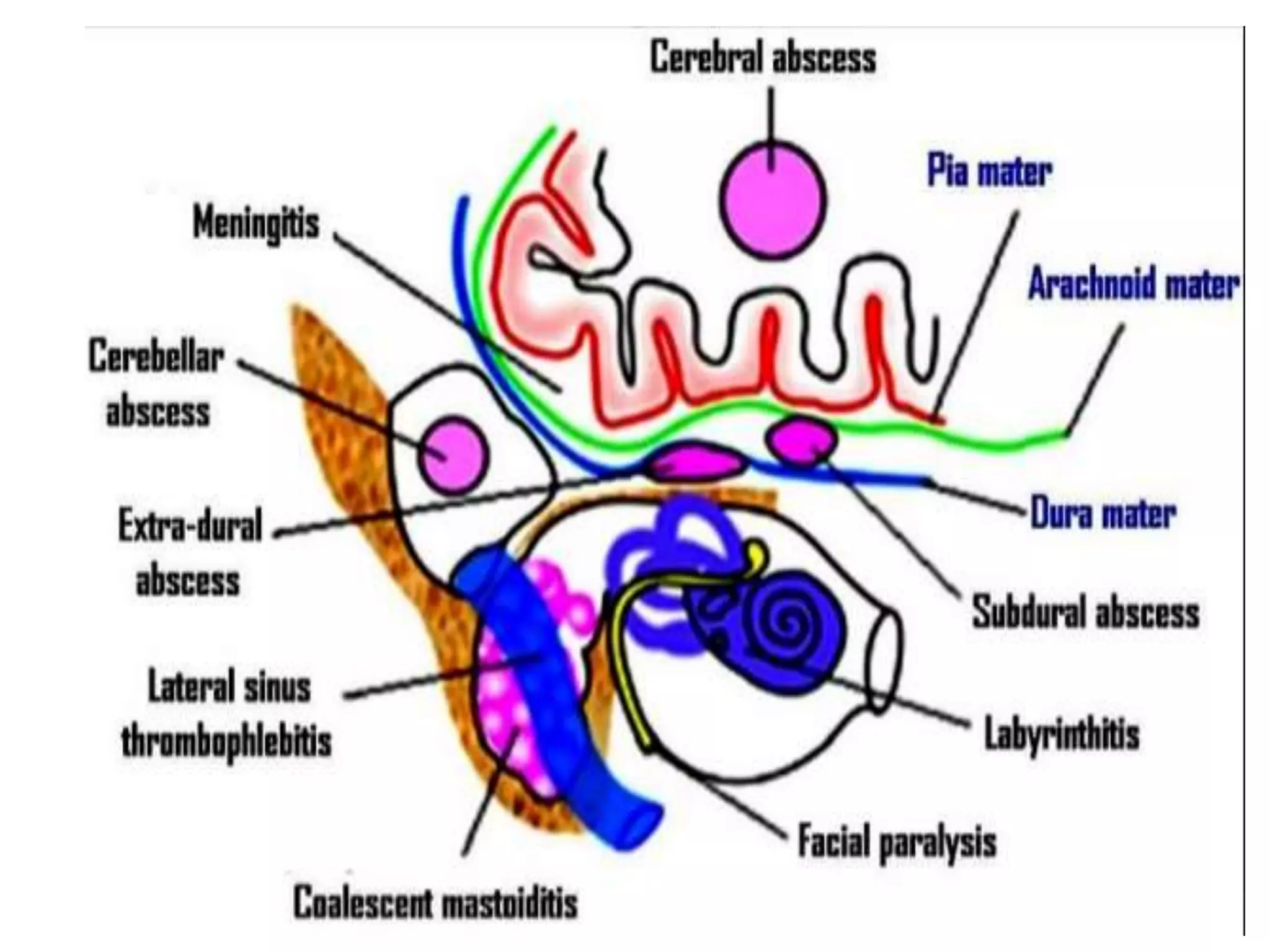
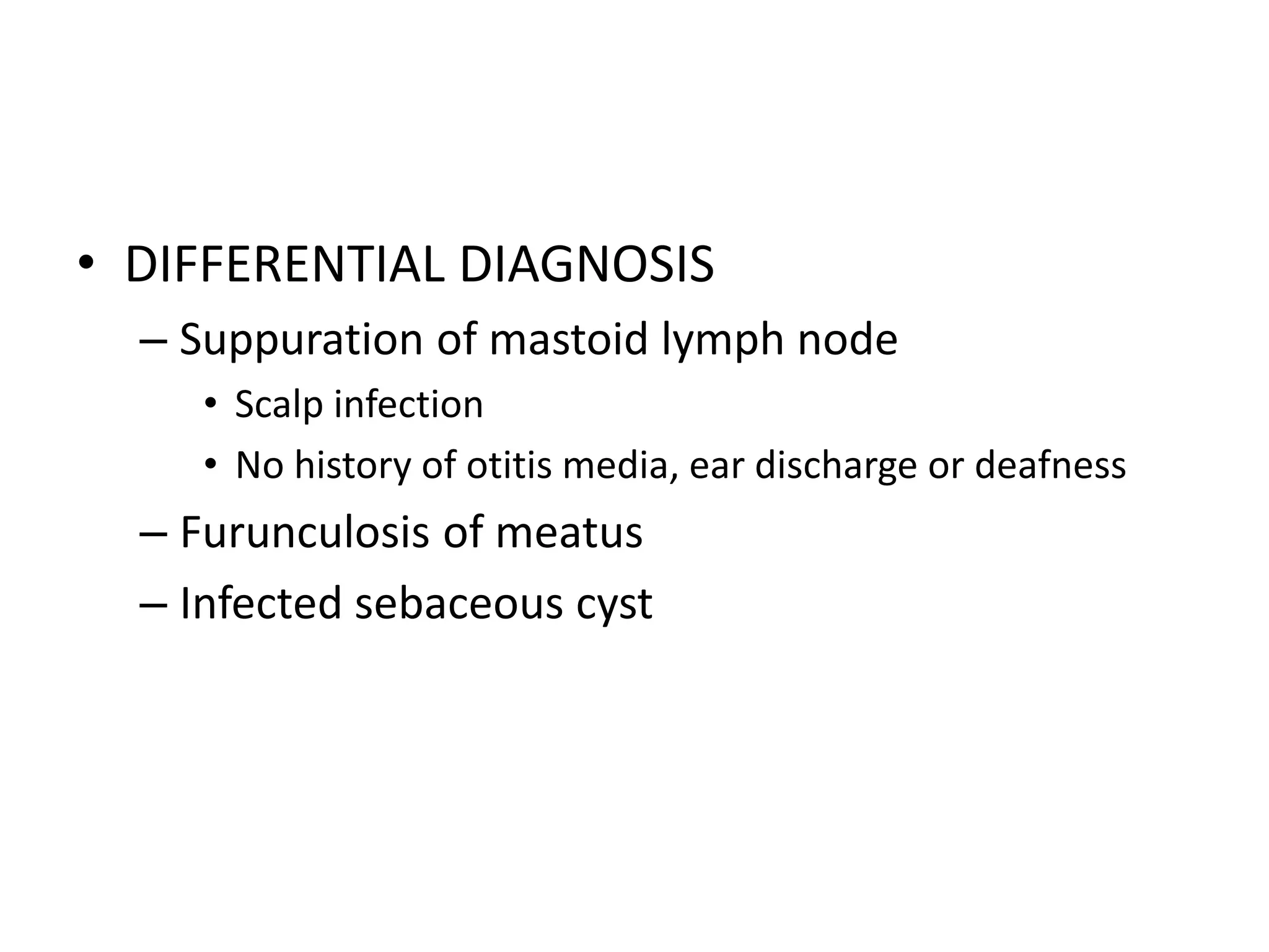
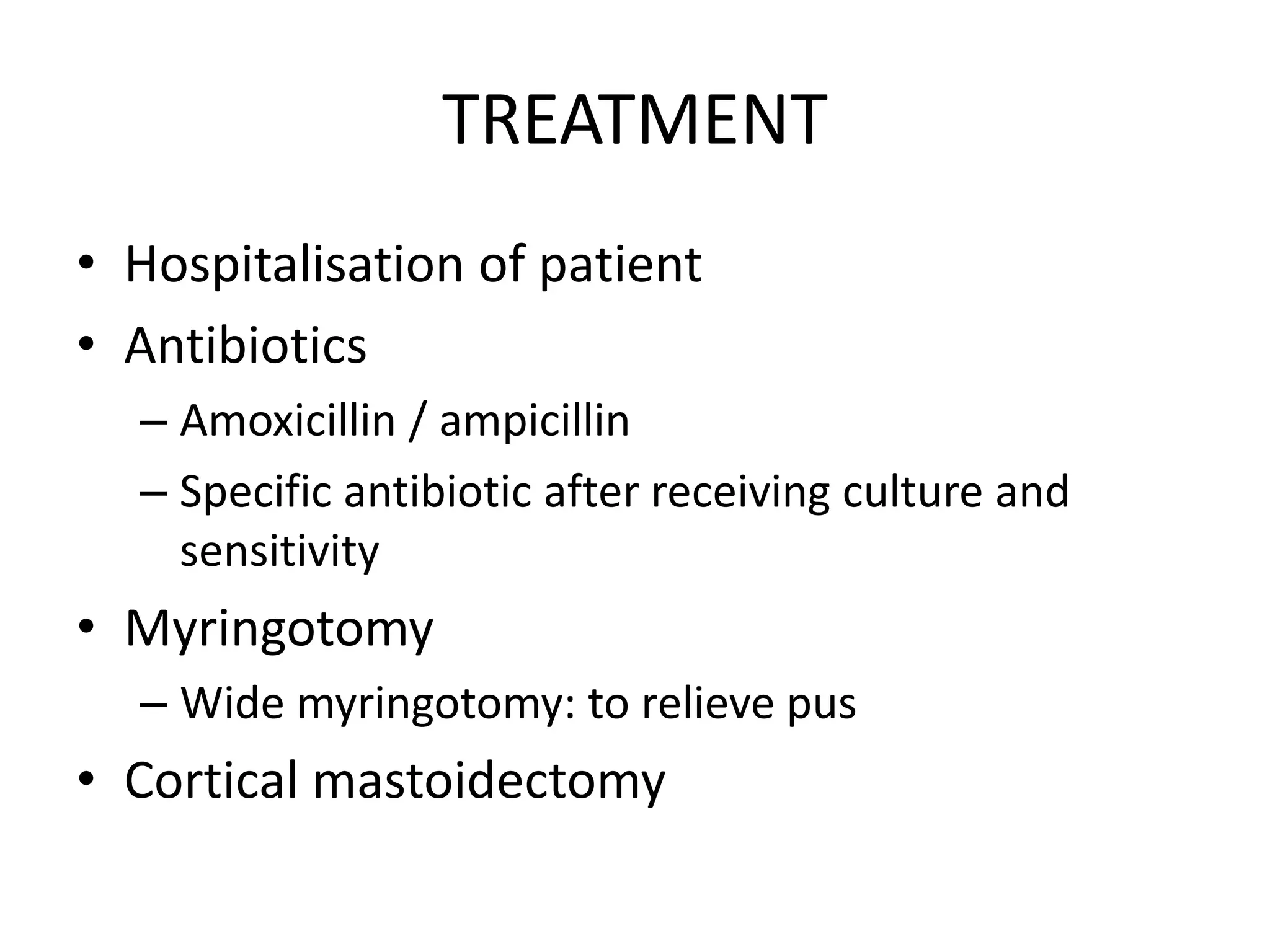
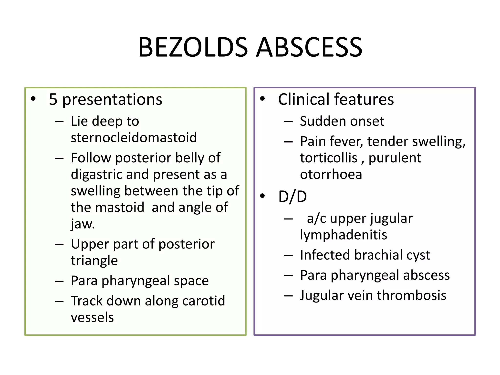
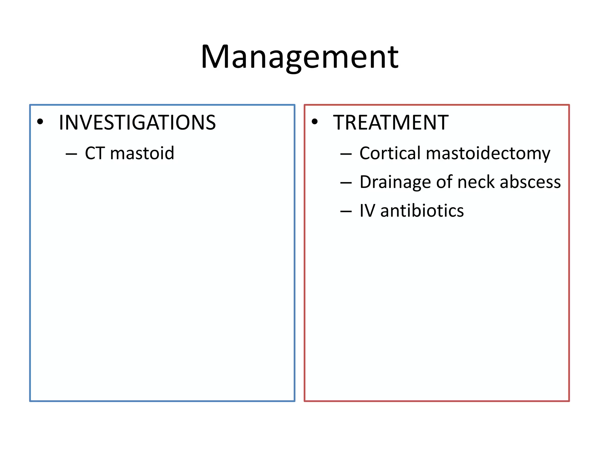
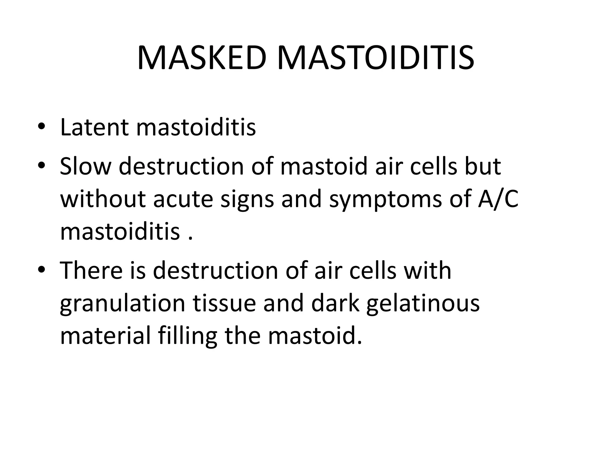
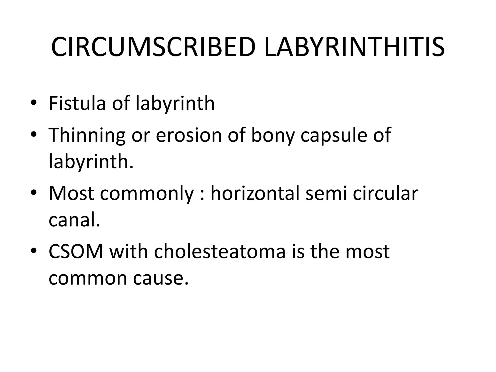
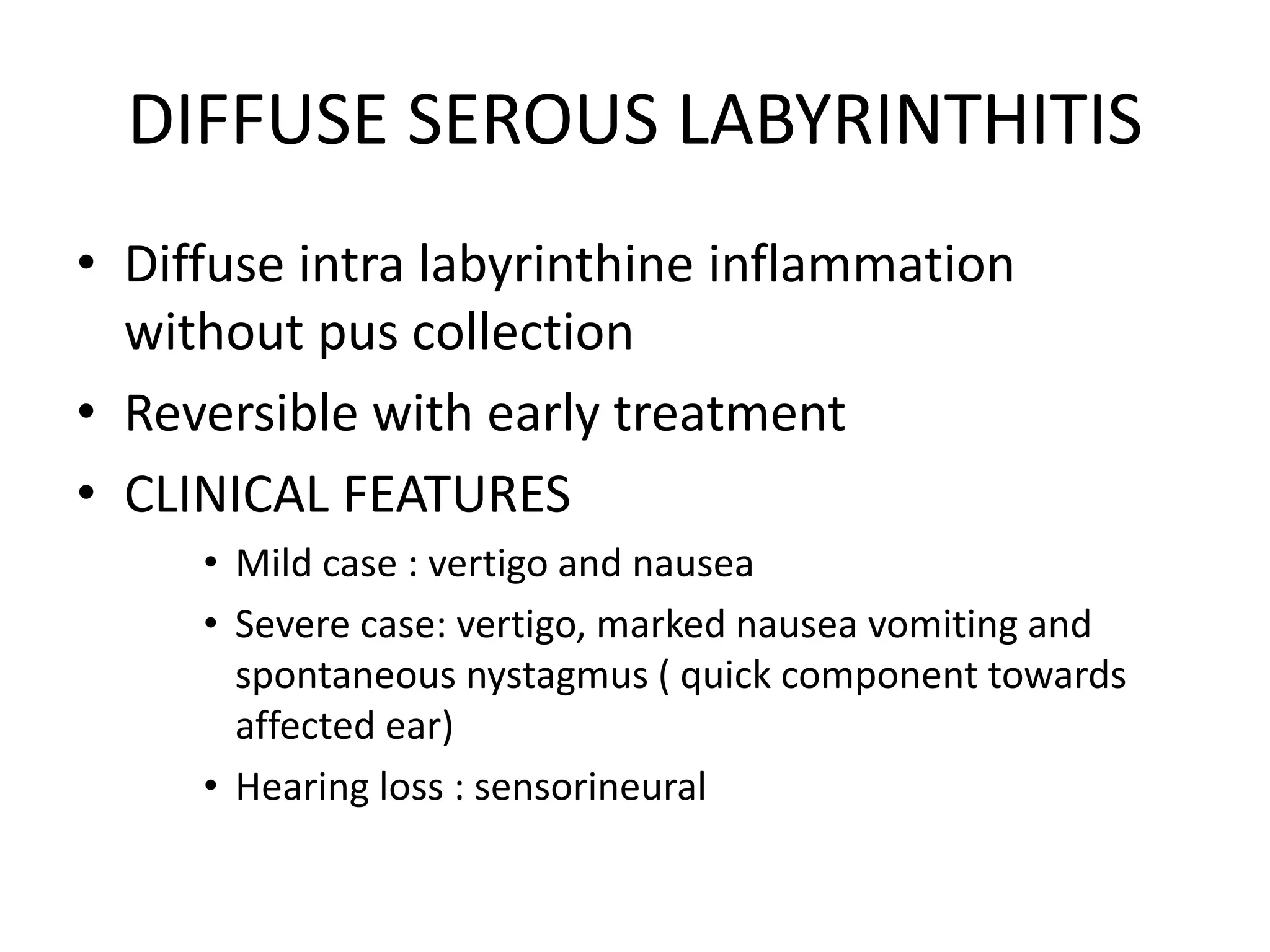
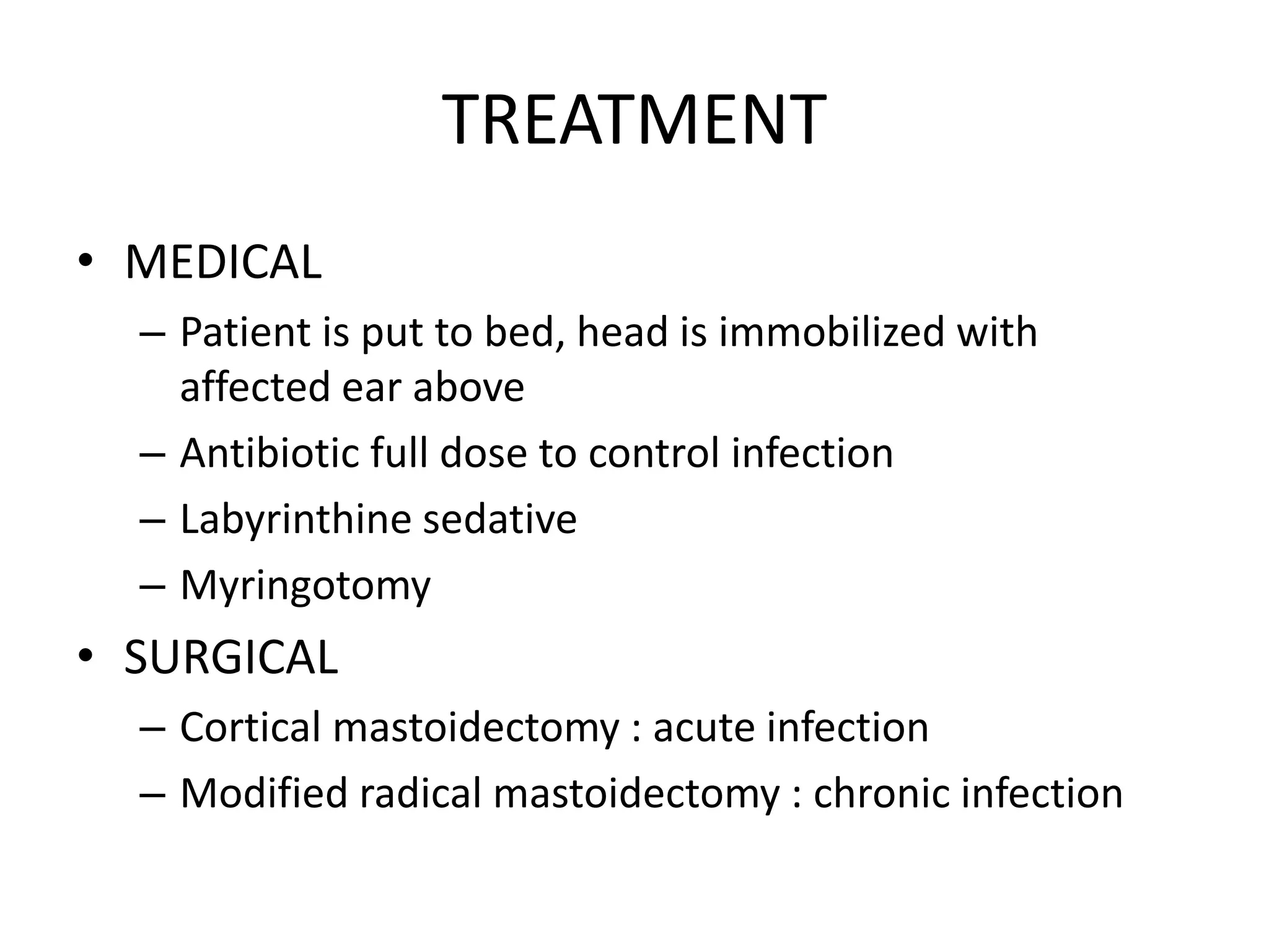
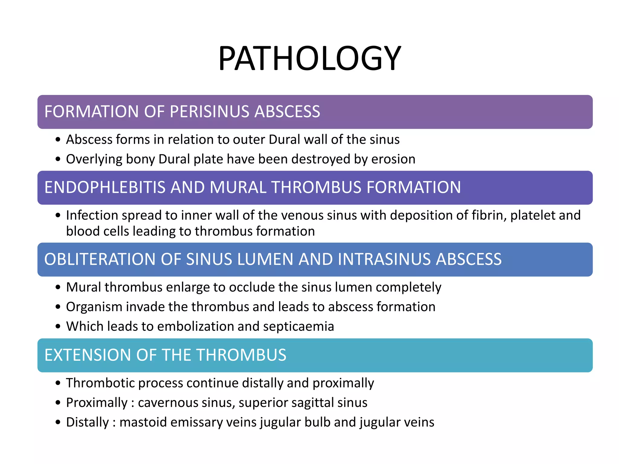
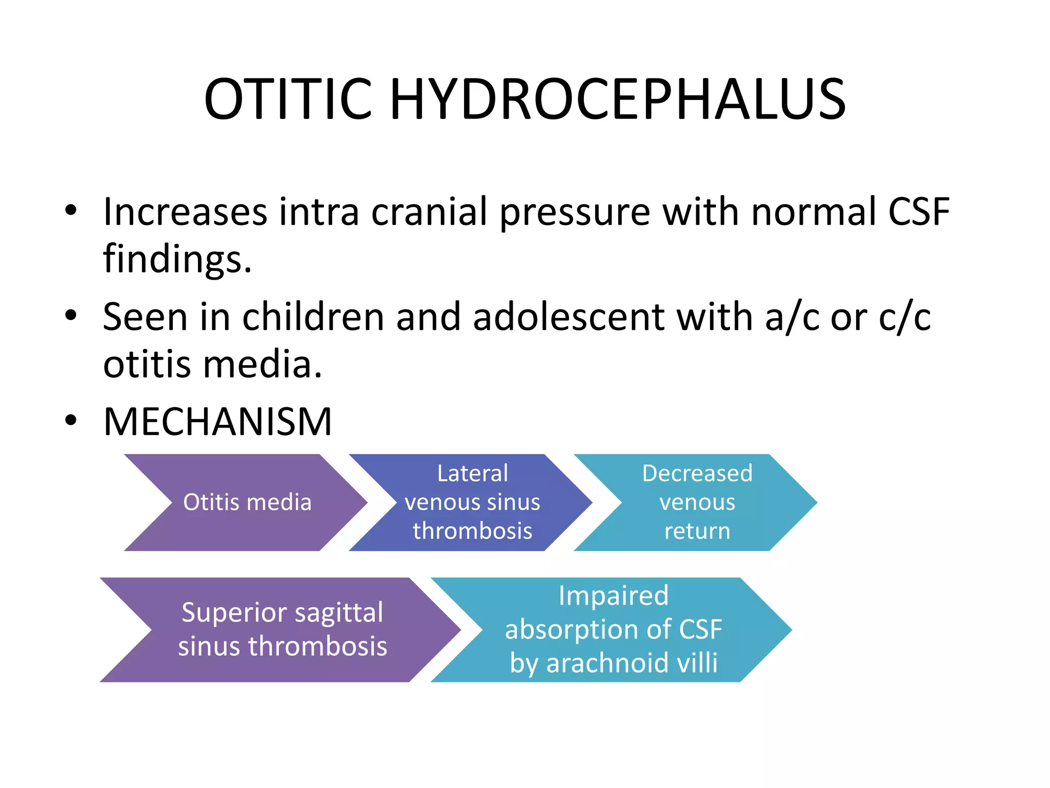
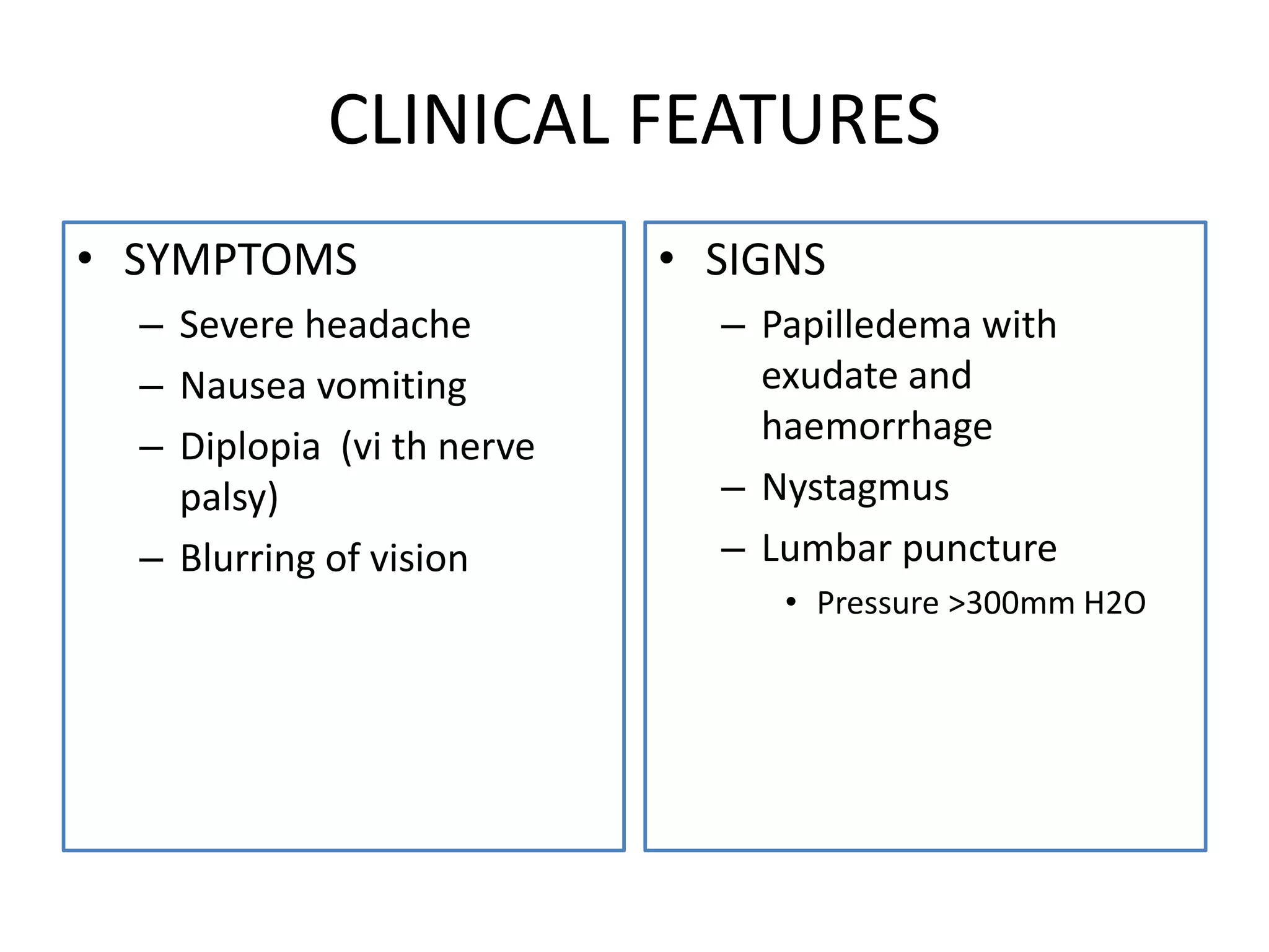
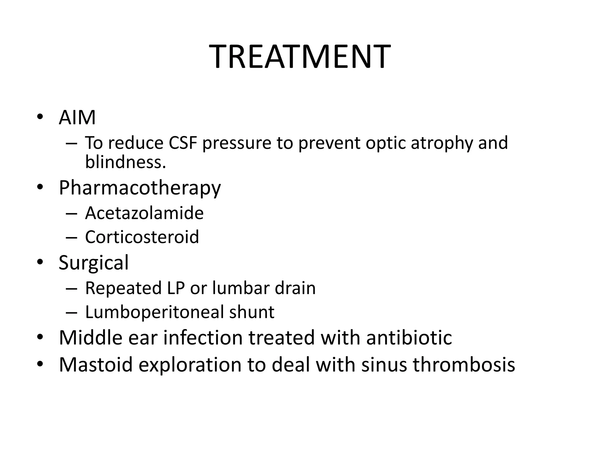
This document summarizes various complications that can arise from otitis media. It discusses both extracranial complications like mastoiditis, mastoid abscesses, and facial nerve palsy, as well as intracranial complications like labyrinthitis, meningitis, brain abscesses, and lateral sinus thrombophlebitis. It provides details on the pathology, clinical features, investigations, differential diagnosis, and management of each complication.
2. • Though there is a general decline in the
incidence of complication, they are still
frequently seen in India
• Decline in the incidence of complications of
CSOM due to
– Early diagnosis
– Better antibiotic therapy
3. • FACTORS INFLUENCING
DEVELOPMENT OF COMPLICATIONS
– Age : extremes of age
– Poor socio economic status
– Virulence of organism
– Immune compromised host
– Preformed pathways
– Cholesteatoma
9. ACUTE MASTOIDITIS
• Infection involving the bony wall of the
mastoid air cell system.
• Aetiology
– Accompanies or follows ASOM
– determining factors are
• High virulence of organism
• Low resistance of patient
• Poor nutrition
• Systemic disease
11. PATHOLOGY
• 2 main pathological process involved are
– Production of pus under tension
– Hyperaemic decalcification and osteoclastic
resorption of bony wall
12. Acute infection of mastoid air cells
Persistent hyperaemia congestion and oedema of mucous membrane
of air cells + osteitis
Purulent exudate in air cells
Venous stasis
Local acidosis
Breaking down of the partition of air cells
Abscess
Post auricular
abscess
Bezolds abscess
Citellis abscess Para pharyngeal
abscess
Zygomatic abscess Lucs abscess
Apical abscess
Obstruction of the aditus
Pressure effect
Absorption of calcium Hyperaemic decalcification
14. – Swelling over mastoid
• Ironed out feeling
– Hearing loss
• Conductive hearing loss
– Tuning fork test
• Rinne’s test –ve
• Weber test lateralised to
diseased side
• ABC - normal
• SIGNS
– Mastoid tenderness
– Ear discharge
• Mucopurulent/purulent
• Light house
effect(pulsatile discharge)
• Mastoid reservoir sign
– Sagging of
posterosuperior meatal
wall
– Perforation of TM
15. MANAGEMENT
• INVESTIGATIONS
– Blood count : polymorphonuclear leukocytosis
– ESR : raised
– X-ray mastoid – clouding of air cells due to
collection of exudate
– CT temporal bone
– Ear swab for culture and sensitivity
16. • DIFFERENTIAL DIAGNOSIS
– Suppuration of mastoid lymph node
• Scalp infection
• No history of otitis media, ear discharge or deafness
– Furunculosis of meatus
– Infected sebaceous cyst
17. TREATMENT
• Hospitalisation of patient
• Antibiotics
– Amoxicillin / ampicillin
– Specific antibiotic after receiving culture and
sensitivity
• Myringotomy
– Wide myringotomy: to relieve pus
• Cortical mastoidectomy
18. CORTICAL MASTOIDECTOMY
• INDICATION
– Sub periosteal abscess
– Sagging of posterior
superior meatal wall
– Positive reservoir sign
– Worsening of condition
of patient in spite of
medical treatment for 48
hrs.
– Mastoiditis leading to
complication
• AIM
– Exenterate all the
mastoid air cell
– Remove any pocket of
pus
20. Abscess in relation to mastoid
infection
POST AURICULAR ABSCESS
• Abscess over the mastoid
• Pinna displaced forwards outwards and downwards
• Infants and children abscess over Mac Ewens triangle
Zygomatic abscess
• Infection of zygomatic air cells
• Swelling in front and above pinna
• Oedema of upper eye lid
LUC ABSCESS / MEATAL ABSCESS
• Pus breaks through the bony wall between antrum and external osseous meatus
• Swelling in deep part of bony meatus
CITELLIS ABSCESS / BEHIND MASTOID
• Abscess of digastric triangle
• Pus at the tip of mastoid
Para pharyngeal or retropharyngeal abscess
21. BEZOLDS ABSCESS
• 5 presentations
– Lie deep to
sternocleidomastoid
– Follow posterior belly of
digastric and present as a
swelling between the tip of
the mastoid and angle of
jaw.
– Upper part of posterior
triangle
– Para pharyngeal space
– Track down along carotid
vessels
• Clinical features
– Sudden onset
– Pain fever, tender swelling,
torticollis , purulent
otorrhoea
• D/D
– a/c upper jugular
lymphadenitis
– Infected brachial cyst
– Para pharyngeal abscess
– Jugular vein thrombosis
23. MASKED MASTOIDITIS
• Latent mastoiditis
• Slow destruction of mastoid air cells but
without acute signs and symptoms of A/C
mastoiditis .
• There is destruction of air cells with
granulation tissue and dark gelatinous
material filling the mastoid.
24. • AETIOLOGY
– Inadequate antibiotic therapy for ASOM
• CLINICAL FEATURES
– SYMPTOMS
• Mild pain behind the ear
• Persistent hearing loss
– SIGNS
• TM : thick and loss of translucency
• PTA : conductive hearing loss
• MANAGEMENT
– INVESTIGATION
• X RAY : clouding of air cells with loss of cell outline
– TREATMENT
• Same as acute mastoiditis
27. MANAGEMENT
• INVESTIGATION
– CT scan temporal bone
• Petrous apex
• Air cells
– MRI
• Helps to differentiate fluid
from pus
• TREATMENT
– Cortical / modified
radical / radical
mastoidectomy
– Fistula tract is found out
then currated and
enlarged to provide free
drainage of pus
– IV antibiotics
28. FACIAL NERVE PALSY
• ACUTE OTITIS MEDIA
– Middle ear infection
spread to epi or
perineurium and causes
facial nerve palsy
– Function can be fully
recovered when ASOM is
controlled by systemic
antibiotics
– Myringotomy and
cortical mastoidectomy
is sometimes needed
• CHRONIC OTITIS MEDIA
– d/t cholesteatoma or
penetrating granulation
tissue
– Facial palsy is insidious
but slowly progressive
– Treatment is urgent
exporation of middle ear
and mastoid
– Granulation tissue is
scrapped
– Nerve grafting
30. CIRCUMSCRIBED LABYRINTHITIS
• Fistula of labyrinth
• Thinning or erosion of bony capsule of
labyrinth.
• Most commonly : horizontal semi circular
canal.
• CSOM with cholesteatoma is the most
common cause.
31. • CLINICAL FEATURES
– SYMPTOM
• Transient vertigo
– SIGN
• Positive fistula sign
– Pressure on tragus
– Siegel's speculum
• TREATMENT
– CSOM is treated
– Antibiotics
32. DIFFUSE SEROUS LABYRINTHITIS
• Diffuse intra labyrinthine inflammation
without pus collection
• Reversible with early treatment
• CLINICAL FEATURES
• Mild case : vertigo and nausea
• Severe case: vertigo, marked nausea vomiting and
spontaneous nystagmus ( quick component towards
affected ear)
• Hearing loss : sensorineural
33. TREATMENT
• MEDICAL
– Patient is put to bed, head is immobilized with
affected ear above
– Antibiotic full dose to control infection
– Labyrinthine sedative
– Myringotomy
• SURGICAL
– Cortical mastoidectomy : acute infection
– Modified radical mastoidectomy : chronic infection
34. DIFFUSE SUPPURATIVE LABYRINTHITIS
• Diffuse pyogenic infection of labyrinth with
permanent loss of vestibular and cochlear
function
• CLINCAL FEATURE
– Severe vertigo nausea and vomiting
– Spontaneous nystagmus( quick component towards
healthy side )
– Total loss of hearing
• TREATMENT
– Same as serous Labyrinthitis
36. EXTRA DURAL ABSCESS
• Collection of puss between bone and dura
• Affected dura will be covered with
granulations and appears unhealthy and
discoloured.
• PATHOLOGY
a/c otitis
media
Hyperaemic calcification of bone
over dura
c/c otitis
media
Bone destroyed by cholesteatoma
Pus comes in
direct contact
with dura
37. CLINICAL FEATURES
• Usually asymptomatic
• SYMPTOMS
– Persisted head ache on side of otitis media
– Severe ear pain
– General malaise with low grade fever
– Pulsatile purulent ear discharge
– Disappearance of headache with free flow of pus
from ear.
38. MANAGEMENT
• INVESTIGATIONS
– Contrast enhanced CT or MRI
• TREATMENT
– Cortical or modified radical or radical mastoidectomy
• Abscess evacuated by removing overlying bone till healthy
dura is reached
• In suspicion of abscess, the intact bony plate ( tegmen
tympani and sinus plate ) is removed deliberately to
evacuate collected pus.
– Antibiotics
• Minimum for 5 days.
39. SUB DURAL ABSCESS
• Collection of pus between dura and arachnoid
• PATHOLOGY
Bone erosion Extra Dural abscess thrombophlebitis
Sub Dural abscess
40. CLINICAL FEATURES
• Signs and symptoms are due to
– Meningeal irritation
• Head ache
• Fever, malaise, neck rigidity, kerning's sign positive
– Thrombophlebitis of cortical veins of cerebrum
• Aphasia , hemiplegia, hemianopia, epilepsy
– Raised intracranial pressure
• Papilledema, ptosis, dilated pupil
41. TREATMENT
• LP is contraindicated due to risk of herniation
of cerebellar tonsil.
• Neurological emergency
• Craniotomy or series of burr hole is done to
drain sub Dural empyema
• IV antibiotic
• Treat underlying ear pathology
• Mastoidectomy in indicated cases.
42. MENINGITIS
• Inflammation of leptomeninges.
• MC intracranial complication of otitis media.
• MODE OF INFECTION
– Blood borne : children and infants
– Thrombophlebitis or bone erosion : adult
43. CLINICAL FEATURES
• SYMPTOMS
– Fever with chills and rigor
– Headache
– Neck rigidity
– Photophobia and mental
irritation
– Nausea and vomiting
– Drowsiness delirium coma
– Cranial nerve palsy,
hemiplegia
• SIGNS
– Neck rigidity
– Positive kerning's sign
– Positive Brudzinski sign
– Tendon reflex
• Initial: exaggerated
• Later : absent or sluggish
– Papilledema
44. MANAGEMENT
• INVESTIGATIONS
– LP and CSF examination
• Turbid
• Cell count : >1000/ml
• Sugar : reduced
• Chloride : diminished
– Culture and sensitivity
– Contrast CT and MRI
• TREATMENT
– MEDICAL
• Antibiotics
• Corticosteroids
– SURGICAL
• ASOM: myringotomy or
cortical mastoidectomy
• CSOM: radical or modified
radical mastoidectomy
•
45. OTOGENIC BRAIN ABSCESS
• Adult : CSOM with cholesteatoma
• Children : ASOM
• ROUTE OF INFECTION
– Direct spread fro middle ear via tegmen
• Associated with extra Dural abscess
– Retrograde thrombophlebitis
• Associated with extra Dural abscess ,perisinus abscess,
sigmoid sinus thrombophlebitis or Labyrinthitis
46. PATHOLOGY
STAGE OF INVASION
• Initial encephalitis
• Usually asymptomatic
• Headache, low grade fever, malaise
STAGE OF LOCALIZATION
• Latent abscess
• No symptoms
• Last for several weeks
STAGE OF ENLARGEMENT
• Manifest abscess
STAGE OF TERMINATION
• Rupture of abscess
• Abscess ruptures into ventricles or sub arachnoid space resulting in fatal meningitis
Enlargement of
abscess
Oedema
around abscess
Aggravation of
symptoms
47. CLINICAL FEATURES
• Due to raised intracranial
tension
– Headache
– Nausea and vomiting
– Lethargy delirium coma
– Papilledema
– Slow pulse and subnormal
temperature
• LOCALIZING FEATURES
– TEMPORAL LOBE ABSCESS
• Nominal aphasia
• Homonymous hemianopia
• Contra lateral motor palsy
• epileptic fits
• Pupillary oedema and
oculomotor palsy
– Cerebellar abscess
• Headache
• Spontaneous nystagmus
• Ipsilateral hypotonia and
weakness ,ataxia
• Past pointing and intention
tremor
• Dysdiadochokinesia
48. MANAGEMENT
• INVESTIGATION
– Skull X ray
• Midline shift
• Gas In the abscess
• Calcification of pineal
glands
– CT scan
• Site and size of abscess
• Complications can be
detected
– X ray mastoid or CT of
temporal
– LP
• TREATMENT
– MEDICAL
• IV antibiotic
• Dexamethasone/ mannitol
– NEUROSURGICAL
• Repeated aspiration
through burr hole
• Excision of abscess
• Open incision of abscess
and evacuation of pus
– Otologic
• Antibiotic for ASOM
• Radical mastoidectomy :
CSOM
49. LATERAL SINUS THROMBOPHLEBITIS
• SIGMOID SINUS THROMBOSIS
• Inflammation of inner wall of lateral venous
sinus with formation of an intrasinus thrombi.
50. PATHOLOGY
FORMATION OF PERISINUS ABSCESS
• Abscess forms in relation to outer Dural wall of the sinus
• Overlying bony Dural plate have been destroyed by erosion
ENDOPHLEBITIS AND MURAL THROMBUS FORMATION
• Infection spread to inner wall of the venous sinus with deposition of fibrin, platelet and
blood cells leading to thrombus formation
OBLITERATION OF SINUS LUMEN AND INTRASINUS ABSCESS
• Mural thrombus enlarge to occlude the sinus lumen completely
• Organism invade the thrombus and leads to abscess formation
• Which leads to embolization and septicaemia
EXTENSION OF THE THROMBUS
• Thrombotic process continue distally and proximally
• Proximally : cavernous sinus, superior sagittal sinus
• Distally : mastoid emissary veins jugular bulb and jugular veins
51. CLINICAL FEATURES
• Hectic picket fence type of
fever with rigor
• Headache
• Progressive anaemia and
emaciation
• Griesingers sign
– Oedema over posterior part of
mastoid
– d/t thrombosis of mastoid
emissary vein
• Tenderness along jugular vein
– Thrombophlebitis extend to
jugular vein
– Enlargement and inflammation
of jugular chain of lymph node
• Papilledema
• Tobey Ayer test
– CSF pressure using manometer
– See the effect of manual
compression of one or more
jugular veins
– Affected side : no change
– Healthy side : rise in pressure
• Crowe Beck test
– Pressure on jugular vein of
healthy side produce
engorgement of retinal or
supraorbital vein
52. MANAGEMENT
• INVESTIGATION
– Blood smear
• Rule out malaria
– Blood culture
– CSF examination
– X ray mastoid
– Imaging studies
• Contrast enhanced CT
scan : delta sign
– Culture and sensitivity of
ear swab
• TREATMENT
– IV antibiotics
– Mastoidectomy and
exposure of sinus
– Ligation of internal
jugular vein
– Anticoagulant therapy
– Supportive treatment
53. COMPLICATIONS
• Septicaemia and pyogenic
abscess in lung bone and
joints
• Meningitis and sub Dural
abscess
• Cerebellar abscess
• Thrombosis of jugular
bulb and jugular vein with
involvement of Ixth, Xth,
XI th cranial nerve
• Cavernous sinus
thrombosis
• Otitis hydrocephalous
54. OTITIC HYDROCEPHALUS
• Increases intra cranial pressure with normal CSF
findings.
• Seen in children and adolescent with a/c or c/c
otitis media.
• MECHANISM
Otitis media
Lateral
venous sinus
thrombosis
Decreased
venous
return
Superior sagittal
sinus thrombosis
Impaired
absorption of CSF
by arachnoid villi
55. CLINICAL FEATURES
• SYMPTOMS
– Severe headache
– Nausea vomiting
– Diplopia (vi th nerve
palsy)
– Blurring of vision
• SIGNS
– Papilledema with
exudate and
haemorrhage
– Nystagmus
– Lumbar puncture
• Pressure >300mm H2O
56. TREATMENT
• AIM
– To reduce CSF pressure to prevent optic atrophy and
blindness.
• Pharmacotherapy
– Acetazolamide
– Corticosteroid
• Surgical
– Repeated LP or lumbar drain
– Lumboperitoneal shunt
• Middle ear infection treated with antibiotic
• Mastoid exploration to deal with sinus thrombosis