
Recommended
More Related Content
What's hot
What's hot (20)
Similar to DLC Guide: Differential Leukocyte Count Interpretation
Similar to DLC Guide: Differential Leukocyte Count Interpretation (20)
More from MohanSinghDhakad1
More from MohanSinghDhakad1 (16)
Recently uploaded
Recently uploaded (20)
DLC Guide: Differential Leukocyte Count Interpretation
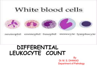
- 1. DIFFERENTIAL LEUKOCYTE COUNT By Dr. M. S. DHAKAD Department of Pathology
- 2. Learning Objectives • Introduction • Identification of WBCs • Differential Counting of WBCs • Pathological variations in DLC
- 3. Introduction • DLC Relative proportion of different leukocytes expressed as percentage. USES: * To support the diagnosis of infectious, inflammatory or allergic disorders. * Diagnosis of malignant blood disorders
- 4. White blood cells Granulocytes are of three types named according to their staining characteristics in blood films. They are • Neutrophils • Eosinophils • Basophils. • Agranulocytes/Mononucle ar cells are divided into • Lymphocytes • Monocytes.
- 6. Granulocytes Polymorph (Neutrophil) • Cell diameter : 12-15 μm • Nucleus : 2-5 lobes, clumped chromatin • Cytoplasm : Pink granules • Normal % : 40-70 • Absolute count per μl : 2000- 7500
- 8. Granulocytes -Eosinophil • Cell diameter : 12-15 μm • Nucleus : Bilobed, clumped chormatin. • Cytoplasm : Coarse grimson red granules. • Normal % : 1-6. • Absolute count per μl : 40-400.
- 10. Granulocytes - Basophil • Cell diameter : 12-15 μm • Nucleus : Bilobed, clumped chormatin. • Cytoplasm : Large, coarse purplish granules obscuring the nucleus. • Normal % : 0-1. • Absolute count per μl : 10-100.
- 12. Agranulocytes Lymphocytes • Cell diameter : Small Lymphocyte: 9-12 μm Large Lymphocyte : 12-16 μm • Nucleus : Large nucleus round to indented fills the cell, clumped with chromatin • Cytoplasm : Peripheral rim of basophilic cytoplasm, no granules • Normal % : 20-40 • Absolute count per μl : 1500-4000
- 14. Agranulocytes Monocytes • Cell diameter : 12-20 μm • Nucleus :Large lobulated,indented, with fine chormatin • Cytoplasm : Light basophilic, may contain fine granules or vacuoles. • Normal % : 2-10 • Absolute count per μl : 200-800
- 16. Blood can be collected from 3 different sources: Capillary blood. Venous blood. Arterial blood.
- 18. FOCUSING 4X to see the general formation of slide. • 10X for WBC counting • For a differential WBC count, an oil-immersion objective with around 100x magnification (1.4NA) is used.
- 19. Choose an area near the junction of body with the tail of the smear tail body head
- 21. Start counting
- 22. Normal Reference Range • White blood cell count 4.0–11.0 x 10⁹ /l • Differential white cell count – Neutrophils – Lymphocytes – Monocytes – Eosinophils – Basophils 2.0–7.0 x 10⁹ /l (40–80%) 1.0–3.0 x 10⁹ /l (20–40%) 0.2–1.0 x 10⁹ /l (2–10%) 0.02–0.5 x 10⁹ /l (1–6%) 0.02–0.1 x 10⁹ /l (<1–2%)
- 23. AUTOMATED COUNTING • It is done by electronic counting method. • Coulter – Automated haemanalyser. • There are 3 types of electronic methods— • by cell size analysis, • by flow cytometry • high resolution pattern recognition. • Automated DLC counters have a differential counting capacity of counting either • 3-part DLC (granulocytes, lymphocytes and monocytes) • 5-part DLC (P, L, M, E, B).
- 24. AUTOMATED COUNTING • Coulter – Automated haemanalyser Advantages Easy and rapid method. Time saving. Provide additional information on cell size, shape, nuclear size and density. Very large number of cells are counted rapidly High level of precision Disadvantages Costly Calibration error Nucleated RBCs/normoblasts are counted as lymphocytes Platelet clumps counted as leucocytes
- 26. Learning Objectives • Introduction • Identification of WBCs • Differential Counting of WBCs • Pathologic variations in DLC
- 27. Neutrophilia Increase in neutrophil count above 7,500/μl. Causes 1. Acute infections (By bacteria, fungi,parasites and some viruses) i. Pneumonia ii. Acute appendicitis iii. Acute cholecystitis iv. Salpingitis v. Peritonitis vi. Abscess and physical agents vii. Acute tonsillitis viii. Actinomycosis ix. Poliomyelitis x. Furuncle xi. Carbuncle
- 28. Neutrophilia Increase in neutrophil count above 7,500/μl. Causes 2. Intoxication i. Uraemia ii. Diabetic ketosis iii. Poisoning by chemicals anaemia iv. Eclampsia
- 29. Neutrophilia Increase in neutrophil count above 7,500/μl. Causes 3. Inflammation from tissue damage i. Burns ii. Ischaemic necrosis iii. Gout iv. Hypersensitivity reaction
- 30. Neutrophilia Increase in neutrophil count above 7,500/μl. Causes 4. Acute haemorrhage i. Acute haemolysis
- 31. Neutrophilia Increase in neutrophil count above 7,500/μl. Causes 5. Neoplastic conditions i. Myeloid leukaemia (CML) ii. Polycythaemia vera iii. Myelofibrosis iv. Disseminated cancers
- 32. Neutrophilia Increase in neutrophil count above 7,500/μl. Causes 6. Miscellaneous conditions i. Administration of corticosteroids ii. Idiopathic neutrophilia
- 33. Neutropenia Fall in neutrophil count below 2,000/μl Causes – 1. Infections i. Typhoid ii. Brucellosis iii. Measles iv. Malaria v. Kala azar
- 34. Neutropenia Fall in neutrophil count below 2,000/μl Causes – 2. Drugs and chemicals and physical agents i. Antimetabolites ii. Benzene iii. Nitrogen mustard iv. Irradiation
- 35. Neutropenia Fall in neutrophil count below 2,000/μl Causes – 3. Haematological and other diseases i. Aplastic anaemia ii. Pernicious anaemia iii. SLE iv. Gaucher’s disease v. Cachexia vi. Anaphylactic shock
- 36. Lymphocytosis Increase in absolute lymphocyte count to more than 4,000/μl Causes – 1. Acute Infections i. Pertussis ii. Infectious mononucleosis iii.Viral hepatitis 2.Chronic Infections i. Tuberculosis ii. Brucellosis iii.Secondary syphilis 3.Haematopoietic Disorders i. CLL ii.NHL
- 37. Lymphopenia absolute lymphocyte count below 1,500/μl Causes – i. Aplastic anaemia ii. High dose of steroid administration iii. AIDS iv. Hodgkin’s disease v. Irradiation
- 38. Monocytosis Rise in absolute monocyte count above 800/μl Causes – 1. Bacterial infections i. Tuberculosis ii. SABE iii. Syphilis 2. Protozoal infections i. Malaria ii Kala azar iii. Trypanosomiasis
- 39. Monocytosis Rise in absolute monocyte count above 800/μl Causes – 3. Haematopoietic disorders i. Monocytic leukaemia ii. Hodgkin’s disease iii. Multiple myeloma iv.Myeloproliferative disorders 4. Miscellaneous conditions i. Sarcoidosis ii. Cancer of ovary, breast, stomach
- 40. Eosinophilia Increase in the absolute esosinophil count above 400/μl Causes – 1. Allergic disorders i. Bronchial Asthma ii. Urticaria iii. Hay fever iv. Drug hypersensitivity 2. Parasitic infestations i. Round worm ii. Hookworm iii. Tape worm iv. Echinococcosis
- 41. Eosinophilia Increase in the absolute esosinophil count above 400/μl Causes – 3. Skin diseases i. Pemphigus ii. Dermatitis herpetiformis iii. Erythema multiforme 4. Pulmonary diseases i. Loeffler’s syndrome ii. Tropical eosinophilia
- 42. Eosinophilia Increase in the absolute esosinophil count above 400/μl Causes – 5. Haematopoietic diseases i. Chronic myeloid leukaemia ii. Polycythaemia vera iii. Hodgkin’s disease iv.Pernicious anaemia 6. Miscellaneous conditions i. Rheumatoid arthritis ii. Polyarteritis nodosa iii. Sarcoidosis iv.Irradiation
- 43. Eosinopenia Fall in the absolute eosinophil count below 40/μl Causes – Steroid administration
- 44. Basophilia Increase in the absolute basophil count above 100/μl Causes – i. Chronic myeloid leukemia ii. Polycythaemia vera iii. Myxoedema iv.Ulcerative colitis v.Hodgkin’s disease vi Urticaria pigmentosa