3. INTRODUCTION
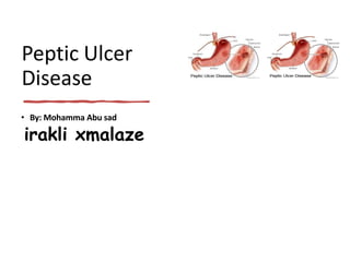
Peptic Ulcer is a lesion in the lining
(mucosa) of the digestive tract, typically in
the stomach or duodenum, caused by the
digestive action of pepsin and stomach acid.
4. • Lesion may subsequently occur into the lamina
• propria and submucosa to cause bleeding. –
Most of peptic ulcer occur either in the duodenum, or in
the stomach – Ulcer may also occur in the lower esophagus due to
reflexing of gastric content – Rarely in certain areas of the small
intestine
6. • Under normal conditions, a physiologic balance
exists between gastric acid secretion and gastroduodenal
mucosal defense. Mucosal injury and, thus, peptic ulcer occur when
the balance between the aggressive factors and the
defensive mechanisms is disrupted. Aggressive
factors, such as NSAIDs, H pylori infection, alcohol, bile salts,
acid, and pepsin, can alter the mucosal defense by allowing back
diffusion of hydrogen ions and subsequent epithelial cell injury.
7. ETIOLOGY/
RISK
FACTORS • Lifestyle
– Smoking
– Acidic drinks
– Medications
•
•
H. Pylori infection
– 90% have this bacterium
– Passed from person to
person (fecal-oral route
or oral-oral route)
Age
– Duodenal 30-40
– Gastric over 50
•
•
•
Gender
– Duodenal: are increasing
in older women
Genetic factors
– More likely if family
member has Hx
Other factors: stress
can worsen but not the
cause
14. DIAGNOSTIC
TEST
• Esophagogastrodeuodenoscopy(EGD)
• Endoscopic procedure
• Visualizes ulcer crater
• Ability to take tissue biopsy
to R/O cancer and diagnose
• H. pylori
• Upper gastrointestinal series
(UGI)
• Barium swallow
• X-ray that visualizes
structures of the upper GI
tract
• Urea Breath Testing
• Used to detect H.pylori
• Client drinks a carbon-
enriched urea solution
• Exhaled carbon dioxide is
then measured
15. In all patients with
“Alarming symptoms”
endoscopy is required.
• Dysphagia.
• Weight loss.
• Vomiting.
• Anorexia.
• Hematemesis or Melena
16. Complications
of Peptic
Ulcers •
•
•
Hemorrhage
– Blood vessels damaged as ulcer erodes into the muscles of
stomach or duodenal wall
– Coffee ground vomitus or occult blood in tarry stools
Perforation
– An ulcer can erode through the entire wall
– Bacteria and partially digested food spill into
peritoneum=peritonitis
Narrowing and obstruction (pyloric)
– Swelling and scarring can cause obstruction of food leaving
stomach=repeated vomiting
21. Indications:
• Failure of medical
treatment.
• Development of
complications
• High level of gastric
secretion and combined
duodenal and gastric ulcer.
• Principle:
• Reduce
acid and
pepsin
secretion.
26. Post-op
Complications
• Bleeding
–
–
Occurs at the anastomosed site
First 24 hours and post-op days
4-7
• Duodenal stump leak
–
–
–
Billroth II
Severe abdominal pain
Bile stained drainage on
dressing
• Gastric retention
– WILL NEED TO PUT NG TUBE
BACK IN
•
• Dumping Syndrome.
– Prevalent with sub total gastrostomies
– Early-30 minutes after meals
– Vertigo, tachycardia, syncope, sweating,
pallor, palpitations
– Late – 90 min-3 hours after meals
• Rx: Decrease CHO intake, Eat slowly, Avoid
fluids during meals, Increase fat, Eat small,
frequent meals
Anemia
– Rapid gastric empyting decreases
absorption of iron
• Malabsorption of fat
– Decreased acid secretions, decreased
pancreatic secretions, increased upper
GI mobility
27. Summary
• H. pylori is the most common cause of PUD and is a risk factor for gastric
cancer
• H Pylori eradication reduces risk of disease recurrence
• Test-and-Treat strategy is recommended for patients with
undifferentiated dyspepsia
• Intial evaluation with endoscopy is recommended for those with alarm
symptoms or those failing treatment
• Optimum treatment regimens are 14d multidrug with antibiotics and
acid suppressants(Triple therapy)