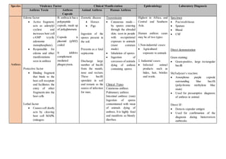
1. Species Virulence Factor Clinical Manifestation Epidemiology Laboratory Diagnosis
Anthrax Toxin Anthrax
Capsule
Animal Anthrax Human Anthrax
Anthrax
Edema factor
Active fragment;
acts as adenylyl
cyclase and
increases host cell
cAMP (cyclic
adenosine
monophosphate).
Responsible for
edema and other
manifestations
seen in anthrax
Protective factor
Binding fragment
that binds to the
host cell receptors
and facilitates the
entry of other
fragments into the
host cells
Lethal factor
Causes cell death;
acts by cleaving
host cell MAPK
(mitogen-
B. anthracis has a
polypeptide
capsule, made up
of polyglutamate
Capsule is
plasmid (pX02)
coded
It inhibits
complement
mediated
phagocytosis.
Zoonotic disease
Horses
Pigs
Ingestion of the
spores present in
the soil.
Presents as a fatal
septicemia
Discharge large
number of bacilli
from the mouth,
nose and rectum.
These bacilli
sporulate in soil
and remain as the
source of infection
for man.
Transmission
Cutaneous mode—
by spores entering
through the abraded
skin; seen in people
with occupational
exposure to animals
(most common
mode)
Inhalation of spores
Ingestion of
carcasses of animals
dying of anthrax
containing spores
Clinical Types
Cutaneous anthrax
Pulmonary anthrax
Intestinal anthrax: (rare)
Ingestion of spores
contaminated with meat
of animals dying of
anthrax. It is highly fatal
and manifests as bloody
diarrhea.
Highest in Africa, and
Central and Southern
Asia.
Human anthrax cases
may be of two types:
1.Non-industrial cases:
Agricultural
exposure to animals
2. Industrial cases:
Infected animal
products such as
hides, hair, bristles
and wools.
Specimen
Pus/swab/tissue
Sputum
Blood
CSF
Direct demonstration
Gram staining:
Gram-positive, large rectangular
bacilli
McFadyean’s reaction:
Amorphous purple capsule
surrounding blue bacilli
(polychrome methylene blue
stain)
Used for presumptive diagnosis
of anthrax in animal.
Direct IF:
Detects capsular antigen
Used for confirmation of the
diagnosis during bioterrorism
outbreaks
2. activated protein
kinases).
These fragments are not
toxic individually, but in
combination, they
produce local edema and
generalized shock. Toxin
synthesis is controlled by
a plasmid (pX01). Loss of
plasmid makes the strain
avirulent. This was
probably the basis of
original anthrax vaccine
prepared by Pasteur.
Agent of Bioterrorism
B. anthracis ( most
common)
Pulmonary anthrax
is the most common
form to cause
bioterrorism
outbreaks
Transmission occurs
via inhalation of
anthrax spores from
contaminated
animal products
Ascoli’s thermo precipitation test :
Ring precipitation test
Performed when specimens are
received in the putrid form
Culture
1. Aerobic
2. Non-fastidious
3. Grows in ordinary media and has
a wide temperature range (12–45)
of growth
Nutrient agar
size, irregular, round, opaque,
grayish white with a frosted glass
appearance
Medusa head appearance colonies
Colonies are viewed under low
power microscope
Edge of the colony which is
composed of long interlacing
chains of bacilli, appears as locks
of matted hair
Blood agar
Dry wrinkled, nonhemolytic
colonies
3. Gelatin stab agar:
Inverted fir tree appearance
growth
Selective media:
Solid medium with penicillin:
String of pearl appearance
PLET medium
Culture smear
Gram-positive rods with bamboo
stick appearance.
Long chain of gram-positive
bacilli with non-bulging spores
Antibodies detection by ELISA
Molecular diagnosis
PCR using BA pX01 primer
targeting gene coding for
protective antigen
PCR BA pX02 primers targeting
capsular gene
4. Species Epidemiological Virulence Factor Laboratory Diagnosis Prevention of Plague
Plague
(Yersinia
Pestis)
Reservoir
Wild rodents, (Tatera indica)
Field mice
Source of infection
Infected wild rodents
Rat fleas
Cases of pneumonic plague
Vector
Rat flea
Feeding on infected wild
Xenopsylla cheopis (the
most efficient vector, found
in North India)
Xenopsylla astia (less
efficient, found in South
India)
Xenopsylla brasiliensis
Fraction 1 (F1) antigen
Capsular protein antigen,
encoded by a plasmid (pFra).
Acts by inhibiting
phagocytosis by macrophage
Highly antigenic and is used
asimmunodiagnostic marker
of infection
Other virulence factors
Phospholipase D (murine
toxin)
Surface proteases
pH 6 antigen
Lipopolysaccharide
(endotoxin)
Pigments (hemin-
containing)
Type III secretion system
Adhesins (help in
attachment)
Siderophore (helps in
acquisition of iron)
Specimen Collection
Bubonic plague - pus or fluid aspirated
from buboes
Pneumonic plague - sputum and blood
Septicemic plague - blood and splenic
aspirate (post mortem).
Transport medium (Cary–Blair medium)
can be used if delay in transportation is
expected.
Direct Microscopy
Gram staining:
Presence of pus cells
Gram negative oval coccobacilli with
rounded ends surrounded by capsule
Wayson stain
Methylene blue staining demonstrates the
bacilli with typical bipolar or safety pin
appearance.
Two ends are darkly stained with clear
central area
Control of cases by early diagnosis,
isolation and treatment of cases
Isolation precaution: Contact precautions
need to be followed for bubonic plague and
droplet precautions for pneumonic plague
(Chapter 21)
Control of fleas by use of effective
insecticides, such as DDT or BHC (β-
hexachloro-cyclohexane)
Control of rodents
Chemoprophylaxis should be given to all
contacts of pneumonic plague. Doxycycline
or levofloxacin is the drug of choice, given
for 7 days
Vaccine
WHO recommends using vaccine only
for prevention of an anticipated
outbreak and not for general use
Formalin killed
Given subcutaneously, two doses 4
weeks apart and a booster given after 6
months. It is contraindicated in infants
5. Mode of transmission
Bite of an infected rat flea
(most common)
Direct contactwith tissues of
infected animal (rodents)
Droplet inhalation (man to
man) from cases of
pneumonic plague „ Bite of
an infected human flea
(Pulex irritans).
Culture
Aerobic and facultatively anaerobic.
Blood agar: Colonies are non-hemolytic
and dark brown pigmented due to the
absorption of the hemin pigment
MacConkey agar: Lactose non-
fermenting colorless colonies are formed
Culture Smear and Motility Testing
Gram staining of culture smear
Pleomorphism coccid
Coccobacillary
Bacillary
Filamentous
Giant forms. Involution forms are seen in
older cultures
Nonmotile both at 25°C and 37°C
Identification
Automated identification systems such as
MALDI-TOF
Conventional biochemical tests
Catalase positive
Oxidase negative
ICUT tests:
Indole test (negative)
6. Citrate test (negative)
Uurease test (negative)
TSI (triple sugar iron agar) test shows
alkaline/acid, gas absent, H2 S absent
MALDI-TOF can be used for rapid
accurate identification of Y. pestis and
also to differentiate its three biotypes.
F1 Antigen Detection
Detected from bubo aspirate or sputum
By direct immunofluorescence test,
ELISA or immunochromatographic test
(ICT) by using monoclonal antibodies.
Antibodies to F1 Antigen
Detection Antibodies may be detected by
ELISA, or passive agglutination.
Antibodies are useful epidemiological
markers, as they remain positive for
several years.
Molecular Methods
PCRis available targeting gene coding F1
antigen
Pesticin gene
Plasminogen activator gene.