
Recommended
Recommended
More Related Content
Similar to A LEVEL PRACTICALS MeetLearn-1.pdf
Similar to A LEVEL PRACTICALS MeetLearn-1.pdf (20)
Recently uploaded
Recently uploaded (20)
A LEVEL PRACTICALS MeetLearn-1.pdf
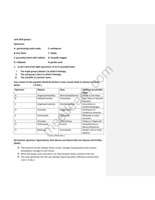
- 1. June 2016 group 1 Specimens: A : germinating maize seeds. E: earthworm B: liver fluke. F: tilapia C: groundnut plant with nodules. G: housefly maggot D: millipede. H: garden snail 1. (a )For each of the eight specimens ( A to H ) provided state: i. The major group ( phylum ) to which it belongs, ii. The sub-group ( class ) to which it belongs, iii. The scientific or common name. Your answer to this question should be written in your answer book in columns as shown below. ( 8 mks ) Specimen Phylum Class Common or scientific name A Angiospermophyta Monocotyledoneae Maize or Zea mays B Platyhelminthes Trematoda Liver fluke or Fasciola hepatica C Angiospermophyta Dicotyledoneae Groundnut or Arachnid hypogea D Annelida Oligochaeta Earthworm or Lumbricus terrestris E Arthropoda Diplopoda Millipede or Julius terrestris F Chordata Osteichthyes Tilapia or Tilapia zilli G Arthropoda Insecta Housefly larva or Musca domestica H Mollusca Gastropoda Garden snail or Helix aspersa ( 1/3 x 24=8 mks ) (b) Examine specimen C (groundnut), then discuss any feature that can improve soil fertility. (4mks) The presence of root nodules which contain nitrogen fixing bacteria that convert atmospheric nitrogen to soil nitrate When the leaves, roots and stems rot, they increase humus content in the soil The roots penetrate into the soil, thereby improving water infiltration and aeration. ( 2x2 = 4 mks ) m e e t l e a r n . c o m
- 2. NB: – The above features of groundnut also go for beans. – 11( c) Make a simple dichotomous key to separate specimens B, C, D, E and F ( excluding colour and size ). ( 4 mks ) Root nodules present C BCDEF bony fins present F No root nodules absent BDEF Clitellum present E Fins absent BDE Clitellum absent BE Many jointed legs E BD No legs B ( d) State two roles of specimens E and G in the land ecosystem. (4 mks) Role of E (earthworm) It creates many channels in the soil, thereby aerating the soil for good root growth. The channels also improve soil drainage for plant growth Their nitrogenase wastes mainly urea increases soil nitrogen level. Mixes up top soil and deep soil which reduces soil acidity and eases farming They drag leaves into soil burrows, thereby increasing soil humus. Their dead body increase soil humus content. They send out carbon dioxide used by plant for photosynthesis. They serve as food to some animals. ( 2 x2=4 mks ) Role of G (housefly) They are vectors of diseases like cholera. They are as scavengers, clearing off debris. They release carbon dioxide used by plants for photosynthesis. They cause food spoilage. (2x2=4 mks) m e e t l e a r n . c o m
- 3. 2. ( a) Make large labelled drawings of the lateral and ventral views of specimen D ( earthworm ). Annotate the parts used for i. Locomotion. ii. Reproduction. ( 12 mks ) m e e t l e a r n . c o m
- 4. (b) ( i) Using a razor blade carefully cut off specimen A longitudinally into two equal halves. Draw the two halves lying side by side and label their parts. ( 6 mks ) ( ii ) Annotate two parts that play a role in germination. ( 2 mks ) 3. ( a) Dissect the thorax and abdomen of the bird to display its digestive system. Make a drawing of your dissection and label fully. ( 15 mks ) ( b ) Annotate m e e t l e a r n . c o m
- 5. i. Two structures concerned with mechanical digestion ii. Three structures concerned with chemical digestion ( 10 mks ) Leave your dissection properly displayed for assessment after the examination. m e e t l e a r n . c o m
- 6. The digestive system of a fowl m e e t l e a r n . c o m
- 7. 4. You are provided with a branch of a mango leaf plant. Carefully remove five fresh leaves from the branch and immediately tie them apart on a string ( thread). Weigh them together and record the weight. Tie the string containing the leaves near a window. Weigh the leaves after every 10 minutes for one hour. Record your result in the following table. X0 X1 X2 X3 X4 X5 X6 Time ( minutes) 0 10 20 30 40 50 60 Initial weight (g) Final weight (g) Amount of water loss ( g ) (a) Describe your procedure and explain your result fully. ( 15mks ) Aim: To investigate the rate of water loss by evaporation from leaves ( the figures are just an example ) X0 X1 X2 X3 X4 X5 X6 Time ( minutes) 0 10 20 30 40 50 60 Initial weight (g) 48 48 40 34 29 25 22 Final weight(g) 48 40 34 29 25 23 22 Amount of water loss (g) 0 8 14 19 23 26 26 ( 6x1/2=3 mks ) Procedure Observation Inference m e e t l e a r n . c o m
- 8. • 5 leaves were detached from a mango plant. • They were immediately tied apart on a string. • The leaves were weighed and the weight noted. • The string containing the leaves was tied near the window. Weighing of the leaves was done every 10 minutes for one hour. • The leaves were becoming weaker. • The leaves were losing water with time • After some time, the weight was no more changing. • Water was being list through the stomata and cuticle of the leaves. • The cells of the leaves were being plasmolysed, causing the leaves to wilt. • The air current near the window increased the rate of transpiration by removing the diffusion shells. • Sunlight and the high temperature in the room also encouraged transpiration. • Transpiration was complete when the weight was stagnant. Stomata were closed. • Wilting was very pronounced. • The cuticle on the upper leaf surface minimized water loss. ( 12x1 mks ) (b) (i) calculate the percentage change in mass of the leaves from the beginning to the end of the experiment. ( 5 mks) Percentage change of weight of leaves= weight at t0 – weight at t60 X 100 Weight at t0 1 = 48 – 22 X 100 = 58.17% 48 1 ( ii) Draw a graph of the amount of water loss per minutes versus time. (5 mks) m e e t l e a r n . c o m
- 10. June 2016 group 4 Specimens: A : grasshopper. E: mushroom B: garden snail. F: spider C: toad. G: fern D: cockroach. H: hibiscus 1. (a )For each of the eight specimens ( A to H ) provided state: i. The major group ( phylum ) to which it belongs, ii. The sub-group ( class ) to which it belongs, iii. The scientific or common name. Your answer to this question should be written in your answer book in columns as shown below. ( 8 mks ) Specimen Phylum Class Scientific or common name A Arthropoda Insecta Grasshopper or Valanga nigricomis B Mollusca Gastropoda Snail or Helix aspersa C Chordata Amphibia Toad or Bufo bufo D Arthropoda Insecta Cockroach or Periplaneta americana E Basidiomycota Basidiomycetes Mushroom or Agaricus campestris F Arthropoda Arachnida Spider or Miranda aurantia G Filicinophyta Filicinae Fern or Dryopteris filix-mas H Angiospermophyta Dicotyledoneae Hibiscus or Hibiscus rosa-sinensis m e e t l e a r n . c o m
- 11. ( b) ( i ) construct a dichotomous key to separate the specimens A, B, C, E, F, G and H. ( 6 mks ) Compound eyes present. A ABCEFGH. Compound eyes absent coil shell present B BCEFGH. Shell absent. Pileus present E CEFGH Pileus absent. CFGH Chelicerae present F CFGH. Chelicerae absent. Flower present H CGH. Sori present G Flower absent. CG Sori absent C ( ii) Why is colour and size not considered when constructing a dichotomous key? (1 mk) m e e t l e a r n . c o m
- 12. This is because these characteristics vary with time, season and the feeding habit of the organism. ( c ) ( i ) Give the habitat of specimen E ( mushroom ) ( 1 mks ) Moist humus soil. Dead organic remains. Rotting tree trunk ( ii ) how is specimen E adapted to its environment? ( 4 mks ) Has permeable hyphae ( Rhizoid) for absorption of soluble nutrients Secretes extracellular enzymes to hydrolyse dead organic matter. Has basidia form the production of haploid basidiospores. Has a stipe for support of pileus. Has gills which support the sterigmata and basidia Basidiospores for asexual reproduction. 2. ( a ) observe specimen A ( grasshopper ) carefully and make a large labelled diagram. ( 7 mks ) ( b ) annotate on the diagram the parts concerned with ( i ) respiration. ( ii ) locomotion. ( iii ) sensitivity. ( 6 mks ) External structure of a grasshopper ( c ) Tabulate 4 differences between specimens D and F ( 4 mks ) m e e t l e a r n . c o m
- 13. Specimen D ( cockroach ) Specimen F ( spider ) Two pairs of wings No wings Has compound eyes Has simple eyes Lacks chelicerae One pair of chelicerae Lacks Pedipalps Has one pair of Pedipalps Has 3 pairs of jointed legs Has 4 pairs of jointed legs Body divided into head, thorax and abdomen Body divided into cephalothorax and abdomen Has trachea for gaseous exchange Has both book lungs and trachea for gaseous exchange Lacks silk gland Has silk glands Lacks spinnerets Has spinnerets ( d ) How can a cockroach be considered a vector of diseases? ( 3 mks) It lives in toilets and other filthy places containing large amounts of decaying organic matter Picks up germs on its appendages like antennae, jointed legs and hairs on the body. It visits and settles on human food. May have carried with it germs for diseases like leprosy 3. ( a ) cut out one operculum to expose the opercular cavity. Make a drawing of the whole organism, showing the opercular cavity. Annotate one structure concerned with gaseous exchange ( 12 mks ) m e e t l e a r n . c o m
- 14. ( b ) Dissect the abdominal cavity and display the digestive system. Annotate two structures concerned with digestion. (13 mks ) m e e t l e a r n . c o m
- 16. 4. Given solution J ( made up of 1% soya beans, 1% sucrose and 1% starch ). Use solution J to test for the presence of ( i ) carbohydrates. (17 mks ) ( ii ) proteins. ( 4 mks ) ( iii ) Fats. ( 4 mks ) Answer ( i ) test for carbohydrates * test for starch Procedure Observation Inferences • 5ml of solution J was put into a clean test tube using a syringe. • 5 ml of iodine solution were added into the test tube, while shaking after each drop. • A blue black colouration was seen • Solution J contains a high concentration of starch *Test for reducing sugars Procedure Observation Inferences • 2ml of solution J were put into a clean test tube using a syringe. • 2ml of Benedict’s solution were added into the test tube. • The mixture was shakened and boiled in a warm water bath for 5 minutes, while shaking at regular interval • The blue colour of the Benedict’s solution / mixture did not change. • No reducing sugar is present in solution J. m e e t l e a r n . c o m
- 17. ( ii ) Test for proteins. Procedure Observation Inferences • 2ml of solution J were put into a clean test tube. • 1ml of dilute NaOH was added. • 1% CuSO4 was added drop by drop into the mixture, while shaking after each drop. • On adding 2 drops of 1% CuSO4,purple or mauve or violet colour was seen. • Much protein is present in solution J ( iii ) Test for fats Procedure Observation Inferences • 2ml of solution J were put into a clean test tube using a syringe. • The floating fat droplets at the top of the mixture picked • Solution J contains fat. Procedure Observation Inferences • 2ml of solution J were put into a clean test tube using a syringe. • 1ml of dilute HCl was added. • The mixture was boiled in a warm water bath for 3 minutes. • The mixture in the test tube was allowed to cooled and Li2CO3 or NaHCO3 powder was added until fizzing stopped, or 1ml of dilute HCl • 2ml of Benedict’s solution were added into the mixture. • The mixture was boiled in a hot water bath for 5 minutes, while shaking at intervals • The colour of the mixture changed from blue to green, to greenish yellow, to orange and then to brick red. • Non- reducing sugars are present in solution J. • The HCl hydrolysed the glyosidic bond holding the two monosaccharides together in the disaccharide. • The resulting monosaccharides later gave the positive reducing sugar test. Commented [1]: m e e t l e a r n . c o m
- 18. • 2ml of Sudan lll stain/solution was added into the test tube. • 2ml of water was added into the mixture. • The mixture was shaken vigorously and then allowed to stand for 5 minutes. up the red stains of Sudan lll solution m e e t l e a r n . c o m
- 19. June 2017 group 1 Specimens: A: woodlouse. E: clam B: bean plant with roots. F: garden snail C: star fish. G: Guinea pig D: housefly. H: chicken 1. . (a )For each of the eight specimens ( A to H ) provided state: i. The major group ( phylum ) to which it belongs, ii. The sub-group ( class ) to which it belongs, iii. The scientific or common name. Your answer to this question should be written in your answer book in columns as shown below. ( 8 mks ) Specimen Phylum Class Common or scientific name A Arthropoda Malacostraca Woodlouse or Oniscus asselus B Angiospermophyta Dicotyledoneae Bean plant or Phasealus vulgaris C Echinodermata Stelleroidea Star fish or Asteria rubens D Arthropoda Insecta Housefly or Apis mellifera E Mollusca Pelecypoda/ Bivalvia Clam or Mytilus edulis F Mollusca Gastropoda Garden snail or Helix aspersa G Chordata Mammalia Guinea pig or Cavia cobaye H Chordata Aves/ Avia Chicken/ domestic fowl ( b ) Give four external features each of specimens G and H that make them to adapt to their respective habitats ( 8 mks ) Adaptations of specimen G ( Guinea pig ) Fur/ hair on the body to maintain a constant body temperature. m e e t l e a r n . c o m
- 20. Pinnae to collect sound waves into the ear drum. Presence of whiskers to feel the environment in dark. Muscular hind limbs for hopping Claws to grip the ground during locomotion Eyes for vision Pair of nostrils for smelling and breathing. Adaptations of specimen H ( chicken ) Fore limbs modified to wings for flight. Keel feathers for flight. Pointed sharp horny beak/beck for picking grains. Horny scale on legs for protection Sharp claws for scratching the ground for food. Feathers for protection and insulation. Nostrils for breathing. Eyes with nictitating membrane for vision. ( c ) How is specimen H economically important? ( 2 mks ) They are reared for sale ( generate income) They are a source of food ( protein) Their eggs are used for food and for sale. They are a source of employment e.g. poultry farming They destroy crops Fowl drop ( their faeces ) is used as manure Feathers are used for decoration. ( d ) Give two structural differences between specimens C and E. ( 2 mks ) Specimen C ( star fish ) Specimen E ( clam ) Pentaradial symmetry Bilateral symmetry Pedicellariae to protect the body Bivalve shells to protect the body Aboral and oral sides Laterally compressed 2. ( a ) Draw the dorsal view of specimen A ( woodlouse ). ( 6 mks ) m e e t l e a r n . c o m
- 21. Dorsal view of wood louse ( b ) ( i ) Why are specimens A ( woodlouse ) and D ( housefly ) grouped in the same major group ( phylum ). ( 3 mks ) Both have segmented bodies. Both have jointed legs Their bodies are covered by chitinous exoskeleton Both have bilateral symmetry Both have jointed antennae ( ii ) Why are specimens A and D grouped in different sub-groups ( classes ). ( 4 mks ) Specimen A ( woodlouse ) Specimen D ( housefly ) No wings Wings for flight Many pairs ( 7 ) of jointed legs 3 pairs of jointed legs Chewing mouth part ( mandible ) Sucking mouth part ( proboscis ) Thorax not conspicuous ( cephalothorax ) Thorax is conspicuous Compound eyes only Compound and simple eyes m e e t l e a r n . c o m
- 22. Haltere or balancer absent Haltere or balancer present ( c ) Draw the head region of specimen F ( garden snail ) . ( 4 mks ). Annotate any two parts to show their functions.( 4 mks ) Head region of garden snail 3. ( a ) Dissect the bird provided and display the digestive, circulatory, respiratory, excretory and reproductive systems. ( 13 mks ) ( b ) Annotate two parts each concerned with the following: ( i ) for respiration. ( ii ) for mechanical digestion. ( iii ) for chemical digestion. ( 12 mks ) m e e t l e a r n . c o m
- 23. Respiratory, circulatory, digestive, excretory and reproductive systems of a fowl m e e t l e a r n . c o m
- 24. 4. ( a ) Macerate the bean seeds provided and form a solution. Use the solution to test for the presence of the following: Starch, protein, reducing sugars and non-reducing sugars. (20 mks) Answer Ten grains of germinating bean seeds were macerated using a mortar and a pestle. 12ml of distilled water was added to the crushed grains to make a solution pool The solution was filtered using a funnel and filter leper. Test aim Procedure Results Inferences Starch • 2ml of solution was put into a clean test tube using a syringe • 3 drops of iodine solution were added • A dark yellow colouration observed • No starch present Protein • 2ml of solution was put into a clean test tube using a syringe • 1ml of NaOH was added • 3 drops of CuSO4 was added, while shaking after each drop. • A violet or purple colouration seen Proteins present Reducing sugars • 2ml of solution was put into a clean test tube using a syringe. • 2ml of Benedict’s solution was added. • The mixture was heated in a hot water bath for 5 minutes, while shaking at intervals. • Greenish yellow or orange colouration observed • Reducing sugars present Non-reducing sugars • 2ml of the solution was put into a clean test tube. 5 drops of HCl was added and the mixture shakened • Greenish yellow to brick red colouration observed. • Non-reducing sugars present m e e t l e a r n . c o m
- 25. • The mixture was heated in a hot water bath for 3 minutes. • The mixture is cooled under running tap water. • Solid Na2CO3 was added to neutralize the acid. 2ml of Benedict’s solution was added and shaken • The mixture was heated in a warm water bath for 5 minutes, while at intervals. ( b ) Critically comment on the results obtained. ( 5 mks ) Seeds are storage organs for food. In beans/ dicots, food is stored in the cotyledons Protein and small amount of starch are stored in the seeds. On germination, starch is converted to sugars. The sugars are oxidized to produce energy for germination Proteins are used for growth. m e e t l e a r n . c o m
- 26. June 2017 group 2 Specimens: A: Toad. E: Pine B: Cockroach. F: Mushroom C: Obelia. G: Moss D: Hibiscus. H: lizard 1. (a) For each of the specimens ( A to H ) provided, state: i. The major group ( phylum ) to which it belongs, ii. The Sub-group ( class ) to which it belongs, iii. The scientific or common name Your answer should be written in your answer book in columns as shown below: ( 8 mks ) Specimen Phylum Class Scientific or common name A Chordata Amphibia Toad or Bufo bufo B Arthropoda Insecta Cockroach or Periplaneta americana C Cnideria Hydrozoa Obelia or Obelia geniculata D Angiospermophyta Dicotyledoneae Hibiscus or Hibiscus rosa-sinensis E Coniferophyta Coniferinae/Coniferae Pine or Pinus sylvestris F Basidiomycota Basidiomycetes Mushroom or Agaricus campestris G Bryophyta Musci Moss or Polytrichum commune H Chordata Reptilia Lizard or Agama agama ( b ) Give one characteristic each common to the phyla A, B, C, D and E. ( 5 mks ) Specimen Characteristic feature of phylum m e e t l e a r n . c o m
- 27. A ( toad ) • Presence of paired limbs B ( cockroach ) • Jointed appendages • Body covered with cuticle • Segmented body. C ( Obelia ) • Radial symmetry. D ( hibiscus ) • Flowers for reproduction. • Net Venation. • Fibrous root system E ( pine ) • Cones for reproduction. • Needles-like leaves ( c ) ( i ) Give the ecological niche of specimen B ( cockroach ). ( 2 mks ) Lives in dark corners like in cupboards and pit toilets. It lays eggs in cupboards or crevices Feeds on variety of organic wastes. Serves are food to many animals. ( ii ) Give the effect of cockroach to its environment. ( 5 mks ) Destroys cloths in cupboards. Serve as food to animals like chicken. Agent of decay. Can transmit diseases like leprosy. Release carbon dioxide used by autotrophs for photosynthesis. 2. ( a ) ( i ) Observe specimen H ( lizard ) carefully and make a large labelled drawing of the lateral view. ( 8 mks ) m e e t l e a r n . c o m
- 28. Lateral view of a lizard ( ii ) How is specimen H ( lizard ) adapted to its environment? ( 4 mks ) Body is covered with horny scale to protect against mechanical injury and water loss. Has eyes with eyelids for vision. Has muscular fore and hind limbs for fast movement. Has ear for sound detection. Has hard and sharp claws for gripping the surface when moving. Has protrusible tongue to capture preys. Has gular fold and nuchal crest for courtship. ( b ) Give three visible structural differences between specimens A ( toad ) and H ( lizard ). ( 3 mks ) Specimen A ( toad ) Specimen H ( lizard ) Tail absent Tail present Wide gaping mouth Mouth is not so wide No scale on body Body covered with horny scales Muscular long hind limbs Hind limbs not so muscular or long m e e t l e a r n . c o m
- 29. No gular fold Gular fold present No nuchal crest Nuchal crest present Webbed feet Feet are not webbed Poison glands on the back No poison glands Ear drum Ear tube Eye with no eyelids Eyes with eyelids ( c ) Make a large labelled and annotated diagram of the reproductive structure of specimen C ( Obelia ) ( 6 mks ) m e e t l e a r n . c o m
- 30. Reproductive structure of specimen C (Obelia) 3. ( a ) Dissect the fish provided and display the digestive and reproductive systems. ( 13 mks ) ( b ) Annotate three structures concerned with ( i ) reproduction. ( ii ) excretion ( 12 mks ) m e e t l e a r n . c o m
- 33. 4. Solution S = fresh palm wine. Answer ( a ) ( i ) Preparation of serial dilutions ( e.g. for a volume of 10ml ) four solutions of concentrations 5%, 25%, 50% and 75% were prepared as follows: 5% = 0.5ml of solution S + 9.5ml of distilled water 25% = 2.5ml of solution S + 7.5ml of distilled water 50% = 5ml of solution S + 5ml of distilled water 75% = 7.5ml of solution + 2.5 of distilled water Precaution: All the volumes should be 10ml ( ii ) Procedure. 1ml of each solution ( 5%, 25%, 50 and 75% ) was put into separate clean labelled test tubes using a syringe. 5 drops of methylene blue was added. The test tubes were allowed for 60 minutes. The time taken for each solution to decolorize was noted. Observations / results. Conc. of solution S ( % dilution ) 75% 50% 25% 5% Time for decolorization 15 minutes 25 minutes 50 minutes 60 minutes Explanation of results. Active palm wine had live yeast cells. The yeasts ferment the wine sugar. Electrons are released. The electrons are picked up by the dye methylene blue. The dye is reduced by reductase enzymes. The dye is decolorized as it is reduced. m e e t l e a r n . c o m
- 34. The highest concentration 75% has more yeast cells, and therefore much reductase for reduction. The least concentration 5% has the least yeast cells, therefore less reductase for reduction. ( b ) Control experiment to show that enzymes are involved. Procedure Observation Inferences • 2ml of solution S was put into a clean test tube. • The test tube was heated for 5 minutes in a hot water bath. • The solution was cooled under running tap water. • 5 drops of methylene blue was added into the test tube. • The mixture was shaken and allowed to stand for 60 minutes • No decolorization took place indefinitely. • The enzyme reductase that would have caused the decolorization of methylene blue or caused the reduction was denatured by heat. m e e t l e a r n . c o m
- 35. June 2017 group 3 Specimens: A: crayfish. E: clam B: elephant grass. F: earthworm C: sea urchin. G: chicken D: grasshopper. H: lizard 1. (a) For each of the specimens ( A to H ) provided, state: iv. The major group ( phylum ) to which it belongs, v. The Sub-group ( class ) to which it belongs, vi. The scientific or common name Your answer should be written in your answer book in columns as shown below: ( 8 mks ) Specimen Phylum Class Scientific or common name A Arthropoda Malacostraca Crayfish or Cambarus affinis B Angiospermophyta Monocotyledoneae Elephant grass or Festuca pratensis C Echinodermata Echinoidea Sea urchin or Echinus esculentus D Arthropoda Insecta Grasshopper or Valanga nigricomis E Mollusca Bivalvia Clam or Mytilus edulis F Annelida Oligochaeta Earthworm or Lumbricus terrestris G Chordata Aves or Avia Chicken or Gallus domestica H Chordata Reptilia Agama lizard or Agama agama ( b ) Give the external features of specimens G and H that make them adapt to their respective modes of life or habitats. ( 8 mks ) * External structural adaptations of specimen G ( Chicken ) m e e t l e a r n . c o m
- 36. Has keel or filoplume feathers for flight. Pointed horny beak for picking grains. Has horny scales on legs for protection. Has sharp claws for scratching the soil for food. Has eyes with eyelids for vision. Has nostrils for breathing and smelling. Body is covered with feathers for protection and temperature regulation. Fore limbs are modified to form wings for flight. * External structural adaptations of specimen H ( Lizard ) Body is covered with horny scales for protection against mechanical injury and water loss. Has eyes with eyelids for vision. Has muscular fore and hind limbs for fast movement. Has ear for sound detection. Has hard and sharp claws for gripping the surface when moving. Has protrusible tongue to capture preys. Has gular fold and nuchal crest for courtship. ( c ) Give 2 structural differences between specimens C and E. ( 2 mks ) Specimen C ( sea urchin ) Specimen E ( clam ) Has pentaradial symmetry Bilateral symmetry Has long spines to protect the body Has bivalve shell to protect the body Has Aboral and oral sides Compressed laterally 2. ( a ) Observe specimen A carefully. Make a large labelled drawing of the dorsal view of specimen A ( 8 mks ) m e e t l e a r n . c o m
- 37. Structure of the Dorsal view of crayfish ( b ) ( i ) Why are specimens A ( crayfish ) and D ( grasshopper ) grouped in the same major group/phylum? ( 3 mks ) Both have segmented bodies. Both have jointed legs/appendages. Their bodies are covered by chitinous exoskeleton. Both have a bilateral symmetry. 1q ( ii ) Why are specimens A and D grouped in different sub-groups ( class ). ( 4 mks ) Specimen A ( crayfish ) Specimen D ( grasshopper ) No wings Wings present for flight m e e t l e a r n . c o m
- 38. Many pairs of jointed legs 3 pairs of jointed legs Pincer present Pincer absent Thorax if not conspicuous, but has cephalothorax Thorax is conspicuous Stalked compound eyes Sessile compound eyes Antennules present No Antennules ( c ) Draw the head region of specimen F ( earthworm ) and annotate two parts concerned with feeding. ( 5 mks ) m e e t l e a r n . c o m
- 39. 3. ( a ) Dissect the bird provided and display the digestive, circulatory, respiratory, excretory and reproductive systems. ( 13 mks ) ( b ) On your diagram, annotate three structures each concerned with ( i ) blood circulation. ( ii ) excretion. ( 12 mks ) Leave your dissection properly displayed for assessment after the examination. m e e t l e a r n . c o m
- 40. Structure of Respiratory, circulatory, digestive, excretory and reproductive systems of a fowl. m e e t l e a r n . c o m
- 41. 4. ( a ) Macerate the germinating maize seeds provided and form solution S. Use the solution S to test for the presence of the following: Starch, protein, reducing sugars and non-reducing sugars. ( 20 mks ) Answer Ten grains of germinating maize seeds were macerated using a mortar and a pestle. 12ml of distilled water was added to the crushed grains to make a solution pool ( solution S ) The solution was filtered using a funnel and filter leper. Test aim Procedure Results Inferences Starch • 2ml of solution S was put into a clean test tube using a syringe • 3 drops of iodine solution were added • A blue black colouration was seen • Solution S contains starch Protein • 2ml of solution S was put into a clean test tube using a syringe • 1ml of NaOH was added • 3 drops of CuSO4 was added, while shaking after each drop. • A violet or purple colouration seen Proteins is present in solution S Reducing sugars • 2ml of solution S was put into a clean test tube using a syringe. • 2ml of Benedict’s solution was added. • The mixture was heated in a hot water bath for 5 minutes, while shaking at intervals. • Greenish yellow or orange colouration observed • Reducing sugars is present in solution S. m e e t l e a r n . c o m
- 42. Non-reducing sugars • 2ml of the solution S was put into a clean test tube. 5 drops of HCl was added and the mixture shakened • The mixture was heated in a hot water bath for 3 minutes. • The mixture is cooled under running tap water. • Solid Na2CO3 was added to neutralize the acid. 2ml of Benedict’s solution was added and shaken • The mixture was heated in a warm water bath for 5 minutes, while at intervals. • Greenish yellow to brick red colouration observed. • Non-reducing sugars is present in solution S ( b ) Critically comment on the results obtained. ( 5 mks ) Seeds are storage organs for food. In maize, food, principally starch is stored in the endosperm. Small amount of proteins are stored in the aleurone layer On germination, starch is converted to sugars. The sugars are oxidized to produce energy for germination Proteins are metabolized for growth and cellular synthesis. m e e t l e a r n . c o m
- 43. June 2013 group 3. Specimens: A: mango. F: earthworm B: toad. G: tilapia C: fruit fly. H: water flea D: selaginella. I: dog fish E: snail. J: maize plant 1. (a) For each of the specimens ( A to J ) provided, state: vii. The major group ( phylum ) to which it belongs, viii. The Sub-group ( class ) to which it belongs, ix. The scientific or common name Your answer should be written in your answer book in columns as shown below: ( 15 mks ) Specimen ( i ) ( ii ) ( iii ) A Angiospermophyta ( Anthophyta ) Dicotyledoneae Mango twig or Mangifera indica B Chordata Amphibia Toad or Bufo regularis C Arthropoda Insecta Fruit fly or Drosophila melanogaster D Lycopodophyta or Lycophyta Lycopodinae Selaginella or Selaginella densa E Mollusca Gastropoda Snail or Helix aspersa F Annelida Oligochaeta Earthworm or Lumbricus terrestris G Chordata Osteichthyes Tilapia or Tilapia zilli H Arthropoda Branchiopoda Water flea or Daphnia pulex I Chordata Chondrichthyes Dog fish or Squalus acanthias J Angiospermophyta Monocotyledoneae or Liliopsida Maize plant or Zea mays ( ½ x 30=15 mks ) ( b) Draw a well labeled diagram specimen D ( selaginella ) Annotate one feature concerned with reproduction. ( 7 mks ) m e e t l e a r n . c o m
- 44. ( c ) How does specimen F improve soil fertility ? ( 8 mks ) Mixes up top soil and deep soil which reduces soil acidity and eases farming. Burrows aerate plant roots and other soil organisms. Borrows help to improve soil drainage. The water is taken up by plants for photosynthesis. Worm casts add humus and increase soil fertility. They carry leaves into the soil through borrows that decay and add to soil fertility. When they die, their bodies decompose and add to humus content of soil. Their nitrogenous wastes mainly urea increase nitrogen level in the soil. 2. Examine specimens E and F closely. a. For each specimen state four external structural adaptations to its mode of life. ( 8 mks ) Specimen E ( snail ) Has tentacles with small eyes for vision. Hard calcareous shell protects the body against mechanical injury and water loss. Has a large muscular foot for locomotion. m e e t l e a r n . c o m
- 45. Show torsion of visceral mass at some stage of development to allow for easy movement Specimen F ( earthworm ) Streamlined body allows easy passage through the borrows. Has chaetae used for locomotion and hold two worms together during copulation. Has clitellum which helps in copulation and also secretes the cocoon in which eggs develop Presence of a terminally located mouth for feeding and anus for sending out of undigested materials. Moist glandular body for gaseous exchange and smooth movement in the soil. b. Draw a well labeled diagram of specimen G ( tilapia ) and annotate clearly 3 features concerned with movement. ( 12 mks ) Structure of tilapia 3. ( a) Draw and annotate the head region of the bird provided to show how this animal is adapted to life in its environment. ( 5 mks ) m e e t l e a r n . c o m
- 46. Structure of the head region of a fowl ( b ) ( i ) Dissect into the perivisceral cavity of the bird provided to display organs concerned with respiration, circulation, digestion, excretion and reproduction. ( 15 mks ) ( ii ) Annotate five structures concerned with Rep. ( 5 mks ) m e e t l e a r n . c o m
- 47. Structure of respiratory, circulatory, digestive, excretory and reproductive systems of a fowl m e e t l e a r n . c o m
- 48. 4. Use four leaves from the branch labeled F to investigate the rate of water loss from the leaves. Detach the leaves and weigh them carefully, and then weigh them for a further three times, allowing 10 minutes between each weighing for 40 minutes. Suspend the leaves on the line provided between the weighings. ( a) Tabulate and plot a graph of your result. ( 9 mks ) ( b) Using the final set of data, calculate the overall percentage change in mass. ( 2 mks )l ( c ) Comment critically on the validity of your method and on the information it gives regarding the rate of water loss from the leaves. ( a) X0 X1 X2 X3 X4 Time ( minute ) 0 10 20 30 40 Weight loss ( g ) 20 16 14 12 11 Amount of water loss ( g ) 0 4 6 8 9 Graph ( b ) calculation of the percentage change in mass of leaves from the beginning to the end of the experiment. Initial weight ( Xo ) = 20g Final weight ( X 4 ) = 11g % change in mass = Xo-X4 X 100 = 20-11 X 100 = 45% X0 1 20 1 ( c ) m e e t l e a r n . c o m
- 50. Short comings: The balance was not very sensitive. There humidity affected the result. The leaves were not kept under water before the start of experiment. There were poor measurements. • Four leaves were detached from the branch F provided . • They were immediately tied apart on a thread and weighed, and the mass noted. • The leaves were tied on a line beside the window. • The leaves were weighed after every 10 minutes for 40 minutes, and the masses noted. • The temperature of the environment was noted for every 10 minutes. • There was a great drop of mass. • The greatest water loss occurred in the first 10 minutes. • As time was passing, the rate of water loss was decreasing. • The leaves were fresh and contained water. • Stomata were opened and the leaves lost water by transpiration. • Transpiration rate was fast in the first 10 minutes. • Light, high temperature and wind increased the rate of transpiration. • At the 40th minutes, the rate of transpiration decreased because the leaves were stressed and the stomata closed. m e e t l e a r n . c o m
- 51. North West regional mock 2013 group 1 Specimens: A: domestic fowl/chicken F: crayfish B: fern. G: tilapia C: Spirogyra H: hibiscus D: star fish I: Chordata E: earthworm J: octopus 1. (a) For each of the specimens ( A to J ) provided, state: i. The major group ( phylum ) to which it belongs, ii. The Sub-group ( class ) to which it belongs, iii. The scientific or common name Your answer should be written in your answer book in columns as shown below: ( 15 mks ) Specimen Phylum Class Scientific or common name A Chordata Aves / Avia Domestic fowl or Gallus domestica B Filicinophyta Filicinae Fern Dryopteris felix- mas C Chlorophyta Chlorophyceae Spirogyra or Spirogyra jogensis D Echinodermata Stelleroidea Star fish or Asteria rubens E Annelida Oligochaeta Earthworm or Lumbricus terrestris F Arthropoda Malacostraca Crayfish or Cambarus affinis J Chordata Chondrichthyes Dogfish Scyliorhinus caniculus H Angiospermophyta Dicotyledoneae Hibiscus or Hibiscus rosa-sinensis I Chordata Osteichthyes Tilapia or Tilapia zilli J Mollusca Cephalopoda Octopus or Octopus vulgaris m e e t l e a r n . c o m
- 52. (b ) Give one diagnostic features each of the members of the class of specimens A, B, F and H. ( 4 mks ) Specimen Diagnostic feature A ( domestic fowl / chicken ) • presence of feathers • presence of beak/ beck • fore limbs modified to form wings • presence of gizzard for mechanical digestion B ( fern ) • presence of sori under the frond F ( crayfish ) • presence of cephalothorax • presence of carapace • has stalked compound eyes H ( hibiscus ) • Presence of flowers for sexual reproduction. • Tap root system • Net venation on leaves ( c ) Construct a food web to show how specimens A to I are related. ( 4 mks ) Fowl Dog fish Star fish Earthworm Tilapia crayfish. Snail Hibiscus Spirogyra Fern m e e t l e a r n . c o m
- 53. ( d ) Draw specimens B ( fern ) and E (earthworm ) and annotate structures concerned with reproduction. ( 7 mks ) 2. ( a ) ( i ) Open the left wing of the bird provided and draw a large labelled diagram of the anterior region of the bird together with the opened wing. Annotate the four types of feathers. ( 11 mks ) m e e t l e a r n . c o m
- 54. Anterior region of a fowl with an opened wing ( b ) Bring out five structural differences between specimens G and I ( 5 mks ) Specimen G ( dogfish ) Specimen I ( tilapia ) Ventrally located mouth Terminally located mouth Heterocercal tail Homocercal tail m e e t l e a r n . c o m
- 55. Placoid scales Cycloid scales No operculum Presence of operculum Flashy fins Fins supported by bony rays Five gill slits Four gill slits External gill slits present No external gill slit ( c ) Give the role of the different fins in movement. ( 2 mks ) The dorsal and ventral fins reduce yawing ( prevent rolling ) The pelvic and pectoral fins function for steering, balance, change of direction, control pitching and serve as brakes. The caudal or tail fin provides a powerful movement ( thrust ) , control direction of movement and keeps the upright. ( d ) Give the habitats of specimens C, E, G and J. ( 2 mks ) Specimen Habitat C ( Spirogyra ) On slow flowing streams / banks of streams / on ponds E ( earthworm ) In moist humus soil / decaying organic matter G ( dogfish ) In sea / ocean ( marine waters ) J ( toad ) Banks of streams / swampy areas / under old damp blocks 3. ( a ) Draw the leg of the bird and annotate the parts. ( 6 mks ) m e e t l e a r n . c o m
- 56. Structure of the leg of a fowl Dissect the bird provided and display the digestive system. Make a large labelled drawing of your dissection. Annotate any four parts concerned with digestion. ( 16 mks ) m e e t l e a r n . c o m
- 58. ( b ) Bring out 3 differences between the digestive system of bird and that of guinea pig. (3mks) Digestive system of bird Digestive system of Guinea pig Gizzard present Gizzard absent Proventriculus present Proventriculus absent Diverticulum present Diverticulum absent Beak present Beak absent Crop present Crop absent Caecum absent Caecum present Teeth absent Teeth present Appendix absent Appendix present Rectum absent Rectum present 4. You are provided with tradescantia leaves and solutions P and Q and also distilled water. Investigate the effect of the three different solutions on the epidermal cells of the tradescantia. Report your results are fully as possible, give the procedure, observations and the inferences. Draw the cells as seen under the electron microscope. ( 25 mks ) Answer. Aim: To investigate the effect of distilled water and epidermal cells of tradescantia Procedure Observation Inferences • A small strip was peeled off from the lower epidermis of the tradescantia leave. • The strip wad placed on a clean virgin slide • 5 drops of solution distilled water was put on the strip and covered with a cover slid. • It wad allowed to stand for 15 minutes. • The prepared slide was then put on the microscope stage and viewed under low power ( X 40 ) • The cells bulged or larger ( turgid ) • The guard cells were more curved • The stomata were widely open • Water entered the cells by osmosis. • The cells became turgid. • Uneven expansion or the outer and inner walls of guard cells caused the guard cells to curve and the stomata to open widely. • The distilled water is hypotonic while the cell sap is hypertonic. m e e t l e a r n . c o m
- 59. * Cells in solution P ( saturated salt solution ) Procedure Observation Inferences • A small strip was peeled off from the lower epidermis of the tradescantia leave. • The strip wad placed on a clean virgin slide • 5 drops of solution P was put on the strip and covered with a cover slid. • It wad allowed to stand for 15 minutes. • The prepared slide was then put on the microscope stage and viewed under low power ( X 40 ) • The cells became smaller ( flaccid ) • The cells looked weak. • The guard cells became less curved. • The stomata were seen closed. • Water leaves the cells by osmosis. • The cells became flaccid. • The uneven expansion of the outer and inner walls of the guard cells led to closing of stomatal pores. • Solution P is a hypertonic solution e.g. saturated salt solution, while the cell sap is hypotonic. * Cells in solution Q ( 0.1M NaOH ) Procedure Observation Inferences • A small strip was peeled off from the lower epidermis of the tradescantia leave. • The strip wad placed on a clean virgin slide • 5 drops of solution Q was put on the strip and covered with a cover slid. • It wad allowed to stand for 15 minutes. • The prepared slide was then put on the microscope stage and viewed under low power ( X 40 ) • The cells became smaller ( flaccid ) • The cells looked disrupted. • The chloroplasts in the guard cells were destroyed and chlorophyll leaked out, staining all the preparation. • The guard cells and other neighbouring cells were seen wavy in nature. • The solution Q is a very corrosive solution. E.g. concentrated NaOH. • The cell walls were disrupted and destroyed. • Chloroplasts membranes and cell surface membranes were destroyed. • Osmosis did not take place because there was no selectively permeable membrane, since the cell membranes were destroyed. m e e t l e a r n . c o m
- 60. JUNE 2014 GROUP 3. Specimens A: Wood louse. F: cypress B: Hydra. G: domestic fowl C: Hibiscus. H: selaginella D: Earthworm. I: dog fish E: cockroach. J: Guinea pig 1. ( a ) For each of the specimens ( A to J ) provided, state: ( i ) the major group ( phylum ) to which it belongs; ( ii) the sub-group ( class ) to which it belongs; ( iii) the scientific or common name. You answer to this question should be written in your answer book in columns as shown below. ( 15 mks ) Specimen Phylum Class Scientific or common name A Arthropoda Malacostraca Wood louse or Oniscus asselus B Cnideria Hydrozoa Hydra or Hydra fusca C Angiospermophyta Dicotyledoneae Hibiscus or Hibiscus rosa-sinensis D Annelida Oligochaeta Earthworm or Lumbricus terrestris E Arthropoda Insecta Cockroach or Periplaneta americana F Coniferophyta Coniferinae Cypress or Cupressus arizonica G Chordata Aves Chicken or Gallus domestica H Lycopodophyta Lycopodinae or Lycopsida Selaginella or Spike moss or Selaginella densa I Chordata Chondrichthyes Dog fish or Squalus acanthias m e e t l e a r n . c o m
- 61. J Chordata Mammalia Guinea pig or Cavia porcellus ( b) Construct a dichotomous key to separate specimens B, F, G, H, I and J, using only visible diagnostic features. ( 5 mks ) Tentacles present B BFGHIJ cones present F Tentacles absent FGHIJ fur present J Cones absent GHIJ Placoid scales present I Fur absent GHI Scales absent G H Feathers present G GH Feathers absent H ( c ) ( i ) make a well labelled drawing of the half flower of specimen C. ( 6 mks ) m e e t l e a r n . c o m
- 62. ( ii ) How is it adapted to its mode of pollination? ( 4 mks ) It has brightly coloured petals to attract insects and birds Petals produces a good scent to attract pollinators like insects and birds. It produces nectar to attract pollinators like insects and birds Stigma is broad on a firm style. Has five broad sticky stigma to trap pollen grains that adhere to it Pollen grains are sticky to adhere on stigma and on pollinators Stigma is at the tip to easily trap pollen grains Nectarines are found at the base of the petals to ensure that pollen is collected before insect reaches there Petals have nectar guide lines that direct proboscis for nectar collection. 2. ( a ) ( i ) Make a large labelled drawing of the vertical view of specimen A ( wood louse ) ( 9 mks ) ( ii ) Annotate your drawing to show structures concerned with sensory perception. ( 4 mks) m e e t l e a r n . c o m
- 63. Structure of the ventral view of wood louse ( b) ( i ) State three structural similarities between specimens A ( wood louse ) and E ( cockroach). ( 3 mks ) Both have a pair of compound eyes Both have a pair of jointed antennae Both have chewing mouth parts ( mandibles) Their bodies are covered by a chitinous exoskeleton Both have segmented bodies Both have spiracles for breathing Both are dorsoventrally flattened m e e t l e a r n . c o m
- 64. ( ii ) Tabulate four differences between specimens A and E. ( 4 mks) Specimen A ( wood louse ) Specimen B ( cockroach ) Body divided into two, that is cephalothorax and abdomen Body divided into three parts that is head, thorax and abdomen Antennules present Antennule absent No wings Two pairs of wings present Has short antennae Has long antennae Has uropod and telson Lacks uropod and telson Lacks cerci and style Cerci and style present Many pairs of jointed legs Three pairs of jointed legs 3. ( a) Pin the fish provided on its back on the dissecting board or dish. Cut through the muscle wall into the perivisceral cavity to display the digestive and Urinogenital systems. Make a fully labelled drawing of your dissection. ( 19 mks ) ( b ) Make annotations on your drawing to indicate three structures concerned with reproduction and three concerned with excretion. ( 6 mks ) m e e t l e a r n . c o m
- 65. Structure of the digestive and female Urinogenital systems of a fish m e e t l e a r n . c o m
- 66. Structure of the digestive and male Urinogenital systems of a fowl m e e t l e a r n . c o m
- 67. 4. ( a ) Catalase is an enzyme which liberates oxygen as shown by effervescence when hydrogen peroxide is added. ( i ) Remove the gills from the fish you have dissected. Macerate in a mortar, and add some water to obtain a concentrated solution of the gill tissue. Prepare the following dilutions of your solution: 100%, 75%, 50% and 25%. Explain your method in each case. ( 7 mks ) ANSWER. The gills of the fish was removed and macerated using a mortar and a pistil 10ml of water was added to the ground tissue. The concentrated solution of the gill tissue was filtered using a funnel and a filter paper. All the solution was 100% concentration Other percentage dilution were prepared as follow 75% = 1.5ml of gill solution + 0.5ml of water 50% = 1ml of gill solution + 1ml of water 25% = 0.1ml of gill solution + 1.5ml of water. All the percentage dilutions were 2ml each ( ii ) Investigate the effect of these solutions on hydrogen peroxide. Give a concise account of your procedure and explain your observations as fully as you can. ( 13 mks ) ANSWER The different % dilutions were put into separate test tubes The total volume of solution in each test tube was 2ml 2ml of hydrogen peroxide was added into each of the test tubes. The test tubes were allowed to a and for 2 minutes Observation. % concentration 100% 75% 50% 25% Rate of fuzzing ++++ +++ ++ + Explanation of result Active tissues produce hydrogen peroxide as a by-product of tissue respiration Hydrogen peroxide is very toxic, and must be converted to non toxic products. Catalase is an enzyme produced by all living cells that helps to convert hydrogen peroxide to water and oxygen Catalase H202 ––––––––––––––> 2H20 + 02 Fizzing indicates the release of oxygen gas m e e t l e a r n . c o m
- 68. The more concentration the enzyme is in solution, the faster the rate of reaction, and the more the rate of fizzing. This explains why 100% solution produced more fizzing because it has more enzyme Catalase. The 25% solution gave the least fizzing because it has the least amount or concentration of enzyme Catalase. (b ) Suggest an experiment that could be carried out to confirm that your solution contains the enzyme. ( 5 mks ) ANSWER NB: This question requires that the candidate design a control experiment. It requires heating of the solution or changing the pH. 2ml of the 100% concentration of gill tissue was transferred into a clean test tube using a syringe. The solution was heated for 5 minute in a hot water bathe 2ml of H202 was added into the test tube. The test tube and content was allowed to stand in a test tube rag for 5 minutes. No fizzing was seen Explanation of result and Conclusion. Enzymes are protein, and are denatured at very high temperature The active sites of the enzyme were destroyed, hence no enzyme-substrate complex was formed. Hydrogen peroxide was not converted to water and oxygen, hence no fizzing was produced. m e e t l e a r n . c o m
- 69. BAMENDA MOCK 2014 GROUP 2 Specimens A: Spirogyra. F: medicinal leech B: Fern. G: crayfish C: lizard. H: cockroach D: butterfly. I: chicken E: Lycopodium. J: morning glory 1. (a) for each of the specimens ( A to J ) provided, state; ( i) the major group ( phylum ) to which it belongs, (ii) the sub-group ( class ) to which it belongs (iii) the scientific or common name You answer should be written in your answer book in columns as shown below. ( 15 mks ) Answer. Specimen ( i ) ( ii ) ( iii ) A Chlorophyta Chlorophyceae Spirogyra or Spirogyra jogensis B Filicinophyta Filicinae Fern or Dryopteris felix-mas C Chordata Reptilia Agama lizard or Agama agama D Arthropoda Insecta Butterfly or Pieris brassicae E Lycopodophyta Lycopodinae Lycopodium or Lycopodium clavatum F Annelida Hirudinae Medicinal leech Nereis diversicolor G Arthropoda Malacostraca Crayfish or Cambarus affinis H Arthropoda Insecta Cockroach or Periplaneta americana I Chordata Aves or Avia Chicken or Gallus domestica m e e t l e a r n . c o m
- 70. J Angiospermophyta Dicotyledoneae Morning glory or Ipomea purpurea ( b ) ( i) For each of the specimens B, C, D and E, state one diagnostic feature pertaining to its sub-group ( class ). ( 4 mks) Answer Specimen Diagnostic feature B Presence of sori Leaves form fronds C Body covered with waterproof scale Forked tactile sticky tongue Five clawed digits on limbs D A pair of compound eyes 3 pairs of jointed legs Two pairs of wings A pair of antennae Body divided into head, thorax and abdomen E Sporangia in strobili Microphyllous and homosporous ( ii ) Using only external visible features, excluding size and colour, construct a dichotomous key to separate specimens A-F. (5 mks) Antennae present D ABCDEF. Sori present B Antennae absent ABCEF. Scales present C Sori absent ACEF. Suckers present F Scales absent AEF Suckers absent AE Strobili present E AE Strobili absent A m e e t l e a r n . c o m
- 71. ( iii ) How are specimens F and I adapted to their mode of lives. ( 6 mks ) Answer Adaptations of specimen F ( medicinal leech ) Muscular pharynx pumps body fluids of the prey into its system Anterior small sucker for attaching to prey Mouth with 3 horny chitinous jaws for cutting flesh of host Large posterior sucker for looping movement Adaptations of specimen I ( domestic fowl ) Short strong and horny beak ( bill ) for feeding Feathers for warmness and protection Forelimbs modified to form wings for flight Large flight pectoral muscles that help in flight Strong hollow light bipedal limbs for movement ( walking and running ) 2. ( a ) Make a large labelled drawing of specimen H ( cockroach ) . On your drawing, annotate one feature concerned with ( i ) Nutrition ( ii ) Movement. ( 10 mks ) m e e t l e a r n . c o m
- 72. Answer Structure of a cockroach ( b ) List two similarities and three differences between specimens E and J. ( 5 mks ) Answer Similarities between specimens E ( Lycopodium) and J ( morning glory) Both have a conspicuous sporophyte Both have true roots, leaves and stems Both contain the vascular bundles Differences between specimens E and J m e e t l e a r n . c o m
- 73. Lycopodium Morning glory Strobili for reproduction Flower for reproduction Rhizophore present No Rhizophore Two types of leaves One type of leaf ( c ) Identify from specimens A to J which of them are producer, primary consumer and decomposer. ( 5 mks ) Answer Producer Primary consumer Decomposer Spirogyra Fern Lycopodium Morning glory Butterfly Cockroach 3. ( a ) Make a labeled diagram of the lateral view of the bony fish provided. Annotate four features that adapt the organism to survive in its habitat. ( 9 mks ) Structure of the lateral view of a tilapia m e e t l e a r n . c o m
- 74. ( b ) ( i ) Dissect the fish to expose clearly the respiratory and digestive systems. Make a fully labelled drawing of your dissection. ( 12 mks ) ( ii ) Annotate two structures concerned with respiration ( 4 mks ) Leave your dissection for grading at the end of the examination. m e e t l e a r n . c o m
- 75. Respiratory and digestive systems of a fish m e e t l e a r n . c o m
- 76. 4. Many tissues contain the enzyme Catalase, that catalyzes the breakdown of hydrogen peroxide to oxygen and water. The release of oxygen produces a slight fizzing ( a ) Remove a sample of ( i ) blood ( ii ) liver and ( iii ) muscle from your dissected specimen. Using only the materials provided, test each sample for the presence of Catalase. Record your procedure, results and conclusions. ( 13 mks ) Answer Procedure Samples of blood, liver and muscle were removed from the dissected organisms, and were crushed separately using a mortar and a pistil 4ml of water was added it each tissue to form a paste The crushed samples were filtered to obtain enzyme pools Using a syringe, 2ml of each tissue solution was transferred into separate test tubes. 2ml of hydrogen peroxide was added into each of the test tubes The test tubes were allowed to stand for 3 minutes. Results or observations Sample Effervescence or length of foaming Blood +++++ or 9cm Liver ++++++++ or 14cm Muscle +++ or 5cm Explanation of result and conclusions Catalase is present in all living tissues. The most metabolically active tissues is the liver, that is why it produces the highest foaming The less metabolically active tissues is the muscle, that is why is produces the least foaming Catalase converts the toxic hydrogen peroxide to harmless products water and oxygen. Catalase 2H2O2 –––––––––––––––––––––> 2H2O + O2 Fizzing indicates the release of oxygen gas. m e e t l e a r n . c o m
- 77. ( b ) Test the content of beaker P for the presence of starch, reducing sugars and protein. Record your procedure, results and conclusions. ( 12 mks ) Answer NB. Solution P is milk solution. Food type Procedure Results, discussion and conclusions Starch • 2ml of solution P was put in a test tube using a syringe • 3 drops of iodine solution were added, while shaking after each drop • No blue black colour was seen, indicating the absence of starch Reducing sugars • 2ml of solution P was put in a clean test tube using a syringe • 2ml of Benedict’s solution was added and the test tube shakened •The test tube was heated for 5 minutes, while shaking at intervals • The blue colour of the solution changed to green, yellow, orange and to brick red. This indicates the presence of reducing sugar Protein • 2ml of solution P was put in a clean test tube. • 1ml of dilute NaOH solution was added. • 3drops of 5% CuSO4 solution was added, while shaking after each drop. • A purple colour was seen, indicating the presence of protein. m e e t l e a r n . c o m
- 78. June 2015 group 2 Specimens: A: bread mould. E: rag worm / Nereis / sand worm B: moss. F: Agama lizard C: fern. G: tadpole D: honeybee. H: spider 1. (a) For each of the specimens ( A to H ) provided, state: ( i ) the major group ( phylum ) to which it belongs; ( ii ) the sub-group ( class ) to which it belongs; ( iii ) the scientific or common name Your answer should be written in your book in columns as shown below. ( 12 mks ) Specimen Phylum Class Scientific or common name A Zygomycota Zygomycetes Bread mould or Rhizopus nigricans B Bryophyta Musci Moss or Polytrichum commune C Filicinophyta Filicinae Fern or Dryopteris felix-mas D Arthropoda Insecta Honeybee Apis mellifera E Annelida Polychaeta Nereis/ ragworm or Nereis diversicolor F Chordata Reptilia Agama lizard or Agama agama G Chordata Amphibia Tadpole or Bufo regularis H Arthropoda Spider Spider or Araneus diadematus ( b ) ( i ) Give four similarities and five differences between specimens D and H. ( 9 mks ) Similarities between specimens D ( honeybee ) and H ( spider ) Both have jointed legs / appendages. Their bodies are covered by chitinous exoskeleton. Both have spiracles for breathing. m e e t l e a r n . c o m
- 79. Both have abdomen. Both have hairs on the body. Differences between specimens D ( honeybee ) and H ( spider ) Specimen D ( honeybee ) Specimen H ( spider ) Three pairs of jointed legs Four pairs of jointed legs Body divided into head, thorax and abdomen Body divided into prosoma and opisthosoma ( abdomen ) Sucking mouth parts Prehensile Pedipalps Wings for flight No wings present Compound eyes Simple eyes Spiracles for breathing Spiracles and book lungs for breathing Antennae present No antennae No spinnerets Spinnerets present ( ii ) How is specimen D adapted to collect pollen grains? ( 9 mks ) Fore legs with comb for cleaning of pollen from the head and wings of the bee. Mid legs with prong for digging pollen out from pollen basket. Hind legs with pollen basket to carry pollen. Hairy body for collecting pollen. Short tongue for collecting nectar. 2. ( a ) Draw the anterior region of specimen E ( Nereis ) and annotate the parts concerned with ( i ) gaseous exchange and ( ii ) irritability. ( 10 mks ) m e e t l e a r n . c o m
- 80. Head region of Nereis ( b ) make a large labelled drawing of specimen B ( moss plant ) and annotate the parts concerned with nutrition and reproduction. ( 10 mks ) m e e t l e a r n . c o m
- 82. 3. ( a ) Dissect the mammal provided and display the Urinogenital system. Make large labelled drawing of your dissection. ( 13 mks ) ( b ) Annotate three parts concerned with ( i ) excretion. ( ii ) reproduction. ( 12 mks ) m e e t l e a r n . c o m
- 83. Female Urinogenital system of a guinea pig m e e t l e a r n . c o m
- 84. Male Urinogenital system of a guinea pig m e e t l e a r n . c o m
- 85. 4. ( a ) Remove the liver, pancreas and rectum and macerate them separately to form three separate solutions. Use this solutions to show the action of the enzyme Catalase in the different organs with hydrogen peroxide. Report your results fully . represent your result on a bar chart. ( 20 mks ) Answer. Title: Action of liver Catalase with hydrogen peroxide Procedure Observation Inferences • A small piece of liver was removed from the dissected animal • It was macerated using a mortar and a pestle. • 5ml of distilled water was added. • The solution was filtered using a filter paper and a funnel. 2ml of the enzyme pool was transferred into a clean test tube using a syringe. • 2ml of hydrogen peroxide was added to the test tube. • The test tube was allowed to stand for 3 minutes. • Foaming or fizzing was seen. • the length of foam was 12cm • liver contains the enzyme Catalase. • The Catalase breaks down toxic hydrogen peroxide to harmless products water and oxygen. Title: Action of pancreas Catalase with hydrogen peroxide Procedure Observation Inferences • A small piece of pancreas was removed from the dissected animal • It was macerated using a mortar and a pestle. • 5ml of distilled water was added. • The solution was filtered using a filter paper and a funnel. 2ml of the enzyme pool was transferred into a clean test tube using a syringe. • Foaming or fizzing was seen. • the length of foam was 8cm • Pancreas contains the enzyme Catalase. • The Catalase breaks down toxic hydrogen peroxide to harmless products water and oxygen. m e e t l e a r n . c o m
- 86. • 2ml of hydrogen peroxide was added to the test tube. • The test tube was allowed to stand for 3 minutes. Title: Action of rectum Catalase with hydrogen peroxide Procedure Observation Inferences • A small piece of rectum was removed from the dissected animal • It was macerated using a mortar and a pestle. • 5ml of distilled water was added. • The solution was filtered using a filter paper and a funnel. 2ml of the enzyme pool was transferred into a clean test tube using a syringe. • 2ml of hydrogen peroxide was added to the test tube. • The test tube was allowed to stand for 3 minutes. • Foaming or fizzing was seen. • the length of foam was 5cm • Rectum contains the enzyme Catalase. • The Catalase breaks down toxic hydrogen peroxide to harmless products water and oxygen. Tissue type Liver Pancreas Rectum Length of foam ( cm ) 12 8 5 Bar chart. m e e t l e a r n . c o m
- 87. Conclusion: The enzyme Catalase is found in all living tissues. Catalase is an enzyme that converts or breaks down toxic hydrogen peroxide produced as a metabolic waste to harmless products that is water and oxygen. The concentration of Catalase in a tissue depends on the metabolic rate of the tissue. Liver produced more foaming because it metabolically very active, followed by pancreas and then the least rectum. Catalase 2H2O2 –––––––––––––––––––––> 2H2O + O2 ( b ) Device another experiment to show that the liver truly contains the enzyme Catalase. ( 5 mks ) Title: Denaturation of the enzyme Catalase Procedure Observation Inferences • A small piece of rectum was removed from the dissected animal • It was macerated using a mortar and a pestle. • 5ml of distilled water was added. • The solution was filtered using a filter paper and a funnel. 2ml of the enzyme pool was transferred into a clean test tube using a syringe. • The test tube was heated for 5 minutes. • It was then cooled under running tap water. • 2ml of hydrogen peroxide was added to the test tube. • The test tube was allowed to stand for 3 minutes. • No foaming or fizzing was seen. • The heat denatured the enzyme Catalase. • Active sites were destroyed and substrate ( hydrogen peroxide ) could not fit in to form enzyme-substrate complex. • No products were formed. m e e t l e a r n . c o m
- 88. June 2015 group 3 Specimens: A: Obelia. E: selaginella. B: cypress. F: ragworm C: grasshopper. G: tilapia D: spider H: mushroom 1. (a) For each of the specimens ( A to H ) provided, state: ( i ) the major group ( phylum ) to which it belongs; ( ii ) the sub-group ( class ) to which it belongs; ( iii ) the scientific or common name Your answer should be written in your book in columns as shown below. ( 12 mks ) Specimen Phylum Class Scientific or common name A Cnideria Hydrozoa Obelia or Obelia geniculata B Coniferophyta Coniferae/Coniferinae Cypress or Cupressus arizonica C Arthropoda Insecta Grasshopper or Dissosteira carolina D Arthropoda Arachnida Spider or Areneus diadematus E Lycopodophyta Lycopodinae Selaginella F Annelida Polychaeta Ragworm or Nereis diversicolor G Chordata Osteichthyes Tilapia or Tilapia zilli H Basidiomycota Basidiomycetes Mushroom or Agaricus campestris ( b ) State the specific habitat of each of specimens A to H. ( 4 mks ) Specimen Specific habitat A ( Obelia ) Sea bed B ( cypress ) Sand uplands / damp humus soil C ( grasshopper ) Grass lawns / farm / bushes m e e t l e a r n . c o m
- 89. D ( spider ) Walls of buildings / gardens E ( selaginella ) Moist humus soil / road sides F ( Nereis or ragworm or sand worm ) Sandy shores G ( tilapia ) Ponds / rivers / streams H ( mushroom ) Farm land / rotting logs of ( c ) Select two visible features in each of the specimens A to H and use them to describe how they are adapted to their respective environments. ( 14 mks ) Specimen Adaptive features A ( Obelia ) • chitinous exoskeleton for support and protection. • finger-like tentacles for feeding B ( cypress ) • needle-like leaves to reduce transpiration. • cones for spore production C ( grasshopper ) • barbed legs for protection. • compound eyes for vision. • exoskeleton for protection. Wings for flight. • antennae for feeling and smelling. D ( spider ) • spinnerets for web construction. •jointed legs for movement. E ( selaginella ) • Rhizophore for support. • Microphyllous leaves for food production. F ( Nereis ) • parapodia for movement in sand. • long tentacles for feeding. G ( tilapia ) • cycloid scales for protection. • homocercal tail for swimming/steering • dorsal fin for balance and stability H ( mushroom ) • cap-like pileus for spore production. • rhizomorph for support and feeding 2. ( a ) Make a large labelled drawing of specimen A ( Obelia ) ( 8 mks ) On your drawing, annotate ( i ) one part for feeding. ( ii ) one part for reproduction. m e e t l e a r n . c o m
- 90. ( iii ) one part for protection. ( 6 mks ) m e e t l e a r n . c o m
- 92. ( b ) Specimens B and E belong to two different phyla. Using a table construct three prominent differences between these specimens. ( 6 mks ) Specimen E ( selaginella ) Specimen B ( Cypress ) Microphylous leaves Scaly leaves Herbaceous stem Woody stem Fibrous root system Tap root system Creeping stem Erect stem Does not produce seeds Produce seeds 3. ( a ) pin the freshly killed fish on its back on a dissecting board. Carefully dissect the fish by cutting into the perivisceral cavity. Display the Urinogenital system, respiratory system and circulatory system. Make a large labelled drawing of your dissection. ( 13 mks ) ( b ) Annotate on your drawing ( i ) One part each concerned with reproduction and excretion of nitrogenous waste products. ( ii ) One visible blood vessel of the animal. ( iii ) Two structures concerned with gaseous exchange. ( 12 mks ) m e e t l e a r n . c o m
- 93. Structure of the respiratory, circulatory and female Urinogenital systems of a fish m e e t l e a r n . c o m
- 94. Structure of the replicatory, circulatory and male Urinogenital systems of a fish m e e t l e a r n . c o m
- 95. 4. Make strips is 2mm and of maximum length as is possible with the tuber provided ( when you choose a length, all the strips should be of that length ). Note the textures of the strips carefully. Immerse the strips in the different solutions P, Q and R provided. After 30 minutes, remove the strips and blot them. Note their new lengths and textures. Record your procedures and results and comment fully on your findings. ( 25 mks ) Answer Procedure • The potato tuber provided was washed and peeled using a knife. • m e e t l e a r n . c o m
- 96. North west regional mock 2015 group 1 Specimens: A: elephant grass. F: liverwort B: fruit fly G: liver fluke C: woodlouse. H: spider D: lizard. I: fern with sori E: crayfish J: sea urchin 1. (a) For each of the specimens ( A to H ) provided, state: ( i ) the major group ( phylum ) to which it belongs; ( ii ) the sub-group ( class ) to which it belongs; ( iii ) the specific microhabitat Your answer should be written in your book in columns as shown below. ( 15 mks ) Specimen Phylum Class Microhabitat A Angiospermophyta Monocotyledoneae Farms/gardens/bushes B Arthropoda Insecta Kitchen/ dust bins/decaying ripe fruits C Arthropoda Malacostraca Inside barks of logs/ under stones/kitchen D Chordata Reptilia Rocks/ walls/ tree trunks/ on blocks E Arthropoda Malacostraca Rivers/streams/ marine waters F Bryophyta Hepaticae Moist humus soil/ along banks of slow flowing streams/ around taps/ moist verandas G Platyhelminthes Trematoda Liver of sheep H Arthropoda Arachnida Gardens/ under stones/corners of the house I Filicinophyta Filicinae Farm/walls of buildings/tree trunks J Echinodermata Echinoidea Sea bottom m e e t l e a r n . c o m
- 97. ( b ) ( i ) State any two features common to specimens B ( fruit fly ) and H ( spider ). (2 mks ) Both have segmented bodies Both have jointed legs/appendages. Both have tracheal system for gaseous exchange. Both have chitinous exoskeleton covering the body. ( ii ) Give any three structural adaptations of H to its environment. ( 3 mks ) It has 4 pairs of jointed legs for movement. Has sticky claws which ease movement upside down on ceilings. Has 8 simple eyes for vision. Has spinnerets to spin webs to trap preys. Has chelicerae with poison glands to paralyze preys. Body covered with chitinous exoskeleton to protect and also reduce water loss. ( c ) Make an annotated drawing of the ventral view of specimen D ( lizard ) to show how it is adapted to its environment. 10 mks ) m e e t l e a r n . c o m
- 98. Structure of the ventral view of a lizard m e e t l e a r n . c o m
- 99. 2. ( a ) Draw and labelled the lateral view of specimen E ( crayfish ) ( 8 mks ) ( b ) Annotate any five appendages to show their functions. ( 5 mks ) Structure of the lateral view of a crayfish ( c ) State 2 similarities and 3 differences between specimens F ( liverwort ) and I ( fern ). ( 5 mks ) Similarities: Their cells have chloroplasts with chlorophyll for photosynthesis. They both produce spores. Both need water for fertilization. Differences: m e e t l e a r n . c o m
- 100. Specimen F ( liverwort ) Specimen I ( fern ) Gemma cup present Gemma cup absent No rhizome Contains rhizome Lacks true leaves Has true leaves Gametophyte phase is dominant Sporophyte phase is dominant Lacks true roots Has true roots So sori Contains sori ( d ) Why are specimens F and l classified under different phyla? ( 2 mks ) F is has a dorsoventrally flattened thallus body, while I does not. F has dichotomous branching, while I does not. I contains sori, while F does not. Gemmae ( spores ) are dispersed by water in F, while in I, spores are dispersed by wind. Rhizome present in I but absent in F. 3. Pin the fish provided on its back on a dissecting board. Cut through the muscle wall and into the perivisceral cavity to display the circulatory system, respiratory system and the alimentary canal. Make a drawing of your dissection and label fully. Make annotations on your drawing to include the role of two structures each concerned with gaseous exchange, blood circulation, nutrition and reproduction. Leave your dissection properly displayed for assessment at the end of the examination. ( 25 mks ) m e e t l e a r n . c o m
- 101. Structure of the circulatory, respiratory and digestive systems of a fish m e e t l e a r n . c o m
- 102. 4. Solution S = 1M NaCl Solution T = 1M NaOH Solution X = Distilled water You are provided with fresh cocoyam stalks and solutions S, T and X. Investigate the effects of immersing strips from cocoyam stalk into S, T and X. ( a ) Describe your method, record your result and plot them on the graph paper provided. ( 15 mks ) ( b ) Explain the changes that have taken place in the strips immersed in the different solutions. What processes are responsible for the changes in shape, texture and length of these strips. ( 10 mks ) Answer (a) Procedure/method. • Three strips of 50mm X 5mm X 5mm were prepared from the cocoyam stalk provided. • The strips were blotted lightly using tissue paper. • There textures were noted with the fingers. • The strips were completely immersed in three separate beakers containing equal volumes different solutions labelled S, T and X. • After 30 minutes, they were removed using a forceps and blotted with tissue paper. • They were re-measured and their new textures felt. Observations. m e e t l e a r n . c o m
- 103. • Before immersion, all the strips were curved outward and were hard. • On removal, Results in solution S ( NaCl ) Results in solution T (NaOH) Results in solution X (water) m e e t l e a r n . c o m
- 104. • The strip was flaccid or softer. • The strip was convex ( or curved inward and straightened ) • The length was shorter and measured 47mm. • Strip was flexible • The strips appeared cooked, slimy or slippery. • The strip was slightly curved. • The strip was shorter and measured 49mm • The strip was hard or turgid • The strip was concave or curves outward • The length of strip increased and measured 53mm. m e e t l e a r n . c o m
- 106. (b) • The sides of strips without epidermis is freely permeable. • The epidermis is hard and more resistant to pressure. It limits water movement, and this causes curvature. Explanation of result of solution S. • Solution S is having a lower water potential compared to the cell sap. It’s a hypertonic solution. • Water moves out of the cells by osmosis ( exosmosis ) • The cells became plasmolyzed and flaccid. • Resistance of epidermis caused the strip to become convex. • Loss of water by the cells caused the decrease in length of the strip. Explanation of result of solution T (NaOH) • The solution is corrosive and destroyed the cells. • Partial permeability of the cell membrane was destroyed. • There was uncontrolled movement of solution. Cytoplasm and cell wall were destroyed. • The cells lost their turgidity and became flaccid. Explanation of result of solution X ( water) • Solution X is hypotonic. It has a higher water potential than the cell sap. • Water moves into the cells by osmosis (endosmosis). • The cells expanded and became turgid. • Expansion of individual cells caused an overall increase in length. • The resistance of the epidermis to expand caused the strip to become concave. m e e t l e a r n . c o m
- 107. June 2018 group 1 Specimens: A:Spirogyra. E: crayfish B:millipede. F: Guinea pig C:mushroom. G: Honey bee D: maize plant. H: bean plant 1. (a) For each of the specimens ( A to H ) provided, state: ( i ) the major group ( phylum ) to which it belongs; ( ii ) the sub-group ( class ) to which it belongs; ( iii ) the scientific or common name Your answer should be written in your book in columns as shown below. ( 8 mks ) A Chlorophyta Chlorophyceae Spirogyra or Spirogyra jogensis B Arthropoda Diplopoda Millipede or Julus terrestris m e e t l e a r n . c o m
- 108. C Basidiomycota Basidiomycetes Mushroom or Agaricus campestris D Angiospermophyta Monocotyledoneae Maize plant or Zea mays E Arthropoda Malacostraca Crayfish or Cambarus affinis F Chordata Mammalia Guinea pig or Cavia cobaye G Arthropoda Insecta Honey bee or Apis mellifera H Angiospermophyta Dicotyledoneae Bean plant or Phasealus vulgaris ( b ) State the habitats of specimens A, B, C, D, E and H. ( 6mks ) Answer Specimen Habitat A ( Spirogyra ) Fresh water pond, ditches, slow flowing stream B ( millipede ) Forest floor, under decaying leaves, under stones C ( mushroom ) Moist humus soil, decaying log of wood, decaying organic matter D ( maize ) Moist humus soil, garden, cultivated farm land E ( crayfish ) Sea, ocean, fresh waters like streams and rivers H ( bean plant ) Moist humus soil, cultivated farm land, garden ( c ) state three ways each in which specimens G and H are useful to their environments. ( 6 mks ) Answer. Usefulness of specimen G ( Honey bee ) They pollinate flowers to ensure sexual reproduction in plants They produced honey used as food for animals They produced wax used by man to produce candles Their dead body add soil nutrients m e e t l e a r n . c o m
- 109. The release carbon dioxide used by plants for photosynthesis They serve as food to other animals Usefulness of specimen H ( bean plant ) They produced nectar used as food by honey bee and butterfly Their leaves, roots and stems decay and add soil humus Their root nodules house nitrogen fixing bacteria which fix nitrogen and increase soil nitrate They release oxygen used for respiration by animals Bean seeds are eaten by man as a source of protein Their leaves serve as hiding and protection sites for some animals. 2. ( a) ( i ) Make a fully labelled drawing of the lateral view of specimen E ( crayfish ) ( ii ) Annotate one feature each concerned with food capture and protection. ( 12 mks ) m e e t l e a r n . c o m
- 110. ( b ) How are specimens B, C, G and H related ecologically? ( 6mks ) Answer. Feeding relationship Honey bee feeds on the nectar and pollen produced by bean plant Millipede feeds on the dead leaves of bean plant The dead remains of honey bee, millipede and bean plant produce organic matter for the growth of mushroom The burrowing action of millipede helps to aerate soil for the growth of bean plant When Millipede, honey bee and mushroom die, they form humus which promotes the growth of bean plant Reproductive relationship Honey bee pollinates the flowers of bean plant Gaseous exchange relationship Millipede, honey bee and mushroom send out carbon dioxide used by bean plant for photosynthesis Bean plant sends out oxygen used by millipede, honey bee and mushroom for aerobic respiration. Habitat/shelter Bean plant serves as shelter for millipede and honey bee ( c ) State two external features of specimen F that are characteristics of its class. ( 2 mks ) Answer The external features of specimen F ( guinea pig ) characteristic of class Mammalia include: The body is covered with fur The presence of mammary glands Presence of external ear flaps called pinnae. Presence of heterodont dentition Presence of large cranial volume Presence of pentadactyl limbs. m e e t l e a r n . c o m
- 111. 3. ( a ) Dissect the bird to display all the organs in the abdominal cavity. Make a well labelled drawing of your dissection. ( 13 mks ) ( b ) Annotate two structures each involved in digestion, reproduction and excretion. ( 12 mks ) m e e t l e a r n . c o m
- 112. Structure of the organs of the abdominal cavity m e e t l e a r n . c o m
- 113. 4. The enzyme Catalase present in both plant and animal tissues catalyzes the breakdown of hydrogen peroxide, with the evolution of oxygen gas shown by effervescence ( fizzing ) Using the potato tuber provided first obtain an enzyme pool. Describe your method, observations and explain your results as fully as possible. ( 25 mks ) Answer Making an enzyme pool. The potato tubers were washed, peeled and chopped into pieces using a knife The chopped pieces were ground in a mortar using a pestle 50ml of water was added into the ground paste and mixed The enzyme was pool was filtered out using a filter paper and funnel Experiment 1 : Reaction of enzyme Catalase with hydrogen peroxide Procedure Observation Explanation and conclusions • 2ml of Catalase was put into a test clean test tube using a syringe • 2ml of hydrogen peroxide was added • The mixture was left to stand for 5 minutes • The mixture started fizzing and produced foams • The foam gave a length of 8cm • The enzyme Catalase works best with the substrate hydrogen peroxide. • The active sites were not affected • The enzyme converted the hydrogen peroxide to water and oxygen. The released of oxygen gave the foaming Experiment 2: Effect of HCl on the reaction of the enzyme Catalase with hydrogen peroxide Procedure Observation Explanation and conclusions • 2ml of Catalase was put into a test clean test tube using a syringe • The mixture didn’t produced any foams • The HCl destroyed the active sites of the enzyme, m e e t l e a r n . c o m
- 114. • Five drops of conc. HCl was added into the enzyme, and the test tube shaken. • 2ml of hydrogen peroxide was added and shaken • The mixture was left to stand for 5 minutes hence the enzyme was denatured • The substrate could not bind to the active sites to form an enzyme-substrate complex. • No reaction took place and no foams were produced Experiment 3. Effect of NaOH on the reaction of the enzyme Catalase with hydrogen peroxide Procedure Observation Explanation and conclusions • 2ml of Catalase was put into a test clean test tube using a syringe • 5 drops of NaOH was added into the enzyme, and the test tube shaken. • 2ml of hydrogen peroxide was added and shaken • The mixture was left to stand for 5 minutes • The mixture started fizzing slowly and produced small quantity of foams • The foam gave a length of 1.5cm • The NaOH interfered with the enzyme and hydrogen peroxide was not completely broken down, hence the reaction produced little foams. • NaOH is an inhibitor of Catalase activity • NaOH slightly affected the enzyme active sites • NaOH didn’t completely interfere with the working of the enzyme. •The enzyme Catalase possibly has a wide range of pH on alkaline media. Experiment 4: Effect of heat on the reaction of the enzyme Catalase with hydrogen peroxide Procedure Observation Explanations and conclusions • 2ml of Catalase was put into a clean test tube using a syringe • The test tube was heated for 3 minutes in a hot water bath • The test tube was cooled under running tap water • The mixture did not produce any foams • Heat destroyed the active sites of the enzyme and the substrate could not fit into the active sites to form an enzyme-substrate complex. • The enzyme was denatured and no reaction took place, hence no foams were produced. m e e t l e a r n . c o m
- 115. • 2ml of hydrogen peroxide was added and the mixture shaken. The mixture was allowed to stand for 5 minutes. Precautions. The test tubes were all clean Test tubes were labelled m e e t l e a r n . c o m
- 116. June 2018 group 2. Specimens: A: Obelia. E: bread mould B: spider. F: morning glory C: fern. G: wood louse D: liverwort. H: Lycopodium 1. ( a ) For each of the specimens ( A to H) provided, state: ( i ) the major group ( phylum) to which it belongs; ( ii ) the sub-group ( class ) to which it belongs; ( iii ) the scientific or common name. Your answer should be written in your book as shown below ( 8mks ) Specimen Phylum Class Scientific or common name A Cnideria Hydrozoa Obelia or Obelia geniculata B Arthropoda Arachnida Spider or Miranda aurantia C Filicinophyta Filicinae Fern or Dryopteris felix-mas D Bryophyta Hepaticae/Hepaticopsida Liverwort or Marchantia polymorpha E Zygomycota Zygomycetes Bread mould or Rhizopus nigricans F Angiospermophyta Dicotyledoneae Morning glory or Ipomea purpurea G Arthropoda Malacostraca Wood louse or Oniscus ascellus H Lycopodophyta Lycopodinae Lycopodium or Lycopodium clavatum ( b ) ( i ) Make a dichotomous key to separate specimens A — H. ( 7 mks ) m e e t l e a r n . c o m
- 117. Tentacles present A ABCDEFGH. Spinneret present B Tentacles absent BCDEFGH. Sori present C Spinneret absent CDEFGH Sori absent DEFGH Gemma cup present D DEFGH. Sporangia present E Gemma cup absent EFGH. Flower present F Sporangia absent FGH. Antennae present G Flower absent GH Antennae absent H ( c ) How is specimen E adapted to its mode of life? ( 5 mks ) Has rhizoids to penetrate the substratum to obtain nourishment. The rhizoids release extracellular enzymes unto the substratum for digestion. Sporangiophores carry and support the Sporangia above the ground. Sporangial sacs in which Sporangia are produced and discharge in the air. The collumala in the Sporangial sac produces a mechanism for spore dispersal. The presence of stolon to extend the hyphae. The spires are light and can be easily dispersed by wind. 2. ( a ) How are specimens A ( Obelia )and B ( spider ) adapted for food capture? ( 4 mks ) A: Nematoblasts carrying stinging threads to sting and capture preys. m e e t l e a r n . c o m
- 118. Tentacles to capture and carry prey into the mouth. B: It spins webs to capture preys. Has fanged chelicelariae with poison to paralyse preys. The Pedipalps help to hold preys to be paralysed. ( b ) ( i ) Draw and label the under surface of the frond of specimen C ( fern ) ( ii ) Annotate a structure concerned with reproduction. ( 8 mks ) Structure of the under surface of the frond of a fern plant ( c ) What is the role of water in the life cycle of specimen C ( fern ). ( 2 mks ) m e e t l e a r n . c o m
- 119. Biflagellate antherozoids from antheridium swims in film of water and through the archegonial neck to fertilize the egg at the base of archegonium. The annulus cells with unequal thickened walls easily dries up when there is no water/moisture in the air, thus bending out and breaking the weak stomial cells , releasing spores. Water is needed for germination of spores. ( d ) Make a labelled diagram of the dorsal view of specimen B (spider). ( 6 mks ) 3. ( a ) Pin the fish on its back and open the perivisceral cavity to display the alimentary canal and respiratory system. Make a labelled drawing of your dissection. ( 13 mks ) ( b ) Annotate the drawing to show the role of three parts in digestion and three in respiration. ( 12 mks ) Leave your dissection properly displayed for assessment at the end of the examination. m e e t l e a r n . c o m
- 120. Structure of the alimentary canal and respiratory system of a fish m e e t l e a r n . c o m
- 121. 4. Solution X = distilled water, solution Y = saturated salt solution and solution Z = 0.1M NaOH ( a ) Strip off three pieces of the lower epidermis from the leaves provided. Mount each piece on a plain slide in a drop of solutions X, Y and Z respectively. Cover each with a cover slid and allow for 15 minutes. Observe under the microscope and in each case draw at least 6 adjacent cell, including a stoma. ( 10 mks ) ( b ) Explain the behavior of the cells in each of the solutions. ( 15 mks ) * Cells in solution X ( distilled water ) Procedure Observation Inferences • A small strip was peeled off from the lower epidermis of the tradescantia leave. • The strip wad placed on a clean virgin slide • 5 drops of solution X was put on the strip and covered with a cover slid. • It wad allowed to stand for 15 minutes. • The cells bulged or larger ( turgid ) • The guard cells were more curved • The stomata were widely open • Water entered the cells by osmosis. • The cells became turgid. • Uneven expansion or the outer and inner walls of guard cells caused the guard cells to curve and the stomata to open widely. • Solution X is a hypotonic solution e.g. distilled water, m e e t l e a r n . c o m
- 122. • The prepared slide was then put on the microscope stage and viewed under low power ( X 40 ) while the cell sap is hypertonic. * Cells in solution Y ( saturated salt solution ) Procedure Observation Inferences • A small strip was peeled off from the lower epidermis of the tradescantia leave. • The strip wad placed on a clean virgin slide • 5 drops of solution Y was put on the strip and covered with a cover slid. • It wad allowed to stand for 15 minutes. • The prepared slide was then put on the microscope stage and viewed under low power ( X 40 ) • The cells became smaller ( flaccid ) • The cells looked weak. • The guard cells became less curved. • The stomata were seen closed. • Water leaves the cells by osmosis. • The cells became flaccid. • The uneven expansion of the outer and inner walls of the guard cells led to closing of stomatal pores. • Solution Y is a hypertonic solution e.g. saturated salt solution, while the cell sap is hypotonic. * Cells in solution Z ( 0.1M NaOH ) Procedure Observation Inferences • A small strip was peeled off from the lower epidermis of the tradescantia leave. • The strip wad placed on a clean virgin slide • 5 drops of solution Z was put on the strip and covered with a cover slid. • It wad allowed to stand for 15 minutes. • The prepared slide was then put on the microscope stage and viewed under low power ( X 40 ) • The cells became smaller ( flaccid ) • The cells looked disrupted. • The chloroplasts in the guard cells were destroyed and chlorophyll leaked out, staining all the preparation. • The guard cells and other neighbouring cells were seen wavy in nature. • The solution Z is a very corrosive solution. E.g. concentrated NaOH. • The cell walls were disrupted and destroyed. • Chloroplasts membranes and cell surface membranes were destroyed. • Osmosis did not take place because there was no selectively permeable membrane, since the cell membranes were destroyed. m e e t l e a r n . c o m
- 123. North West Regional mock 2018 group 1 Specimens A: maize. E: grasshopper B: paramecium. F: toad C: earthworm. G: mushroom D: crayfish. H: fern 1 a). For each of the specimens (A to H) provided, state; (i) The major group or phylum to which it belongs, (ii) The sub-group or class to which it belongs, (iii) The habitat where it is commonly found Your answer should be written in your answer book in column as shown below (12 mks) Specimen (i) (ii) (iii) A Angiospermophyta Monocotyledoneae Farm land / damp humus soil B Ciliophora Ciliata Fresh stagnant eater, some in marine water C Annelida Oligochaeta Moist humus soil D Arthropoda Malacostraca Fresh water / marine water E Arthropoda Insecta Field/ on green vegetation/ farm F Chordata Amphibia Damp areas /under stones / swampy areas m e e t l e a r n . c o m
- 124. G Basidiomycota Basidiomycetes Moist humus soil/ decaying tree trunks H Filicinophyta Filicinae Damp shady soil/ on the bark of palm trees b) Group specimens A-H under the following trophic levels ( 4 mks) Primary producers: maize, fern plant Primary consumers: grasshopper, crayfish Secondary consumers: toad, paramecium, crayfish Decomposers : earthworm, mushroom c) Make a fully labelled drawing of specimen E (grasshopper. Annotate two structures ( 10 mks ) concerned with movement Structure of a grasshopper d) Explain how specimens A, E, F and G are ecologically related.( 4 mks) Grasshopper feeds on the leaves of maize m e e t l e a r n . c o m
- 125. Maize releases oxygen which is used by grasshopper, toad and mushroom for respiration Grasshopper, toad and mushroom release carbon dioxide which is used by maize for photosynthesis Mushroom decompose the dead bodies of maize, toad and grasshopper, and improve soil fertility for maize to grow healthy Maize serves as shelter and hiding place for grasshopper and toad Grasshopper can accidentally pollinate the glowers of maize 2. a) (i) State three features of biological importance of specimen G ( mushroom) ( 4 mks) Bears fruiting body called pileus, which is the reproductive part Mycelium grow saprophytically on organic matter in the soil Some species are edible Posses sheet-like gills that bear basidia Has septate hyphae Asexual reproduction is by spore formation (ii) Make a fully labelled drawing of this specimen (mushroom) (6 mks) m e e t l e a r n . c o m
- 126. b) Examine specimen F(toad) carefully (i) State the functions of five external features which adapt it to life in its habitat (5 mks) Strong hind limbs for hopping Mucous gland on the skin to keep it moist for gaseous exchange and to prevent drying Protective colouration for camouflage Large bulging eyes to sport danger, food and sex mates Eardrums to perceive croaking sounds made by sex mates and various insects (ii) Tabulate five differences between the hind and fore limbs of specimen F(toad) Fore limb Hind limb Short Long Digits lack webs Webbed digits Less muscular Very muscular Four toes/ digits Five toes/ digits 3. Dissect the bird provided to display the Urinogenital system and its associated organs. Make a drawing of your dissection and label as fully as you can. Annotate any two labels concerned with (i) Reproduction (ii) Excretion m e e t l e a r n . c o m
- 128. 4. a) Macerate and form a solution of each of the following: liver, ileum and muscle of the bird in question 3 above. Place in three separate test tubes. Investigate the presence of the enzyme Catalase in each of the tissues. Make a report of your experiment. ( 20 mks) Answer: see June 2019 group 2 b) Use any of the tissues above, devise a simple experiment to show the effect of heat on the enzyme Catalase. (5mks) see June 2019 group 2 m e e t l e a r n . c o m
- 129. North West regional mock 2018 group 2. Specimens: A: toad E: wood louse B: mushroom F: moss plant C: pine G: hibiscus twig D: Guinea pig H: honey bee 1. ( a ) For each of the specimens ( A to H) provided, state: ( i ) the major group ( phylum) to which it belongs; ( ii ) the sub-group ( class ) to which it belongs; ( iii ) the scientific or common name. Your answer should be written in your book as shown below ( 8mks ) Specimen Phylum Class Scientific or common name A Chordata Amphibia Toad B Basidiomycota Basidiomycetes Mushroom C Coniferophyta Coniferinae Pine D Chordata Mammalia Guinea pig m e e t l e a r n . c o m
- 130. E Arthropoda Malacostraca Wood louse F Bryophyta Musci or Bryopsida Moss plant G Angiospermophyta Dicotyledoneae Hibiscus twig H Arthropoda Insecta Honey bee ( b ) His are specimens B and E. ( 6 mks) ( i ) similar Both carry out heterotrophic nutrition Both feed on dead organic matter ( ii ) different nutritionally? In mushroom, digestion takes place outside the body (extracellular digestion) while in wood louse, digestion is internal along the digestive tract. Mushroom is a saprotroph, while wood louse is a detritivore ( c ) Using only external visible features, excluding colour and size, make a dichotomous key to separate the specimens A — H. ( 8 mks ) Wing present H ABCDEFGH. Flower present G Wing absent ABCDEF. Pileus present B Flower absent ABCDEF. Capsule present F Pileus absent ACDEF Capsule absent ACDE Pinnae present D ACDE. Cone present C Pinnae absent ACE. Antennae present E Cone absent AE Antennae absent A m e e t l e a r n . c o m
- 131. ( d ) How are specimens ( i ) A and ( ii ) C structurally adapted to their respective habitats? (8 mks) ( i ) A (toad) Long sticky tongue to capture preys Muscular hind limbs for jumping Moist glandular skin for seat diffusion of gases Webbed toes for swimming while in water Poison glands to scare predators ( ii ) C (cypress) Scaly leaves tiny reduces surface area for transpiration Leaves are covered with thick waxy cuticle to prevent water loss Have cone for reproduction Light pollen grains for easy dispersal by wind Trunk and branches are covered with scales to prevent water loss 2. ( a ) Observe specimen E (wood louse) carefully and make a large labelled drawing of the ventral view. Annotate the parts used for ( i ) sensitivity. ( ii ) locomotion. ( 10 mks ) m e e t l e a r n . c o m
- 133. ( b ) How is specimen H ( Honey bee) structurally adapted for food collection? ( 4 mks ) Compound eyes to locate flowers Membranous wings for flight Jointed legs to craw into the flower Limbs armed with pollen combs and prongs for collection of pollen Hind limbs with pollen basket for storage of pollen ( c ) ( i ) Using a razor blade, carefully cut the flower of specimen G longitudinally. Make a labelled diagram of your dissection. ( ii ) on your diagram annotate any structure concerned with pollination. ( 6 mks ) 3. ( a ) Draw the lateral view of the fish provided to show its external features. Annotate two feathers which are typical of the class to which it belongs. ( 5 mks ) m e e t l e a r n . c o m
- 134. ( b ) Pin the fish on its back on a dissecting board. Cut into the muscle wall and into the perivisceral cavity to display the respiratory, alimentary and Urinogenital systems. Make a fully labelled drawing of your dissection. ( 12 mks ) ( c ) Annotate two structures concerned with each of the following. ( i ) digestion ( ii ) excretion. ( 8 mks ) m e e t l e a r n . c o m
- 137. 4. ( a ) Remove a strip of the lower ( coloured ) epidermis from the leaf P. Mount a small piece of it in distilled water. Observe under low power of a microscope. Draw and label six adjacent cells including a stoma. ( 4 mks ) ( b ) You are provided with a potato tuber and solutions S and T. Investigate the effects of immersing strips of potato tuber in solutions S and T. Describe your methods, record your results and plot them on the graph paper provided. ( 12 mks ) m e e t l e a r n . c o m