
Recommended
More Related Content
What's hot
What's hot (20)
Similar to Anatomi dan-fisiologi-perkemihan-ppt
Similar to Anatomi dan-fisiologi-perkemihan-ppt (20)
Recently uploaded
Recently uploaded (20)
Anatomi dan-fisiologi-perkemihan-ppt
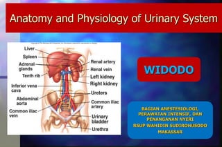
- 1. Anatomy and Physiology of Urinary System WIDODO BAGIAN ANESTESIOLOGI, PERAWATAN INTENSIF, DAN PENANGANAN NYERI RSUP WAHIDIN SUDIROHUSODO MAKASSAR
- 2. Introduction Organ system that produces, stores, and carries urine Humans produce about 1.5 liters of urine over 24 hours, although this amount may vary according to the circumstances. Increased fluid intake generally increases urine production. Increased perspiration and respiration may decrease the amount of fluid excreted through the kidneys. Some medications interfere directly or indirectly with urine production, such as diuretics.
- 4. Kidney Location and External Anatomy
- 5. Functions of the Kidney: Maintaining balance Regulation of body fluid volume and osmolality Regulation of electrolyte balance Regulation of acid-base balance Excretion of waste products (urea, ammonia, drugs, toxins) Production and secretion of hormones Regulation of blood pressure
- 6. A. Renal Vein B. Renal Artery C. Ureter D. Medulla E. Renal Pelvis F. Cortex 1. Ascending loop of Henle 2. Descending loop of Henle 3. Peritubular capillaries 4. Proximal tubule 5. Glomerulus 6. Distal tubule The Kidney and the Nephron
- 7. The Nephron Functional unit of the kidney (1,000,000) Responsible for urine formation: – Filtration – Secretion – Reabsorption
- 8. •Glomerulus •Afferent and Efferent arterioles •Proximal Tubule •Loop of Henle •Distal Tubule •Collecting Duct Components of the nephron
- 9. From http://www.emc.maricopa.edu/faculty/farabee/BIOBK/BioBookEXCRET.html Overview of nephron function
- 10. Filtration
- 11. THE GLOMERULUS
- 12. •Components of plasma cross the three layers of the glomerular barrier during filtration •Capillary endothelium •Basement membrane (net negative charge) •Epithelium of Bowman’s Capsule (Podocytes –filtration slits allow size <60kD) •The ability of a molecule to cross the membrane depends on size, charge, and shape • Glomerular filtrate therefore contains all molecules not contained by the glomerular barrier - it is NOT URINE YET! Plasma is filtered through the glomerular barrier
- 13. Glomerular Filtration Rate (GFR) Measure of functional capacity of the kidney Dependent on difference in pressures between capillaries and Bowman’s space Normal = 120 ml/min =7.2 L/h=180 L/day!! (99% of fluid filtered is reabs.)
- 14. The Response to a Reduction in the GFR
- 16. Reabsorption Active Transport –requires ATP – Na+, K+ ATP pumps Passive Transport- – Na+ symporters (glucose, a.a., etc) – Na+ antiporters (H+) – Ion channels – Osmosis
- 17. Factors influencing Reabsorption Saturation: Transporters can get saturated by high concentrations of a substance - failure to resorb all of it results in its loss in the urine (eg, renal threshold for glucose is about 180mg/dl). Rate of flow of the filtrate: affects the time available for the transporters to reabsorb molecules.
- 18. What is Reabsorbed Where? Proximal tubule - reabsorbs 65 % of filtered Na+ as well as Cl-, Ca2+, PO4, HCO3 -. 75-90% of H20. Glucose, carbohydrates, amino acids, and small proteins are also reabsorbed here. Loop of Henle - reabsorbs 25% of filtered Na+. Distal tubule - reabsorbs 8% of filtered Na+. Reabsorbs HCO3-. Collecting duct - reabsorbs the remaining 2% of Na+ only if the hormone aldosterone is present. H20 depending on hormone ADH.
- 20. Secretion Proximal tubule – uric acid, bile salts, metabolites, some drugs, some creatinine Distal tubule – Most active secretion takes place here including organic acids, K+, H+, drugs, Tamm-Horsfall protein (main component of hyaline casts).
- 21. Countercurrent exchange The structure and transport properties of the loop of Henle in the nephron create the Countercurrent multiplier effect. A substance to be exchanged moves across a permeable barrier in the direction from greater to lesser concentration. Image from http://en.wikipedia.org/wiki/Countercurrent_exchange
- 22. Loop of Henle – Goal= make isotonic filtrate into hypertonic urine (don’t waste H20!!) – Counter-current multiplier: Descending loop is permeable to Na+, Cl-, H20 Ascending loop is impermeable to H20- active NaCl transport Creates concentration gradient in interstitium Urine actually leaves hypotonic but CD takes adv in making hypertonic
- 23. Hormones Produced by the Kidney Renin: – Released from juxtaglomerular apparatus when low blood flow or low Na+. Renin leads to production of angiotensin II, which in turn ultimately leads to retention of salt and water. Erythropoietin: – Stimulates red blood cell development in bone marrow. Will increase when blood oxygen low and anemia (low hemoglobin). Vitamin D3: – Enzyme converts Vit D to active form 1,25(OH)2VitD. Involved in calcium homeostasis.
- 24. Renin, Angiotensin, Aldosterone: Regulation of Salt/Water Balance
- 25. Aldosterone Secreted by the adrenal glands in response to angiotensin II or high potassium Acts in distal nephron to increase resorption of Na+ and Cl- and the secretion of K+ and H+ NaCl resorption causes passive retention of H2O
- 26. Anti-Diuretic Hormone (ADH) Osmoreceptors in the brain (hypothalamus) sense Na+ concentration of blood. High Na+ (blood is highly concentrated) stimulates posterior pituitary to secrete ADH. ADH upregulates water channels on the collecting ducts of the nephrons in the kidneys. This leads to increased water resorption and decrease in Na concentration by dilution
- 27. Ureters Slender tubes that convey urine from the kidneys to the bladder Ureters enter the base of the bladder through the posterior wall – This closes their distal ends as bladder pressure increases and prevents backflow of urine into the ureters
- 28. Ureters Ureters have a trilayered wall – Transitional epithelial mucosa – Smooth muscle muscularis – Fibrous connective tissue adventitia Ureters actively propel urine to the bladder via response to smooth muscle stretch
- 29. Chapter 25: Urinary System 29 Urinary Bladder Smooth, collapsible, muscular sac that temporarily stores urine It lies retroperitoneally on the pelvic floor posterior to the pubic symphysis – Males – prostate gland surrounds the neck inferiorly – Females – anterior to the vagina and uterus Trigone – triangular area outlined by the openings for the ureters and the urethra – Clinically important because infections tend to persist in this region
- 30. Chapter 25: Urinary System 30 Urinary Bladder The bladder wall has three layers – Transitional epithelial mucosa – A thick muscular layer – A fibrous adventitia The bladder is distensible and collapses when empty As urine accumulates, the bladder expands without significant rise in internal pressure
- 31. Chapter 25: Urinary System 31 Urinary Bladder Figure 25.18a, b
- 32. Urethra Muscular tube that: – Drains urine from the bladder – Conveys it out of the body
- 33. Urethra Sphincters keep the urethra closed when urine is not being passed – Internal urethral sphincter – involuntary sphincter at the bladder-urethra junction – External urethral sphincter – voluntary sphincter surrounding the urethra as it passes through the urogenital diaphragm – Levator ani muscle – voluntary urethral sphincter
- 34. Chapter 25: Urinary System 34 Urethra The female urethra is tightly bound to the anterior vaginal wall Its external opening lies anterior to the vaginal opening and posterior to the clitoris The male urethra has three named regions – Prostatic urethra – runs within the prostate gland – Membranous urethra – runs through the urogenital diaphragm – Spongy (penile) urethra – passes through the penis and opens via the external urethral orifice
- 35. Chapter 25: Urinary System 35 Urethra Figure 25.18a. b
- 36. Micturition (Voiding or Urination) The act of emptying the bladder Distension of bladder walls initiates spinal reflexes that: – Stimulate contraction of the external urethral sphincter – Inhibit the detrusor muscle and internal sphincter (temporarily) Voiding reflexes: – Stimulate the detrusor muscle to contract – Inhibit the internal and external sphincters
- 37. Chemical Composition of Urine Urine is 95% water and 5% solutes Nitrogenous wastes include urea, uric acid, and creatinine Other normal solutes include: – Sodium, potassium, phosphate, and sulfate ions – Calcium, magnesium, and bicarbonate ions Abnormally high concentrations of any urinary constituents may indicate pathology
- 38. ACUTE KIDNEY INJURY WIDODO RSUP WAHIDIN sUDIROHUSODO MAKASSAR
- 39. Pendahuluan Salah satu kondisi yang paling sering terjadi pada kasus-kasus trauma dan penyakit kritis. Gagal ginjal akut (ARF) Sistem scoring keparahan penyakit seperti APACHE III dan SOFA, memberi bobot yg cukup besar terhadap disfungsi ginjal
- 40. Pendahuluan Disfungsi Ginjal • Berat • Memerlukan RRT • Ringan Perubahan kecil nilai kreatinin atau produksi urin Mempengaruhi morbiditas dan mortalitas pasien ARF paling sering terjadi ICU dan sering merupakan bagian dari disfungsi organ lainnya
- 41. Pendahuluan Jaringan Kolaborasi berbagai kelompok : ADQI= the Acute Dialysis Quality Initiative ASN = American Society of Nephrology NKF = the National Kidney Foundation dan European Society of Intensive Care Medicine AKIN the Acute Kidney Injury Network AKI
- 42. DEFINISI Belum ada konsensus terhadap berapa besar disfungsi ginjal yg dsb AKI. Acute Kidney Injury klasifikasi Risk, Injuri, Failure, Loss and End Stage Kidney RIFLE ADQI
- 43. DEFINISI mendefenisikan 3 tingkatan keparahan Risk ( kelas R ) Injuri ( Kelas I ) Failure ( Kelas F ) Loss dan End Stage Kidney Disease Risiko disfungsi ginjal Sdh terjadi injuri pd ginjal Gagal ginjal kelas outcome Kelas Tingkatan
- 44. Gambar 1. Skema klasifikasi AKI berdasarkan kriteria RIFLE (dikutip : Belomo A, Ronco C,Kellum JA,et al.ARCritical care 2004,8:R204-R212 )
- 45. DEFINISI Pasien Masuk RS Asumsi GFR awal normal Tidak ada data awal fungsi ginjal Gunakan Nilai Kreatinin Serum Usul ADQI Rumus MDRD untuk perhitungan GFR Modification of Diet in Renal Disease 75-100 ml/menit per 1,73m2 Rumusan MDRD ini hanya dipakai untuk memperkirakan kreatinin serum baseline GFR perkiraan 75(ml/min per 1.73 m2) = 186 x (Scr) - 1.54 x (umur) - 0.0203 x (0.742 Perempuan )x(1.210 Kulit hitam )
- 46. Tabel 1. Perkiraan kreatinin serum baseline Age (years) Black males (mg/dl [μmol/l]) Other males (mg/dl [μmol/l]) Black females (mg/dl [μmol/l]) Other females (mg/dl [μmol/l]) 20–24 1.5 (133) 1.3 (115) 1.2 (106) 1.0 (88) 25–29 1.5 (133) 1.2 (106) 1.1 (97) 1.0 (88) 30–39 1.4 (124) 1.2 (106) 1.1 (97) 0.9 (80) 40–54 1.3 (115) 1.1 (97) 1.0 (88) 0.9 (80) 55–65 1.3 (115) 1.1 (97) 1.0 (88) 0.8 (71) >65 1.2 (106) 1.0 (88) 0.9 (80) 0.8 (71)
- 47. DEFINISI AQDI AKIN Berkurangnya fungsi ginjal secara mendadak (dlm 48 jam) yg didefenisikan sebagai peningkatan kreatinin serum lebih dari atau sama dengan 0,3mg/dl (≥26,4 umol/l),atau peningkatan persentase kreatinin serum lebih dari atau sama dengan 50% (1,5 kali base line) atau berkurangnya urin output (oligurio kurang dari 0,5 ml/kg per jam selama lebih dari 6 jam
- 48. DEFINISI AKIN Mengusulkan penyempurnaan kriteria RIFLE Penelitian Terbaru Perubahan Kecil Kreatinin Serum Berhubungan dengan ↑ mortalitas < 48 jam Termasuk AKI Kreatinin ≥ 26,2umol/l Memerlukan RRT Termasuk AKI Stadium I AKI Stadium III
- 49. DEFINISI Tabel 2: Perbandingan Definisi dan Skema Klasifikasi AKI berdasarkan RIFLE dan AKIN Risk Injury Failure Peningkatan Cr serum≥1,5x baseline atau penurunan GFR≥25% Peningkatan Cr serum≥ 2 x baseline atau penurunan GFR≥50% Peningkatan Cr serum≥ 3 x baseline atau penurunan GFR≥ 75% atau Cr ≥ 354umol/L dengan peningkatan akut sekurangnya 44umol/L <0,5 mL/kg/h ≥ 6 jam <0,5 mL/kg/h ≥12jam <0,5 mL/kg/h ≥24jam atau anuria ≥ 12 jam. AKIN Kriteria Kriteria kreatinin serum Kriteria Urin Output Stage 1 Stage 2 Stage 3 Peningkatan Cr serum ≥ 26,2umol/L atau ≥150-199%(1,5-1,9kali)baseline Peningkatan Cr serum 200-299%(>2-2,9 kali) baseline Peningkatan Cr serum ≥354umol/L dengan peningkatan sekurangnya 44umol/L atau dimulainya RRT <0,5 mL/kg/h ≥ 6 jam <0,5 mL/kg/h ≥12jam <0,5 mL/kg/h ≥24jam atau anuria ≥ 12 jam RIFLE Kriteria Kreatinin Serum Kriteria urin output
- 50. pasien yg memenuhi defenisi AKI memiliki 3 kali kecenderungan mati selama perawatan di RS. Mereka secara bermagna memerlukan dialisis dan lama perawatan lbh lama dibandingkan pasien tanpa AKI DEFINISI Perbandingan Kriteria AKIN & RIFLE penelitian multisenter terhadap 120.123 pasien sakit kritis oleh Bangshaw dkk AKIN tdk lbh sensitif dari pd RIFLE dlm mendiagnosis AKI dlm 24 jam pertama di ICU penelitian secara kohor pd 471 pasien yd dirawat di ICU selama 1 thn oleh Barrantes dkk
- 51. EPIDEMIOLOGI AKI berat & perlu RRT 5% di ICU ARF 20 tahun terakhir ARF yang memerlukan RRT 20 tahun terakhir 61 288 per 100.000 populasi 4 27 per 100.000 populasi AKI di USA periode penelitian 15 tahun 4 kali lipat dari 610 menjadi 2880 pasien AKI di Australia 18% AKI di AS 12,4% masuk kategori RIFLE Risk, 26,7% RIFLE Injury dan 28,1% RIFLE Failure
- 52. ETIOLOGI Bersifat fungsional dan secara definisi tidak disertai perubahan histopatologi. Jika sdh terjadi kerusakan pada struktur nefron sprti: glomerulus,tubulus,pembuluh darah dan interstisial. Terjadi pd obstruksi traktus urinarius.
- 53. Tabel 3. Penyebab AKI Pre Renal Volume responsive Intrinsik Post renal Hipovolemia - Muntah dan diare - Perdarahan Berkurangnya volume sirkulasi efektif - Gagal jantung - Septic shock - Sirosis Obat - ACE inhibitors Glomerular - Glomerulonefritis Glomerular endothelium - Vaskulitis - HUS - Hipertensi maligne Tubular - ATN - Rhabdomyolisis - Myeloma Intersisial - Nefritis intersisial Obstruksi - Batu ginjal - Fibrosis retroperitoneal - Hypertrophy prostat - Carcinoma - Striktur uretra - Neoplasma bladder - Neoplasma pelvis - Neoplasma retroperitoneal ETIOLOGI
- 54. OUTCOME Mortalitas 19 – 83%. Kematian di RS dgn RIFLE •Klas R 8,8%, •Klas I 11,4%, •Klas F 26,3% •Pasien tanpa AKI 5,5% Lama Perawatan ICU dan RS Pasien dengan AKI memiliki lama perawatan di ICU dan rumah sakit yang lebih lama jika dibandingkan dengan pasien tanpa AKI Morbiditas End Stage • Biaya yang mahal • Menurunnya kualitas kesehatan seseorang, • Mortalitas yang lebih besar dari populasi secara umum ( 28,1%) • Pemulihan fungsi ginjal menjadi salah satu outcome Yang penting untuk dievaluasi.
- 55. PENATALAKSANAAN Konsensus Mengenai Terapi AKI Yang Efektif Belum ada karena: 1. Penyebab AKI yang multifaktorial 2. Bervariasinya definisi AKI. 3. Penilaian penurunanGFR yang tergantung pada perubahan kreatinin serum. 4. Tingginya angka mortalitas AKI 5. Tidak ada konsensus kapan dan jenis dialisis apa yang tepat untuk penderita AKI.
- 56. PENATALAKSANAAN Penelitian pd Hewan agent yg terbukti efektif utk AKI Loop diuretik Low-dose dopamin ANP Hormon tyroid IGF-1 Penelitian secara klinis tidak ada yg terbukti efektif
- 57. PENATALAKSANAAN Renal Replacment Therapy (RRT) ◦ Pengganti ginjal ( Renal Replacement) ◦ Pendukung fungsi ginjal/organ lainnya (Renal/multi-organ support ◦ Berdasarkan mekanisme pengeluaran cairan/solud dan Intermitten atau Kontinyu ◦ Semua RRT kecuali PD dicapai dengan Ultrafiltrasi • Gradient tekanan akan mendorong cairan melewati membran semipermiabel. • Laju UF dipegaruhi oleh: gradien tekanan trensmembran, permeabiltas air membran, dan luas permukaan membran.
- 58. PENATALAKSANAAN Renal Replacment Therapy (RRT) Berdaarkan mekanisme utama removal solute difusi dan konveksi Removal solute yang Predominan pada masing-masing jenis RRT 1. Intermittent haemodialysisi ( IHD) – difusi 2. Continous venovenous haemofiltration (CVVH) – konveksi 3. Continous venovenous haemodialysis ( VVHD) – difusi 4. Continous venovenous haemodiafiltration (VVHDF) – difusi dan konveksi
- 59. PENATALAKSANAAN Renal Replacment Therapy (RRT) Inisiasi:1. Oliguria (UO < 200 ml/12 jam 2. Anuria ( UO : 0-50 ml/12jam) 3. Urea > 35 mmol/l 4. Creatinin > 400 umol/l 5. K > 6,5 mmol/L atau peningkatan yang cepat 6. Udem pulmo yang refrakter dengan diuretik 7. Asidosis metabolik yang tak terkompensasi ( pH<7,1) 8. Na < 110 dan > 160 mmol/l 9. Temperatur > 40C 10. Komplikasi uremia : ( ensefalopati,miopati, neuropati dan perikarditis) 11. Overdosis obat/ toksin yang dialyzable Jika ada satu kriteria, RRT harus dipertimbangkan. Jika ada dua kriteria secara bersamaan, RRT sangat dianjurkan Tabel 4.Indikasi modern (R.Bellomo ) untuk memulai RRT pada AKI
- 60. PENATALAKSANAAN Renal Replacment Therapy (RRT) Penggunaan kriteria konvensional untuk memulai RRT Grade D RRT seharusnya dimulai sebelum terjadi komplikasi Grade E Laju perubahan urea dan kreatinin lebih bermakna daripada nilai absolutnya Grade C Tetapi pada kebanyakan kasus, RRT dimulai sebelum urea mencapai 20-30 mmol/L). RRT harus dimulai berdasarkan balans cairan, jumlah urin, kadar kalium ataupun derajat asidosis tergantung kondidi klinis pasien.
- 61. PENATALAKSANAAN Renal Replacment Therapy (RRT) Pilihan Metode RRT IHD CRRT SLED Mekanism removal cairan Ultrafiltrasi Ultrafiltrasi Ultrafiltrasi Mekanisme removal solute Difusi Difusi dan atau konveksi Difusi Blood Flow rate ≥ 200 ml/menit < 200 ml/menit 200 ml/menit Dialysate flow rate ≥ 500 ml/menit 17-34 ml/menit 300 ml/menit Durasi 3-4 jam 24 jam/ hari 6-12 jam/hari
- 62. PENATALAKSANAAN Renal Replacment Therapy (RRT) Keuntungan dan pertimbangan khusus IHD CRRT SLED Removal cairan yang cepat √ Bersihan solute cepat √ Hiperkalemia berat √ Hempdinamik tak stabil √ √ Kontrol cairan lebih baik √ √ -High nutritional Support -Removal solute MMW √ √ ?
- 64. In the United States, there is a rising incidence and prevalence of Kidney Disease. Nearly 350,000 of these are on dialysis. Also, there is an increasing prevalence of earlier stages of chronic kidney disease which unfortunately is “under- diagnosed” and “under-treated” in the United States. In 2000, the National Kidney Foundation (NKF) Kidney Disease Outcomes Quality Initiative (K/DOQI) Advisory Board approved development of clinical practice guidelines to define chronic kidney disease and to classify stages in the progression of chronic kidney disease. Chronic Kidney Disease
- 65. Stages of Chronic Kidney Disease Stage 1 Kidney damage with normal or ↑ GFR GFR ≥ 90 ml/min/1.73 m2 Stage 2 Kidney damage with mild ↓ GFR GFR 60-89 Stage 3 Moderate ↓ GFR GFR 30-59 Stage 4 Severe ↓ GFR GFR 15-29 Stage 5 Kidney failure GFR <15 (or dialysis)
- 66. Causes of End Stage Renal Disease 0% 10% 20% 30% 40% 50% 60% 70% 80% 90% 100% All W hite Black Asian Am erIndian % Other Interstit N Cystic KD GN BP Diabetes USRDS Annual Data Report
- 67. Chronic Kidney Disease Many terms are used to describe states of reduced glomerular filtration (GFR) not requiring renal replacement therapy; – Chronic Renal Insufficiency – Chronic Renal Failure – Renal Insufficiency – Pre dialysis renal disease – Pre uremia – Renal dysfunction They are imprecise & poorly defined.
- 68. Measurement of GFR – Gold standard is Inulin Iothalamate. – Creatinine Clearance calculated by timed (24h) urine collection along with serum collection for Creatinine. – Overestimate GFR when CKD is severe due to an increase in tubular secretion of creatinine. – This factor can be corrected by cimetidine. Estimation of GFR – More than 10 formulae for estimation of GFR. – MDRD most widely accepted now. Chronic Kidney Disease
- 69. CKD – Risk Factors Diabetes Mellitus Hypertension Cardiovascular Disease Obesity Metabolic Syndrome Age and Race Acute Kidney Injury Malignancy Family history of CKD Kidney Stones Infections like Hep C and HIV Autoimmune diseases Nephrotoxics like NSAIDS
- 70. CKD - Causes Diabetic Non Diabetic – Glomerular Nephritic: PIGN, IgA, MPGN Nephrotic: FSGS, Membranous, Amyloidosis – Tubulointerstitial: Analgesic, Reflux, Ch. Obs – Vascular: Vasculitis, HTN, RAS – Cystic: ADPKD – CKD in transplantation
- 71. CKD - Causes
- 72. CAUSES OF DEATH IN ESRD 39% 5%26% 11% 15% 4% Cardiac Cerebrovascular Other known Unknown Infection Malignancy U.S. Renal Data System: USRDS 2002
- 73. Abnormal Sodium-Water metabolism – Edema, Hypertension Abnormal Acid-base abnormalities – Metabolic Acidosis due to uremia or RTA Abnormal hematopoesis – Anemia of CKD Cardiovascular Abnormalities – LVH, CAD, Diastolic Dysfunction Abnormal Calcium-Phosphorus metabolism – Hyperphosphatemia, pruritus, arthralgia – Hyperparathyroidism – Renal Osteodystrophy CKD - Manifestations
- 74. CKD - Management Diagnostic work up to decide underlying etiology Treatment of Hypertension and Dyslipidemia Treatment of Anemia Treatment of Hyperphosphatemia Avoidance of Dehydration & Nephrotoxic agents Proper Dosing of Drugs Preparation for Renal Replacement Therapy
- 75. CKD - Evaluation
- 76. Serum electrolytes Urine spot protein analysis (24 hour no longer recommended). ANA, C3, C4 SPEP, UPEP Kidney Ultrasound Urine sediment analysis Biopsy – Evidence of glomerular disease without diabetes – Sudden onset of nephrotic syndrome or glomerular hematuria CKD - Evaluation
- 77. CKD - Hypertension Anti-Hypertensive Agents – Single most important measure could be adequate BP control – Target BP <130/80 with minimal proteinuria and BP<125/75 with significant proteinuria (>1g). – ACEIs and ARBs have been demonstrated to slow both diabetic and non-diabetic renal disease in both experimental and human studies. – Decrease the sodium intake to 2.5 g /day – Usually requires more than 2 medications. – Diuretics enhance the antihypertensive and antiproteinuric effects of other agents..
- 78. CKD - Dyslipidemia Dyslipidemia and Cardiovascular morbidity – Several studies like the 4D study showed no benefit of statins in dialysis patients. – However, post hoc analysis of this data does suggest that the management of dyslipidemia in CKD 2 – 4 improves cardiac mortality and morbidity. – Dyslipidemia is frequently seen in glomerular disease with proteinuria (nephrotic syndrome) and its control reduces atherosclerosis related morbidity and mortality.
- 79. CKD - Anemia Decreased quality of life with anemia. Diagnosis of exclusion. Mostly apparent in the stage 4 and 5 of CKD. Due to decrease in EPO production in the kidney.
- 80. CKD - Anemia Erythropoietin – Epoetin alfa :Procrit ® , Epogen® – Darbepoietin Alpha: ARANESP ® Target Hg levels between 11g and 12g but not exceeding 13g. Greater than 13g showed increased mortality as per the CHOIR study. Sufficient Iron should be administered to correct iron stores.
- 81. CKD - Hyperphosphatemia Control of Hyperphosphatemia – Due to decreased excretion in urine. – Control of hyperphosphatemia by dietary measures slow progression in experimental models of CKD. – Hyperphosphatemia leads to pruritus, calcification in synovial membranes, blood vessels and even cardiac valves. – Therapy includes Phosphorus restriction to 800mg/day and use of phosphrous binders with food. Calcium Carbonate (TUMS), Ca-acetate (PHOSLO) Lanthanum Renagel
- 82. CKD – Bone and Mineral disease Hyperparathyroidism: – High phosphorus and low Vitamin D causing low calcium. – Monitor Intact PTH levels and keep between 100 and 500. – Maintain Phosphorus and Calcium within normal ranges. – Vitamin D analog paricalcitol. – Calcimimetic agents like cinacalcet.
- 83. CKD - Nephrotoxics Avoidance of Dehydration/Nephrotoxic Agents – Drugs such as Aminoglycosides, NSAIDs – Avoiding exposure to Radio contrast agents. – In presence of dehydration, even in absence of renovascular disease, ACEIs or ARBs can aggravate renal dysfunction – Dehydration is frequent in tubulo-interstitial disorders where urinary concentration is impaired. – Proper Dosing of Drugs eg. Allopurinol
- 84. CKD – Medication Dosing Proper Dosing of Drugs – Uremia affects GI absorption; eg Iron. – Impaired plasma protein binding of drugs; eg Dilantin. – Increased volume of distribution; – Excretion of many drugs depends upon the kidney; Some drugs used in normal dose will lead to nephrotoxic effects eg. Allopurinol Other drugs when used in normal dose will lead to other toxic effects eg. Vancomycin. Dose Reduction or Interval Extension
- 85. CKD - RRT Preparation for Renal Replacement Therapy – Education for Options of Dialysis & Renal Transplantation for Renal Replacement – Hemodialysis Vs Peritoneal Dialysis – Avoidance of Veni-puncture & insertion of catheters in peripheral veins once GFR < 60mls. – Timely placement of vascular access or PD catheter.
- 86. CKD - RRT Indications (Absolute): – Uncontrolled hyperkalemia and acidosis – Uncontrollable hypervolemia (pulmonary edema) – Pericarditis – AMS and somnolence (advanced encephalopathy) – Bleeding diathesis Indications (Relative): – Nausea, vomiting and poor nutrition – Metabolic acidosis – Lethargy and Malaise – Worsening kidney function <10 ml or <15 ml in diabetics
- 87. CKD - RRT Transplantation: – Preemptive transplant carries both patient and graft survival advantage. – Graft survival better with living donor kidneys. – Immunosuppresion is almost always a must.
- 88. CKD - RRT Transplantation: – Diseases like FSGS may reccur early in the transplanted kidney. – Increased risk for infection, bone loss, cardiovascular disease. – Contraindications: Malignancy (recent or metastatic) Current infection Severe extra renal disease Active use of illicit drugs