
Recommended
More Related Content
What's hot
What's hot (20)
Similar to Virology - Microbiology
Similar to Virology - Microbiology (20)
More from MBBS Help
More from MBBS Help (20)
Recently uploaded
Recently uploaded (20)
Virology - Microbiology
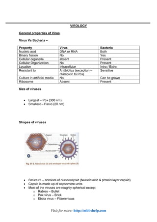
- 1. Visit for more: http://mbbshelp.com VIROLOGY General properties of Virus Virus Vs Bacteria – Property Virus Bacteria Nucleic acid DNA or RNA Both Binary fission No Yes Cellular organelle absent Present Cellular Organization No Present Location Intracellular Intra / Extra Resistant to Antibiotics (exception – rifampicin to Pox) Sensitive Culture in artificial media No Can be grown Ribosome Absent Present Size of viruses Largest – Pox (300 nm) Smallest – Parvo (20 nm) Shapes of viruses Structure – consists of nucleocapsid (Nucleic acid & protein layer capsid) Capsid is made up of capsomere units Most of the viruses are roughly spherical except o Rabies – Bullet o Pox virus – Brick o Ebola virus – Filamentous
- 2. Visit for more: http://mbbshelp.com o Tobaco mosaic virus – Rod shaped Nucleic acid – Made up either DNA or RNA DNA viruses- Herpes, HBV, Adeno, Papova, Parvo & Pox RNA viruses – others All the RNA viruses are single stranded except Reoviruses (double stranded RNA) All the DNA viruses are double stranded except Parvoviruses (single stranded DNA) Symmetry – Icosahedron – All DNA, most of the RNA virus possess icosahedron symmetry Helical – Few RNA viruses (Bunya, Myxo, Rhabdo, Filoviridae) Pox – complex symmetry Envelope : Enveloped Virus: Made up lipoprotein subunits called peplomere Lipid part is host cell membrane derived & protein part is virus derived Envelop provides chemical, physical & biological properties to cell. Ether sensitive, heat labile, pleomorphic Example – all, other than non enveloped virus are enveloped virus Non-Enveloped virus : Ether resistant, heat stable & non-pleomorphic DNA – Parvo, Adeno, Papova (PAP) RNA – Picorna, Astrovirus, Calcivirus, Reovirus (PARC) Segmented RNA : Bunya, Influenza Rota Arena Replication 1. Adsorption- Most specific step requires respective receptors If bypassed then any virus can attack any cell 2. Penetration 3. Uncoating – 4. Biosynthesis- DNA Viruses – Replicates in nucleus (except – Pox) RNA Virus – Replicates in Cytoplasm (expect Myxo & Retro virus) Viral protein is synthesized only in cytoplasm
- 3. Visit for more: http://mbbshelp.com +ve sense RNA virus Viral RNA itself acts as mRNA Infectious & translated directly to protein E.g. – Picoma, Togaviruses -ve Sense RNA virus Have polarity opposite to mRNA Non infectious & possess their own RNA polymerase for transcription to form mRNA E.g. Myxo, Rabies Retroviruses – Viral reverse transcriptase converts viral ssRNA to dsDNA Then dsDNA integrates with host DNA 5. Maturation 6. Assembly 7. Release Bacteriophage – by host cell lysis Animal virus – usually without lysis (Myxo – by budding) Exception – Picoma – by host cell lysis Viral Cultivation Animal inoculation Coxsackie – A- flaccid paralysis, B-spastic paralysis Arbovirus Egg inoculation Chorioamnicotic membrane – Produce pocks E.g. Vaccinia, Variola, HSV Yolk sac – arbovirus, Chlamydia, Rickettsia Amniotic membrane – Influenza culture Allantoic cavity – vaccine preparation for Influenza, Yellow (17D), Rabies (Flury) Tissue culture Organ culture – tracheal ring (corona) Explant culture – adenoid (Adeno) Cell line – Primary cell line – Undergo limited division (5-10), diploid karyosome o E.g. – Rhesus Kidney cell line, Human amniotic cell line, chick embryo fibroblast
- 4. Visit for more: http://mbbshelp.com Secondary cell line – Undergo moderate cell division (10-50), diploid karysome o E.g. – Human fibroblast used for CMV Continuous cell line – indefinite divisions haploid karysome o E.g. – HeLa, Hep2, BHK Inclusion bodies Intracytoplasmic Negri body – rabies Guarnier body – Vaccinia, Paschen body – Variola Bollinger body – Fowl pox Molluscum body – Molluscum contagiosum virus Intranuclear – Cowdry A – Herpes , Yellow fever Cowdry B – Adeno, Polio Latent virus Herpes – HSV I, II, VZV – nerve CMV – kidney, secretory gland EBV – lymphoid HIV – CD4 T cell Slow virus – neuron Teratogenic virus CMV Rubella Herpes VZV Transfer through Placenta – Coxsackie B Hepatitis B, C Parvo B19 HIV Measles, Mumps VIRAL VACCINE Inactivated Vaccine – Salk Influenza Japanese B – (Nakayama) formalinized mouse brain
- 5. Visit for more: http://mbbshelp.com Hepatitis B (submit – HBs Ag cloned in yeast) Rabies – BPL, Semple and non neural (PVC, PCEC, HDC) Live Vaccine – Sabin Polio – avirulent MKD Influenza – egg Japanese B – (14-14-2) MMR Yellow (17D) – chick embryo Small pox Oka Strain – Varicella Towne & AD – CMV Jerry Lynn – Mumps Edmonston – Zagreb : Measles RA 27/3 – Rubella
- 6. Visit for more: http://mbbshelp.com HERPES HSV 1 & 2 – differentiated by HSV 1 : lesion around mouth, HSV 2 : lesion around genitalia, Transmission – HSV1 – contact droplet inhalation, HSV2 – sexual mode HSV 2 larger pocks in Chick embryo HSV 2 replicates well in Chick embryo fibroblast HSV 2 more drug resistance Antigen detection & PCR also can differentiate Clinical feature
- 7. Visit for more: http://mbbshelp.com Mucosal – MC site – buccal mucosa Most frequent primary lesion – gingivostomatitis, pharyngitis Most frequent recurrent lesion – herpes labialis MC cause of ulcerative stomatitis – HSV Secondary bacterial infection is common with – Streptococcus, Pneumococcus CNS- MC sporadic acute viral encephalitis Meningitis Most imp diagnosis – brain biopsy GBS Cutaneous – MC site – Face Herpetic whitlow – seen in doctor, nurse Erythema multiforme – MC cause – HSV Ophthalmic – Acute keratoconiuncctivitis Dendritic ulcer – Rx – Topical acyclovir, IFN C/I steroid Genital – HSV II MC than HSV I Diagnosis – TZANCK smear of keratinocyte Toludine blue staining of base of vesicle reveals multinucleated giant cell with faceted nuclei & homogenously stained ground glass appearance. Type A intranuclear inclusion body in Giemsa stain Isolation = CAM, primary human kidney cell line Varicella Zoster Varicella (Chicken pox) MC site – Spinal cord One attack gives life long immunity Source – patients Portal of entry – respiratory tract or conjunctiva Incubation period – 2 weeks
- 8. Visit for more: http://mbbshelp.com Infectious during initial stage - 2 to 5 days of onset of rash Rash – usually start in trunk, rapid evolution, centripetal distribution Chicken pox is a disease of childhood When occur in adult, it is more severe with bullous & hemorrhagic rash Complication- o Varicella pneumonia o Myocarditis, nephritis, encephalitis, cerebellar ataxia o Reye’s syndrome – fatty liver after salicylate Vaccine – Oka strain (live attenuated) & VZIG (immunoglobulin) Treatment – acyclovir, Steroid is contraindicated Zoster o Reactivation of latent virus o Trigeminal N (Ophthalmic branch) o Segmented & unilateral o Affect Geniculate ganglia – Ramsay hunt syndrome characterized by – Tetrad – facial N palsy + vesicle on tympanic membrane, External auditory meatus & tongue Chicken pox Herpes Zoster (Shingles) Primary infection Reactivation of latent virus MC site – Spinal cord Trigeminal N (Ophthalmic branch) Generalized & bilateral Segmented & unilateral Child > adult (severe) Old age Localization in skin capillary endothelial cells Confined to segment of sensory nerve Persist and reactivate as zoster Act as source of chicken pox CMV Largest member of Herpes virus family Enlargement of the virus infected cell Intra nuclear & cytoplasmic inclusion body – owl’s eye Spread slowly & requires close contact Route – via secretions, sexual, Blood transfusion Exhibit strict host specificity
- 9. Visit for more: http://mbbshelp.com Congenital – Cytomegalic inclusion disease Hepatosplenomegaly (MC) Microcephaly, Mental retardation 3Cs – chorioretinitis, cerebral calcification, convulsion Infants are – highly infectious Transmit the virus in urine for 3-5 year Acquired Mononucleosis like syndrome- Occurs in adult (following Blood Transfusion) Atypical lymphocytosis seen Paul bunnel test (heterophile antibody) is negative Post kidney transplant infection Lab diagnosis – Specimen – urine, saliva, cervix secretion, semen Culture – human fibroblast cell line Growth occurs in 2-3 wk, can be improved by shell vial technique IgM or fourfold rise of IgG Pp65 antigenemia DNA PCR Transplacental CMV infection- o At Birth – IgG antibody +ve, IgM just appearing o At 1 month – IgG antibody +ve, IgM peak Post natal CMV infection Birth IgG antibody +ve, IgM –ve o 1 month – igG antibody +ve, IgM just appearing o 3 month – IgG+, IgM peak Treatment – Gancyclovir, o Foscarnet, o Valgancyclovir, o Cidofovir, o Miribaviris EBV Attach to CD21/CR2 receptor on B cell But atypical lymphocytosis occurs with T cell B cell become immortalized, polyclonally activated leading to hypergammaglobulinemia Not highly contagious, Spread slowly Intimate oral contact required
- 10. Visit for more: http://mbbshelp.com Source – Saliva (Kissing disease) Commonly found in hyperendemic malaria areas Disease – o Hodgkin lymphoma o Burkit lymphoma o Nasopharyngeal Ca- risk factor- genetic, salted fish (nitrosamine), herbal snuff (phorbolester) o Infectious Mononucleosis Infectious Mononucleosis o Glandular fever o Seen in nomimmune young adults following primary infection with EBV o Incubation period 4-8 weeks o Characterized by atypical T cell (IM) LN Hepatosplenomegaly & Rash (after ampicillin) o Heterophile antibody to sheep RBC – Paul bunnel test o Confirmed by monospot test / differential absorption test HHV 6- Sixth disease Exanthem subitum, roseola infantum HHV 8- Kaposi sarcoma, OTHER DNA VIRUSES Small pox Variola – largest virus, possess ds DNA, Brick shaped Only DNA virus which replicates in cytoplasm Small pox –Eradicated from world – 1983 Still can be a potential agent of bioterrorism Paschen body (Variola), Guarneri body (Vaccinia) Vaccinia pocks on CAM – larger necrotic & hemorrhagic than Variola
- 11. Visit for more: http://mbbshelp.com Small pox Eradication is successful because- Exclusively human pathogen no reservoir Source – patient only, No carriers Highly affective Live vaccine o Prepared from Vaccinia Molluscum contagiosum Seen in Children & young adult Pearly white wart like nodule on skin composed of eosinophilic inclusion bodies Humans are the only host Cannot be grown in egg or tissue culture & animal Sexually transmitted Adenovirus DNA, non enveloped, space vehicle shaped Manifestation – o Hemorrahgic cystitis– Adenovirus type 11 & 21 o Infant diarrhea – Adeno 40,41 o STD – Adenovirus type 37 o Epidermic conjunctivitis – Adenovirus type 8,19,37 o Swimming pool conjunctivitis – Adenovirus type 3,7,14 o Pharyngitis & Pneumonia – Adenovirus type 3 & 7 Parvovirus Smallest, ss DNA 5th disease Cause Erythema infectiosum – characterized by rash, arthralgia, lymphadenopathy Slapped cheek appearance – rash 1st on cheek Causes aplastic crisis in sickle cell anemia patients Pregnancy causes non-immune fetal hydrops Transmission – respiratory route / blood Papovavirus Human Papillomavirus Small Pox Chicken Pox Rash-palm & sole & extensor surface Rash-Axilla & flexor surface Fever subsides with appearance of rash Fever rises with each crop of rash
- 12. Visit for more: http://mbbshelp.com Ca cervix – Low risk – type 6, 11 –CIN High risk – type 16,18,31,33 – CaCx Risk factor – early sex, multiple sex partners, multiparous, Condyloma acuminata / genital wart – by Type6,11 Common wart / verruca vulgaris – by Type 1,2,3,4 Vaccine Prophylactic – using HPV 16/18 late structural protein L1 Therapeutic – using HPV 16/18 Early non-structural protein E6/E7 Other Papova viruses- JC virus – Hodgkin disease & PML (progressive multifocl leukoencephalopathy) BK virus renal infection Polyoma virus Simian virus Bacteriophage Viruses that attack bacteria Ds DNA surrounded by protein coat Bacteriophage with ssDNA or RNA –also identified Consists of head, neck & tail 2 cycles – Lytic & lysogenic phage Uses: o Phage typing – staphylococcus, Vi antigen typing of S. Typhi, V. cholera o Transduction o Used as a cloning vector o Used in diagnosis – e.g. Mycobacteriophage o Codes for Toxin – Cholera toxin, VT of EHEC, Botulinum toxin C,D, Diphtheria toxin & Streptococcal pyrogenic toxin A,C
- 13. Visit for more: http://mbbshelp.com MYXO VIRUS Orthomyxo Influenza A,B,C Segmented RNA, unstable Both hemagglutinin (HA)& Neuraminidase (NA) spikes present Paramyxo Para influenza, Measles, Mumps, RSV, HA Spike present in – Parainfuenza, Mumps, Measles, NA spike present in – Parainfuenza, Mumps Single RNA, Inclusion body – intracytoplasmic (only for measles it is both intracytoplasmic & intranuclear) INFLUENZA Influenza – A(MC), B, C Antigenic Shift – Results in Pandemic – MC seen in Type A Antigen drift – Results in Epidemic – MC seen in A,B MC manifestation – URTI MC complication – o Bacterial pneumonia, viral pneumonia o Reye’s syndrome – with Type B (following aspirin) Diagnosis – Egg inoculation – Amniotic cavity – by A,B,C Allanotic cavity –by only A
- 14. Visit for more: http://mbbshelp.com Hemagglutination with Fowl & Guinea pig, RBC Type A – agglutinates with Guinea pig, B-both, C-agglutinates with fowl RBC at 4c Ag detection form nasopharyngeal cells by Immunofluorescence Vaccine – Killed, Live, Recombinant Avian Flu H5N1 Seen from 2003 onwards Only bird to human transmission seen, but no human-human transmission seen Highly virulent H1N1 2009 Flu April 2009 Pandemic – Including India Recombination of 4 strain – (1 Human + 2 Swine + 1 Avian) Swine influenza strain causing pandemics so far- H1N1 classical (1918), H1N2, H3N2, currently 2009 H1N1 recombinant strain Human to human transmission seen Diagnosis – by RT PCR detecting HA & NA genes Treatment – NA Inhibitor – Oseltamivir (Tamiflu), Zanamivir Resistant to Amantadine Vaccine- o Injectable killed – HA Protein o Live nasal spray – HA Protein MUMPS MC cause of Parotitis (Non suppurative enlargement) Complication
- 15. Visit for more: http://mbbshelp.com Resolve except deafness Secondary attack – 85% Human – only host, Source – only patients, no carriers MC seen in Children Once infected, gives lifelong immunity Transmission – Droplet, saliva, Direct contact, Fomite borne, urine Saliva is infectious from -1 to +2 wk of parotitis Specimen – urine, saliva, CSF Lab diagnosis- o Isolation – Egg, MKC, o IgM Antibody by ELISA o CFT using S Antigen o Antibody to S antigen (internal Ag) appear early, goes early – indicate acute infection o Antibody to V antigen (surface Ag) appear late, goes late Vaccine – Jeryl Lynn strain (at 9 months) & MMR MEASLES Rubeola MC childhood rash Source – cases only no subclinical / carrier stage Highest secondary attack rate – 90% Period of communicability - -4 to +5 d of rash Incubation Period – 10 days Koplik’s spot – pathognomic, appear before rash
- 16. Visit for more: http://mbbshelp.com Diagnosis o Warthin finkeldy giant cell o Isolation from nose, throat, conjunctiva & blood- Amniotic route. Complication o Diarrhea o Pneumonia o Otitis media o Encephalitis, o SSPE – (subacute sclerosing panencephalitis) – o high titer antibody in CSF is diagnostic o Suppressed delayed hypersensitivity (false –ve Mantoux test) Vaccine o Prepared – chick embryo or human diploid cell line (no egg vaccine available) o Age – given at 9 months (maternal antibody disappears) o Can be given at 6 months if measles outbreak seen (2nd dose to be given at 9 month) o Type – live attenuated – Edmonston – Zagreb strain. o Side effect – toxic shock sybdrome (d/t contamination of vial), mild measles like illness o Immunoglobulin – can also be given 3 days (In this case live vaccine is given after 8-12 wk) RESPIRATORY SYNCYTIAL VIRUS
- 17. Visit for more: http://mbbshelp.com Only HA, No NA spike MC cause pneumonia & otitis media Age – 6wk – 6 months, new borne are protected due to maternal antibody RSV – MV precipitating factor for Acute asthma Rhino – MC precipitating factor for Acute exacerbation of chronic asthma Reinfection – milder illness Causes epidemics in winter (temperate) & rainy (tropics) Culture – giant cell & Syncytial formation T/T – Ribavirin PARA INFLUENZA VIRUS Para influenza type 1&2 – causes Croup (acute laryngotracheobronchiolitis) Para influenza type 3 – LRTI Type 3 – more Endemic & affects < 1 yr Type 1&2 – affect preschool children RUBELLA German measles, Belongs to family Togaviridae, Not a Myxovirus Identified by interference with Echovirus Source – cases Transmission droplet, contact, sexual Rash on day 1 (face), Lymphadenopathy Forchheimer spots seen. Con genital Rubella
- 18. Visit for more: http://mbbshelp.com o 1st Trim – risk is maximum, after 5th month – risk negligible o Classical Congenital Rubella syndrome – Triad – cataract (MC), deafness, cardiac (PDA) o Expanded Congenital Rubella syndrome – myocarditis, hepatosplenomegaly, bone lesion Vaccination – RA 27/3 live attenuated Prepared from Human diploid cell line Given aft 1 yr, Rash Day wise appearance of rashes: 1st – Rubella 2nd – Chicken pox 3rd – Small pox 4th – Measles 5th Parvo B19 – Exanthem infectiosum 6th – HHV6 – Exanthem subitum / Roseola infantum Vaccine storage : Deep Freezer – Polio Measles Vaccine stored at 4c : DPT, Typhoid, TT, DT, BCG Incubation Period 1wk – Diphtheria (2-6d) 1-2 wk – Measles (10d), small pox – (12d), Pertussis (7-14), Polio (7-14),
- 19. Visit for more: http://mbbshelp.com Tetanus (6-10d) 2-3 wk – chicken pox (14-16d), Mumps (18d), Rubella (18d) PICORNAVIRIDAE Include two major groups of human pathogens: Enteroviruses – Polio & Coxsackie A,B ECHO Virus,Enterovirus (68-71) Rhinoviruses Poliovirus 3 subtypes – Type 1 – MC type Type-2 Type -3 – MC cause of vaccine associated paralysis CFT detects – o C Ag (coreless), o D Ag (dense Ag, type specific) Risk factors – o Following Tonsillectomy o Pregnancy o IM injection o Muscular activity, o Coxsackie A, o Genetic predisposition
- 20. Visit for more: http://mbbshelp.com Pathogenesis Mode if transmission – Faeco oral > Inhalation > conjunctiva Spread – hematogenous spread (MC), also direct neural spread (Following Tonsillectomy) Site of action – Anterior horn of spinal cord Pathological changes always more extensive than distribution of paralysis Clinical types – In apparent infection – 90-95% Abortive infection – 5-10% Aseptic meningitis (non paralytic) -0.1% Paralytic (<0.1%) – Ascending flaccid paralysis (AFP) Lab diagnosis Blood, throat swab, CSF, feces (till 6 wk) Isolation – Monkey kidney tissue culture
- 21. Visit for more: http://mbbshelp.com CFT Antibody – Anti C – comes early, goes early, Anti D- comes late, goes late Polio vaccine Salk (Injectable) Sabin (Oral) Preparation Formalin killed preparation of all 3 types in MKC Each dose contains – Type 1-10 lakh, Type 2-2lakh Type 3-3 lakh of TCID50, Safety Relatively more safer Safer except in immunocompromised pt Efficiency 80-90% by full course 90-100% by 1 or 2 doses Efficacy decreases by Interference by other enteroviruses Frequent diarrheal disease Economy Relatively expensive Economical Duration of protection Need booster doses Periodically Long lasting In epidermics Cannot be used Can be used Herd immunity Not provided Provided Local mucosal immunity Not provided Provided (IgA antibody) Coxsackie virus Group A Coxsackie virus Group B Coxsackie virus
- 22. Visit for more: http://mbbshelp.com Suckling mouse inoculation Flaccid paralysis Generalized myositis Manifestations; Herpangina(Vesicular pharyngitis) Hand foot and mouth disease(Also by Enterovirus 71) Acute hemorrhagic conjunctivitis(Cox: A and Enterovirus 70) Pneumonitis of infant Diarrhoea URTI Fever with Rash Suckling mouse inoculation – Spastic paralysis Focal myositis Necrosis of brown fat Pancreatitis, hepatitis, myocarditis, encephalitis Manifestation: Pleurodynia(Epidemic myalgia)/Bornholm disease: B1,B5 Myocarditis,Pericarditis DM-B4 Pneumonia Both Cox A and B: Aseptic meningitis Encephalitis,Cold,Hepatitis RABIES VIRUS Bullet shaped, -ve sense RNA virus Reservoir- Urban Rabies – Dog 99%, cat Wild life Rabies – Fox, Jackal, Wolf Source – Salvia of Rabid animal Mode of transmission - Bite (MC), Lick on abrasion, corneal transplant, air borne Earliest symptom – Neuritic pain at bite site
- 23. Visit for more: http://mbbshelp.com Sensory N → UMN → sympathetic → mental system Incubation period 1-3 month, IP is shorter in children & upper limb bite (than leg bite) Mechanism – Neural apoptosis, ↓ Acetyl choline Street virus Fixed virus Freshly isolated After serial passage Produce Negri body Doesn’t Produce Negri body Affect salivary gland Doesn’t affect salivary gland Incubation Period -1-12wk IP-5-6 days Used for vaccine Furious Rabies Dumb Rabies 80% of all cases 20% of all cases Hydrophobia, No hydrophobia Absent-fever, Fasciculation, Present – Fever, Fasciculation, Paralytic, Proximal muscle Paralytic, Proximal muscle Seen in unvaccinated individuals Associated with partial vaccine course Antemortein diagnosis : Direct Florescent Antibody test – o Corneal impression smear o Hair follicle of nape of neck Isolation – mice cell line followed by immunofluorescence. Antibody in CSF RNA detection by RT-PCR Post mortem diagnosis : Negri body- Mouse inoculation, Isolation Vaccine – Neural – Semple, BPL, Infant brain vaccine. Poor Immunogenic, encephalogenic Non neural Purified chick embryo cell (PCEC) Purified vero cell (PVC)
- 24. Visit for more: http://mbbshelp.com Human diploid cell (HDC) Protection – for 6m Pre Exposure – 3 dose- at day 0,7,28 Post exposure – 6 dose, at day 0,3,7,14,28 & 90 days ARBOVIRUSES Mosquitoes Japanese encephalitis, dengue, yellow fever, St. Louis encephalitis, EEE, WEE, VEE etc. Ticks Crimean-Congo haemorrhagic fever,KFD,RSSE virus,Colorado Tick fever virus, Sandflies Sicilian sandfly fever, Rift valley fever 1.Togaviridae e.g. EEE, WEE, and VEE,Chikungunya 2. Bunyaviridae e.g. Sandfly Fever, Rift Valley Fever, 3. Flaviviridae e.g. Yellow Fever, dengue, Japanese Encephalitis 4.Reoviridae – Colorado tick fever virus 5.Rhabdoviridae - Chandipura virus 6.Orthomyxoviridae Arboviruses common in India – Hemorrhagic – Dengue, Chiungunya, KFD Encephalitis – Japanese B, Westnile, Sindbis Rara – Ganjam, Vellore, Chandipura, Bhanja Yellow fever NOT found in India Animal Reservoirs In many cases, the actual reservoir is not known Birds: Japanese encephalitis, St Louis encephalitis, EEE, Pigs: Japanese encephalitis Monkeys: Yellow Fever Rodents: VEE, Russian Spring –Summer encephalitis Clinical features Fever and rash with athralgia – non - specific, Encephalitis – e.g. EEE, WEE, St Louis encephalitis, Japanese encephalitis. Hemorrhagic fever – e.g. yellow fever, dengue, hemorrhagic fever, Chikungunya, Hemorrhagic fever with shock – Dengue
- 25. Visit for more: http://mbbshelp.com Lab Diagnosis Serology- CFT Antibody, ELISA Isolation – Suckling mice brain, Mosquito inoculation Culture – mosquito cell lines : C3/36 cell line Detection of antigen – NS 1 Antigen (ELISA & ICT) Detection of RNA (rt-PCR) Japanese Encephalitis 1st seen in Japan as “Summer encephalitis” epidemics – but now uncommon in Japan Called ‘B’ – to distinguish from encephalitis A Affect from Korea to India & Malayasia India – vellore, TN-AP-KA border, now world wide Reservoir: Ardeid (wading) birds Amplifying hosts – Pigs, bats Incidental hosts – Horses, humans, others The most common cause of epidemic encephalitis. Seasonal variation – Live attenuated vaccine (14-14-2) Inactivated vaccine (Nakayama strain) Dengue DEN 4 serotypes – 1,2,3,4 – Type3 MC Vector – Aedes aegypti mosquito Mixed infection : Antibody dependent enhancement (ADE) Clinical features- Dengue fever: (DF) : Break bone fever, LN↑, Maculopapular rash Dengue Hemorrhagic Fever (DHF) Dengue Shock Syndrome (DSS) Chikungunya Fever, rash, LN↑, arthralgia (name derived – doubled up due to joint pain) Differ from Dengue Vector – Aedes aegypti 1964 outbreak – Africa, India (vellore, Pondicherry, Chennai) 1973-2005 – No outbreaks
- 26. Visit for more: http://mbbshelp.com Re-emerged in 2005 – Hyderabad, Karnataka, Maharastra Reason of re-emergence- o New vector – Aedes albopictus o Viral factor – El glycoprotein mutation of virus o More rural involvement Yellow fever virus Endemic – West Africa and Central South America Reservoir – o Forest – Monkey & forest mosquito o Urban – cases urban mosquito o 2 major forms : o Jungle YF – cycle involving primates and forest mosquitoes o Urban YF is – cycle involving human to human by Aedes aegypti mosquito o Don’t Exits In India o Aedes aegypti is present in East cost area in India (where as YF is endemic in West Africa) o Strict vigilance & Quarantine for the travelers o Cross reacting Dengue antibody provides protection o But YF immunization doesn’t protect from Dengue o Incubation period – 3 – 6days Clinical feature – Hemorrhages, Fever, Platelet dysfunction, Relative bradycardia, Jundice Liver – midzonal necrosis, Councilman bodies Torres bodies (intranuclear inclusion body) Darkar vaccine – mouse brain vaccine, risk of encephalitis 17D Live attenuated vaccine Prepared in India (CRI, Kasuli) Chicken embryo, no risk of encephalitis Single dose given sc 95% effective within 10 days of inoculation
- 27. Visit for more: http://mbbshelp.com Reimmunization required every 10 years for travelers Kyasanur Forest Disease Hemorrhagic fever Tick borne Seen in Kyasanur Forest in Shimoga District, Karnataka Hemorrhagic virus (Non Arthropod borne) Hantavirus: HF with renal syndrome Rodent borne (MC route – inhalation from rodent excreta) Hanta pulmonary syndrome – by Sin Nombre virus HEPATITIS VIRUSES Viral causes of hepatitis Hepatitis A,B,C,D,E,G Yellow fever virus Cytomegalovirus Epstein bar virus Herpes simplex virus Rubella virus Entero virus Other – Leptospira, Toxoplasma, Coxiella HAV HBC HCV HDV HEV Common name Infectious Serum Non A non B Post transfusion Delta agent Non A non B Enteric transmitted Family Entero 72 Hepadna Flavi Viroid like Calci Onset Abrupt Insidious Insidious Abrupt Abrupt Age Children Any Adult Any Young adult Route Feco-oral Blood, sexual, vertical Blood, sexual, vertical Blood sexual Feco-oral I.P. (days) 15-50 50-150 15-150 15-50 15-50 Mortality <0.5% 1-2% 0.5 – 1% High Usually 1- 2% & Pregnancy - 20-40% Chronic carrier No Yes Yes Yes No
- 28. Visit for more: http://mbbshelp.com Oncogenic Nil Present (neonate) Present Nil Nil Lab diagnosis of Hepatitis HAV HBV HCV HDV HEV IgM HAV HAV RNA HBs Ag – Acute, chronic, carrier HBeAg – active infection Anti HBc- o IgM – Acute, window period o IgG – chronic infection o Only Anti HBs – vaccination HBV DNA – active infection, viral load (monitoring infection) Anti HBc – epidemiological marker Antibodi by 3rd generati on ELISA using NS5 Antigen HCV RNA Genotyp ing 7 sub types HBs Ag Anti HBc o IgM – Coinfecti on o IgG – Superinf ection HDV Ag HDV RNA Anti HD IgM Anti HBE Antib ody EM of stool HEV RNA HBV HBV – ds DNA virus (double strand is incomplete) DNA polymerase has double action – DNA dependent DNA polymerase + RT activity Super carriers – High HBsAg, HBeAg, HBV, DNA, DNA Polymerase Simple carrier – Low HBsAg, No HBeAg Transmission- Blood (MC route in developing) – highly infectious than HIV Vertical- o During delivery (MC) o In-utero o Breast feeding o HBeAg +ve mother – high risk Sexual (MC route in developed country) Direct skin contact with open skin lesion High risk occupation – paramedical workers, sex workers
- 29. Visit for more: http://mbbshelp.com Though HBV can survive in Mosquito, but no transmission seen Age- Developed country (Young adults), Developing country (younger age) Vaccine – HBs Ag subunit – prepared in Baker’s yeast 3 dose – 0,1,6 months Booster after 5yrs if Anti HBS < 10 IU/ml HCV Belongs to Flaviviridae, Ss RNA, enveloped 50-80% of patients develops – chronic infection Carrier rate- 1-20% Affect Only human MC cause of post transfusion hepatitis Mode of transmission – BT, IV drug abuser, sexual, vertical Hence, affective vaccination is difficult 3rd generation ELISA detecting antibody against NS5 Ag RNA PCR HGV Flavivirus Also known as GB virus Ss RNA Mode of transmission – BT, sexual, vertical Associated with – Acute, chronic, fulminant hepatitis HIV
- 30. Visit for more: http://mbbshelp.com Antigen Envelope antigens o Spike antigen – gp 120 (principal antigen) o Transmembrane pedicle protein – gp41 Shell antigen o Nucleocapsid protein – p18 o Core antigen o Principal core antigen –p24 o Other core antigens – p15, p55 Polymerase antigens o P31, p51, p66 Types of HIV o HIV – I & HIV – 2 o HIV – 1 – divided to 3 groups – M (major), O (outlier), N (new) o M group -10 subtypes or clades (A to J) o India – Type 1 – clade C more common and both type 1 & 2 are seen o 90% HIV cases in AP, TN, Karnataka, Maharashtra, Nagaland and Manipur Mode of transmission Type of exposure - risk % Sexual - 0.1-1% Blood transfusion - >90% Tissue or donation of organ - 50-90% Injection and injuries - 0.5-1% Mother to baby - 30% High viral load found in – blood, genital secretion, CSF Receptors – binds to gp 120 of HIV o All cells expressing CD4 molecules like T helper cells o Macrophages o Dendritic cells and o Glial cells Co-Receptors – binds to gp -41 of HIV o CXCR4 – T tropic strains o CCR5-M tropic strains HIV-related infections most frequently encountered in India Bacterial Viral Fungal Parasitic Other illnesses
- 31. Visit for more: http://mbbshelp.com Tuberculosis Herpes simplex virus infection Candidiasis Cryptosporidiosis AIDS dementia Bacterial respiratory infections Oral hairy leukoplakia Cryptococcosis Microsporidiosis Invasive cervical cancer Varicella zoster virus diseases Pneumocystis jiroveci pneumonia Isosporiasis Non- hodgkin lymphoma Salmonella infection Cytomegalovirus disease Penicilliosis Giardiasis Stongyloides Toxoplasmosis Human papillomavirus infections other infections includes those due to Bartonella henselae, atypical mycobacterioses and human herpesvirus HHV-8 infections Lab Diagnosis of HIV- Screening o ELISA Confirmatory – o Antibody detection – Western blot - uses whole virus lysates Immunofluorescence assay Radioimmunoprecipitation assay (RIPA) More specific o HIV RNA o Viral culture Surrogate markers – o CD4 count o Altered CD4 : CD8 ratio Prognosis / monitoring o CD4 T cell count – most commonly used o HIV RNA – Most consistent o P24 antigen detection NACO Guidelines to prevent neonatal HIV: Single dose NVP to mother during labor and to the baby within 72 hours after birth Diagnosis in window period o Initial time when the antibody detection methods are negative o p24 antigen detection (30% sensitive) o HIV RNA - best Guidelines for post exposure prophylaxis (PEP) o Body fluid considered risk-blood genital secretion, CSE & other body fluid
- 32. Visit for more: http://mbbshelp.com o Body fluid not considered at risk – tear, sweat, saliva feces o PEP should be started within 2hr Basic regimen – Zidovudine or Lamivudine for 4 weeks Expanded regimen – Basic + Indinavir for 4 weeks MISCELLANEOUS VIRUSES ROTA VIRUS Belong to family Reoviridae Double walled virus – looks like a wheel with short spokes Segmented ds RNA virus (11 segments) Serologic types – A to G (MC – group A, Adult rotavirus strains belongs to Group B) Commonest cause of diarrhea in infant & children (6-24 months) Seasonal variation – MC in Winter Route Faeco – oral Incubation period 2-3 days Lab diagnosis – o Electron microscopy – Detection limit 106 particle / ml o ELISA detecting antigen in stool o Culture – difficult o RT – PCR Causes of Viral Gastroenteritis Rotavirus, adenovirus, type 40, 41 Calcivirus o e.g. Norwalk, o Outbreaks associated with uncooked shellfish in older children o Calci-means 32 cup shaped depression on virus surface Astroviruses Coronavirus H1N1 SLOW VIRUS DIEASES
- 33. Visit for more: http://mbbshelp.com Definition : group of viruses which cause slow progressive, neuro degenerative disease of CNS with long incubation period and high mortality rate Character – o Long Incubation periods ranging from months to years o Predilection to CNS o Immune response is either absent or contributes to pathogenesis o Fatal termination o Slow growth rate o Genetic predisposition o Group A Slow viral disease o Slowly progressive infections of sheep, o Caused by lentivirus o Visna – Demyelinating disease of sheep o Maedi – hemorrhagic pneumonia of sheep o Group B – Prion disease of the CNS Prions are proteins not virus (protein without nucleic acid) Animal o Scrapie (sheep) o Mink encephalopathy o Bovine spongiform encephalopathy (BSE) (MAD Cow Disease) Human Prion disease o Kuru – tremor, due to cannibalism o Creutzfeldt – Jakob disease Symptoms are related to site- o Cerebral cortex – Loss of memory and mental acuity, and visual imparement o Thalamus – Insomnia o Cerebellum – Problems to coordinate body movement and difficulties to walk Group C Slow viral disease o Two unrelated CNS disease of human o Subacute sclerosing panencephalitis (defective measles virus)
- 34. Visit for more: http://mbbshelp.com o Progressive multifocal leucoencephalopathy (JC virus)