SlideShare a Scribd company logo
1 of 81
Download to read offline
Visit for more: http://mbbshelp.com
Viruses Bacteria Fungi Parasites
1.Acellular Prokaryotes Eukaryotes Eukaryotes
2.No nucleus Nucleiod region
but nuclear
membrane absent
Nucleus with
nuclear membrane
Nucleus with
nuclear membrane
3.Can be DNA &
RNA
DNA & RNA DNA & RNA DNA & RNA
4.Replicates only in
host cell
6.No Ribosomes 70s Ribosomes
(50s+30s)
80 ribosomes
(60s+40s)
80 ribosomes
7.Few viruses are
enveloped
Cell membrane
does not have
sterols.
Cell membrane
contains
ergosterol
Cell membrane
have cholesterol
8.Do not have cell
wall
Bacteria cell wall is
made of
peptidoglan
Cell wall made up
of chitin
No cell wall in
parasites & human
cells
Viruses exists in 2 forms
Virions - have naked nuclear material without protein coat.
Prions - only protein without nucleic acid.
Carrier: person having a pathogen without presence of disease.
Bacteremia: presence of bacteria in blood without clinical signs & symptoms.
Septicemia: bacteria in blood with clinical signs & symptoms.
Visit for more: http://mbbshelp.com
Normal flora in body
 On Skin - Staph epidermidis
 In Nose – Staph Aureus
 Oropharynx -Strep Viridens like strep mutans
 In Stomach – No bacteria
 In Blood – No bacteria
 Colon – In babies - Bifido bacterium
o In adult - Bacteroids, Ecoli
 Vagina: Lacto bacillus
Major pathogenic mechanisms of bacteria:
 Adherence to cell surface
o Pili /Fimbriae are seen in most Gram –ve
o Techoic acid is seen in most Gram +ve bacteria
 Strep Pneumonia, Neisseria, and H.Influenza produce IgA proteases
 Biofilm produced by Strep mutans, Staph epidermidis
Anti-phagocytic Nature of bacteria:
 Capsule / Slime layer inhibits phagocytic uptake
 Strep pyogens has M-proteins that act as anti phagocyte component
 Pili of Neisseria gonorrhea inhibits phagocytosis
 Protein A of Staph Aureus inhibits phagocytosis by binding to FC fragment
of Ig prevents phagocytosis.
 Intracellular bacteria like Mycobacterium Tuberculosis inhibit phagosome &
lysosome fusion.
 Listeria escapes from phagosome before phagosome & lysosome fusion.
Capsulated Organisms:
Strep pneumonia
Klebsiella pneumonia
Haemophilus influenza
Visit for more: http://mbbshelp.com
Pseudomonas aeroginosa
Pasturella
Neisseria meningitides
Cryptococcus neoformans
Bordetella pertussis
 Most capsules are made of polysaccharides.
 Capsule of Bacillus anthracis is made up of d-glutamate.
Toxins:
Exotoxin Endotoxin
1.Exotoxin are protein toxin Lipopolysaccharides (LPS)
2. Secreted by gram –ve & +ve Secreted by gram –ve bacteria only
[Gram +ve bacteria- Listeria]
3. Secreted outside cell wall 3. Endotoxin are structural components
of outermember of cell wall, released
only when cell is dead or during lysis.
[Neisseria Meningitidis overproduces
endotoxin and can secrete it]
4. Heat labile 4. Heat stable
5. Immunogenic 5. Not Immunogenic
6. Toxic component are
A- active component
B- Component helps to bind to
receptors
6. Active component is lipid A
Mechanism of Action of Toxins:
Protein synthesis inhibitors
 Diptheria Toxin has ADP ribosyl transferase activity and inactivates
elongation factor ii Major targets heart, nerves, epithelium
 Pseudomonas Aeroginosa also acts by ADP ribosyl transferase activity and
inhibits EF ii
o Primary target is Liver.
Visit for more: http://mbbshelp.com
 Shiga toxin produced by shigella dysentery interferes with 60s ribosomal
subunit and inhibits protein synthesis.
 EHEC [Entero haemorrhagic E.coli] produce verotoxin called as “ shiga like
toxin” & interferes with 60s ribosome.
Neurotoxins
 Clostridium botulinum produces Botulinum toxin that inhibit release of Ach.
 Clostridium tetani produce tetanus toxin that block or inhibit the release of
inhibitory neurotransmitters.
C-AMP inducers
 C - vibrio cholera produces cholera toxin, acts by stimulating Gs, increases
activity of adenylate cyclase that stimulate cyclic AMP
 A - Bacillus Anthracis produces anthrax toxic that makes adenylate cyclase
& increases C-AMP causes Edema
 M – ETEC [Entero Toxic E.coli] causes traveller’s diarrhoea, its heat labile
toxin stimulate Gs, increases activity of adenylate cyclase that stimulate
cyclic AMP
 P- Pertussis toxin acts by inhibiting Gi prtn & C-AMP
Cyto toxins
 Clostridium Perfringens produces (alpha) toxins, which has lecithinase
activity that causes myonecrosis.
 Staph Aureus produces (alpha) toxins, which damages the cells
Super antigens
 Toxic shock syndrome toxin of Staph Aureus (TSST1)
 Erythrogenic toxin Strep pyogenes
 They act as superantigens producing prolonged stimulation of T cells &
decrease endotoxin clearance by the liver & cause profound endotoxin
shock so also called as Endotoxin enhancers.
Gram Staining
1.Crystal violet is reagent used that stains gram+ve and gram –ve into purple or
blue colour.
Visit for more: http://mbbshelp.com
2.Grams iodine acts as a fixative giving purple or blue colour to both gram +ve and
gram –ve.
3. De-colourising agent used is alcohol or acetone.
Gram +ve remains same, gram –ve becomes colourless.
4. Countor stain used is safranin.
Gram +ve remain same purple or blue colour but gram –ve becomes or turns
red/pink colour.
Acid fast staining
1.Regent used is carbol fuchsin
2. Heat as a fixative
3.Decolourising agent used is acid alcohol.
4. Counter stain used is methylene blue.
Acid fast stains red or pink
Non-acid fast stains blue or purple.
At the end of gram staining gram +ve is purple or blue, gram –ve is red or pink.
At the end of acid fast staining acid fast pink, non-acid fast blue.
Acid-fast bacilli:
1. Mycobacterium tuberculosis
2. Mycobacterium leprae
3. Legionella
4. Oocyst of cryptosporidium
5. Oocyst of isospora
Nocardia is partially acid fast.
Organism not staining with gram stain:
1. Treponema pallidum
2. Rickettsia
3. Chlamydia
Visit for more: http://mbbshelp.com
4. Mycobacterium
5. Mycoplasma
Special Stains:
1. Giemsa stain for Chlamydia, Plasmodium, Borellia
2. Zeihl neelsen staining for Mycobacteria tuberculosis
3. India ink staining for Cryptococcus neoformans.
4. Silver staining used for Legionella, Pneumocystis jerovosi
Gram +ve Gram –ve
Cocci
 Staphylococcus
 Streptococcus
Cocci
Neisseria meningitidis/gonorrhoea
Moraxella
Gram +ve Bacilli Gram -ve Bacilli
 Actinomyces
 Bacillus
 Corynebacterium
 Clostridium
 Listeria
 Mycobacterium
 Nocardia
E.coil
Shigella
Haemophilus
Salmonella
Chlamydia
Klebsiella
Spirochetes
Rickettsia
Bacterial growth curve:
Log phase: there is high metabolic activity without cell division
Visit for more: http://mbbshelp.com
No of cells before log phase = no of cells after end of log phase
Log phase/Exponential phase: rapid cell growth & rapid cell division.
Stationary phase: No of new cells= no of dying cells
Nutrients start getting used up & spore formation begins in stationary phase.
Death phase: its due to depletion of nutrients & build up of waste products.
Organisms Culture medium
Haemophilus influenza Chocolate agar with factor V (NAD)
factor X (Haematin)
Neisseria Gonorrhoea Thayermartin medium or VPN medium
Nesseria from sterile site Chocolate agar
Corynebacterium diptheria Tellurite agar & loffler’s serum
Mycoplasma pneumonia Eatons agar (requires cholestrol)
Legionella Charcoal yeast extract & cystein
Fungus Sabouroud’s dextrose agar with 10%
KOH
Enteric bacteria Eosin methyleve blue and mac conkeys
agar
Enteric pathogens Xylose lysine deoxychocolate agar
Vibrio cholera
Mycobacterium tuberculosis
Clostridium
TCBS
Lowenstein Jenson medium, Bactec
medium.
Robertson’s cooked meat broth
 Mac-conkeys differentiates lactose fermenters from lactose non-
fermenters
Lactose fermenters converts mac-conkeys agar into pink.
Lactose non-fermenters converts mac-conkeys agar colourless.
Visit for more: http://mbbshelp.com
Cholesterol used in the culture of Mycoplasma.
Cystein is used in culture of Legionella, Brucella, Pasturella, Francisella
Protoporphyrin is used in culture of Haemophillus
Obligate aerobes:
 Bacillus Anthracis
 Mycobacterium Tuberculosis
 Pseudomonas Aeroginosa
Obligate anaerobes:
 Actinomyces
 Bacteroides
 Clostridium
Obligate Intracellular bacteria:
Rickettsia, Chlamydia
Micro Aerophilic Bacteria:
 Helicobacter Pylori
 Campylobacter jejuni
Requires low O2 tension
Quellung Reaction:
Swelling up of capsule when specific anti serum is added, seen with all capsulated
bacteria.
Urease +ve bacteria:
 Proteus
 Ureoplasma
 Klebsiella
 H.pylori
Visit for more: http://mbbshelp.com
 Cryptococus neoformans
 Nocardia
Spore forming bacteria:
 Bacillus Anthracis
 Clostridium
 Coxiella burnetii
Pigment Producing Bacteria:
1. Staph Aureus produces golden yellowish pigment.
2. Pseudomouas Aerugiuosa produces pyocyanin & fluorescin.
3. Serratia prod red pigment.
4. Actinomyces Israeli produces yellow sulphur granules.
Types of Molility:
 Tumbling motility: Listeria [culture medium]
 Actinogenic motility: Listeria in the cells
 Swaming motility: Proteus
 Dartiling motility: Vibrio cholera, Campylobacter jejuni
 Shooting star motility: Vibrio cholera
 Cork- screw motility: Treponema pallidum
 Falling leaf motility: Giardia
Catalase +ve organisms:
Produces infection in patients with chronic granulomatous disease (CGD)
 Staph Aureus
 Pseudomonas aeroginosa
 Aspergillus
 Candida
 E.coli
Phage coded toxins:
 ShigA like toxin
 Botulinum
 Cholera
Visit for more: http://mbbshelp.com
 Diphtheria
 Erythrogenic toxin of Strep Pyogenes
Bacteriology
Gram +ve cocci.
Catalase (+) Catalase (–)
Steaphylococus Streptococcus
Coagulase (+) Coagulase (-)
Staph Aureus Novobiocin test
Novobiocin Novobiocin
Sensitive Resistance
Staph Epidermidis Staph Saprophyticus
Staphylococcus Aureus:
 Catalase (+), coagulase (+)
 It ferments mannitol, produces Beta-hemolysis.
 It produced yellow color colonies in blood agar.
 Its normal flora in the nasal mucosa.
 M/C route of infection is case or carrier.
 M/C route of infection is skin.
 It is the M/C cause of acute endocarditis
 It is the M/C cause of acute osteomyelitis
 It is the M/C cause of Nosocomial pneumonia
 It is the M/C cause of Surgical wound infection
 It is the M/C cause of Paronychia
 It is the M/C cause of Spiral epidural abscess
It also causes Botryomycosis and Mastitis.
Visit for more: http://mbbshelp.com
Transmission of Staph Aureus:
1. By hands 2. Sneezing 3. Surgical wounds 4. Canned foods
Virulence:
Protein A of Staph Aureus binds to Fc component of IgG and inhibits phagocytosis.
Extracellular enzymes:
1. Coagulase converts to fibrinogen to fibrin
2. Hyaluronidase
3. Heat stable Nuclease or DNAase
Toxins:
1. Cytotoxins: S.Aureus produces alpha Toxins which damages cells
2. Exterotoxin: Produced by them are heat stable and fast acting. Causes
Gastroenteritis within 2-5 hrs after consuming salted canned foods and
custards.
3. Toxic shock syndrome toxin (TSST1)
1. Super antigen
2. Common in female using tampons presents with
 Hypotension
 Fever
 Erythematous rash
 Multi organ failure
 TSST1 enchances the endotoxin shock by decreasing the clearance of
lipopolysacchasides.
4.Exfoliative toxin: responsible for staphylococcal-scalded skin Syndrome [SSSS]
Neonates: Ritter’s Disease
Elders: Toxic Epidermal necrosis [TEN]
Milder forms: Pemphigus neonatorum
Bullous impetigo
Visit for more: http://mbbshelp.com
Botryomycosis is a skin infection of staph aurcus:
Rx of Staph Aureus:
 Methicillin
 Naphicillin
 Oxacillin
Methicillin resistance Staph Aureus [MRSA]
 Vancomycin
 Teicoplanin
Vancomycin Resistance Staph Aureus [VRSA]
 Linezolid
 Quinpristine
 Dalphopristine
Staph Aureus diseases:
 Skin: Surgical wound infection, Spiral epidural abscess, Paronychia, Bullous
impetigo, Botryomycosis, Folliculitis, Furuncle, Cellulitis, Hordeolum.
 Musculoskeletal: Osteomyelitis
 Respiratory: Nosocominal Pneumonia
 CNS: Meningitis
 Endovascular: Endorcarditis [Acute]
Staphylococcus Epidermidis:
 Normal Flora: skin
 Coagulase –ve, Novobiocin Sensitive.
 It has predilection on growth and implanted foreign bodies like skin grafts,
cathelers, and prosthetic valves.
Visit for more: http://mbbshelp.com
 It produces a biofilm and is M/C pathogen producing endocarditis,
complicating patients of i.v catheters and exogenous implants.
 It produces stitch abcess
Staphylococcus Saprophyticus:
 It novobiocin resistance, Coagulase –ve
 It produces honeymoon cystitis [infection of urinary blader] in sexually
active young women
Streptococcus [catalase –Ve] on blood agar:
Alpha α -hemolysis [partial] (green color due to biliverdin):
Optochin sensitive Optochin resistant
Strep Pneumonia/pneumococcus Strep viridans
Beta β -hemolysis (complete):
Bacitracin sensitive Bacitracin resistant
Strep pyogenes (Group A Streptococcus) Strep Agalactiae
(group B Step)
Gamma γ-hemolysis (no hemolysis):
6.5% Nacl sensitive 6.5% Nacl resistant
Strep bovis Enterococcus
Visit for more: http://mbbshelp.com
Streptococcus Pneumoniae/Pneumococcus:
 Diplococci [they occur in pairs] lancet shaped, bile soluble.
 Alpha hemolytic, Optochin sensitive.
 Transmitted by resp droplets and is responsible for community acquired
pneumonia & produces typical Pneumonia with rusty brown sputum.
 M/C cause of adult meningitis.
 M/C cause of otitis media & sinusitis in children.
 Predisposing factors asplenia [Splenectomy]
 Influenza
 Measles
 Alcoholism
 Sickle cell anemia
 COPD
 CHF
 It’s a capsulated organism, produces IgA proteases
 Quellung’s reaction +ve
 Produces lobar Pneumonia with productive cough.
Visit for more: http://mbbshelp.com
 M/C complication of pneumococcal pneumonia is Empyema.
Rx: Macrolides for bacteria pneumonia
Ceftriaxone for adult meningitis
Amoxicillin in adults – otitis media, sinusitis
Erythromycin in children
Prevention:
 Done by antibodies in capsule.
 Paediatric vaccine consists of seven serotypes.
 Adult pneumococcal vaccine consists of 23 serotypes and is recommended
for all adults > 65 years and high risk individuals like Asplenia, Sickle cell
anemia, DM, HIV, Chronic lung, renal, heart diseases.
 Latex agglutination particle test produces pneumococcal capsule antigen in
CSF.
Streptococcus Viridens:
 Catalase –ve, alpha hemolytic, optochin resistant, bile insoluble
Eg: Strep mutans, Strep sanguis.
 They are nomal flora of oropharynx.
 Strep mutans is responsible for dental plague, dental caries & produces
biofilm.
 Strep viridens grows on damaged heart valves & is the m/c cause of sub–
acute bacterial endocarditis after dental extraction/procedures.
 Infective endocarditis presents with malaise, weight loss, splinter h’ages,
night sweats, fever, heart murmurs, and Janeway’s lesion.
Rx: Pencillin G with aminoglycosides for endocarditis
Always give prophylactic antibiotic before dental produces for individuals with
damaged heat valves.
B-hemolytics:
Visit for more: http://mbbshelp.com
Streptococcus Pyogenes:
 Also called as group-A streptococci.
 It is Beta-hemolytic, bacitrian sensitive, PYR+ve.
 They are divided into groups based on carbohydrate antigen of cell wall
 It is divided into 20 lance field groups A-V (w/o I-J)
 Most pathogenic streptococci is group A streptococci
 Group A-strep are further divided into different types based on M-proteins
[80 griffithe types]
Pathogenesis:
 Produces hyaluronidase, which is non-immunogenic. M-protein is
antiphagocytic.
 Streptolysin O:
o Immunogenic
o Hemolysis/cytolysin
o O2 labile
o Cardiotoxic
o Poor prognostic factor
 Streptolysin S:
o Non-immunogenic
o O2 stable
o Causes hemolysis on surface only
 Streptokinase is produced that breaks down fibrin clots.
 They produce DNA are NADase
 Strep pyogenes also produces exotoxin [A-C] also called pyrogenic toxin or
erythrogenic toxin that acts as super antigen, which is responsible for
scarlet fever.
 They inhibit clearance of endotoxin by liver, thus acting as endotoxin
enchancers.
Diseases caused:
Visit for more: http://mbbshelp.com
Scarlet fever:
 It is caused by erythrogenic/pyrogenic toxin
 Type A is most common causative agent.
 M/c infection caused is sore throat.
 Streptococcal pharygitis accompanied by sandpaper rash, strawberry
tongue, circum-oral pallor.
Skin involvement in the form of:
o Impetigo
o Cellulititis
o Erysipelas
o Necrotising fasciitis
Acute Rheumatic fever:
 It develops only after throat infection by Strep pyogenes.
 ASO titres are always raised.
 Antibody produces against heart tissue.
 It is type II hypersensitivity.
 Jones criteria is used for diagnosis
 Penicillin prophylaxis is indicated.
Acute glomerulo nephritis [PSGN]:
 Occurs after streptococcal pharygitis & streptococcal skin infection.
 M/c its due M12 proteins.
 ASO titre may or may not be raised.
 Pencillin prophylaxis is not indicated.
 Immune complex deposition producing smoky cocoa coloured urine.
 Type III hypersensitivity.
Dx:
Visit for more: http://mbbshelp.com
 Throat swab culture, Pike’s medium [transport medium for strep pyogenes]
 ASO titre >200 indicate prior streptococcal infection mainly useful for
rheumatic fever.
 Aso titre may be low after skin infection.
Rx:
 Pharyngitis, Impetigo, Cellulitis - Penicillin
 Necrotising fasciitis - surgical debridement, penicillin, clindamycin
 Endocarditis - Pencillin G+ Aminoglycosides.
Clindamycin reduces toxin synthesis & is used in toxic shock syndrome & Scarlet
fever.
Streptococcus Agalactiae:
 It is group B, Beta hemolytic, Bacitracin resistant, CAMP +ve.
 Hydrolyes hippurate.
 Reservoir is vagina.
 Risk factor is preterm rupture of membranes.
 Its M/C cause of neonatal septecemia worldwide.
 Neonatal GBS infection is acquired in utero.
 Rx:
 Pencillin
 Treat the mother prophylactically intrapartum if vaginal cultures are +ve for
GBS.
γ -Hemolytic streptococci:
Group D Streptococci: 1) Enterococcus
2) Non-entercoccal Group D Streptococci
Enterococcus
 Important species are enterococcus faecalis, Enterococcus faecium.
Visit for more: http://mbbshelp.com
 Enterococcus grows in 40% bile, hydrolyzes aesculin in bile, grows in 6.5%
Nacl, PYR +ve.
 Reservoir: human intestines
 Usually survive in bowel & gall bladder.
 It causes urinary & biliary tract infection and also associated with
endocarditis.
Rx:
 Enterococci are resistance to Penicillin, so treated by Vancomycin.
 In vancomycin Resistance Linezolid is used against all entercocci.
 Quinupristin, Dalfopristin are effective against Vancomycin resistant
enterococcus faecium.
 E-Faecium all 3 drugs are used 1. Linezolid, Quinupristin, Dalfopristin
 E.Faecalis only linezolid
Mechanism of resistance:
 Vancomycin resistance is due to change of D-ala, D-ala of cell wall NAM (N-
acetyl muramic acid) with D-ala, D-lactate.
Non Enterococcal Group D Streptococcus
 Grow in bile, do not grow in 6.5% Nacl, PYR –ve
Eg: Streptococcus Bovis
 Strep Bovis associated c endocarditis after bowel surgeries/GI/GU
surgeries.
GRAM +ve Bacilli
 Most bacillus are motile except Bacillus Anthracis
 Branching bacilli are Actinomyces & Nocardia
 Spore producing bacilli are Bacillus & Clostridium
 Nocardia is partially Acid fast
 Mycobacterium is acid fast
Visit for more: http://mbbshelp.com
 Gram +ve anaerobes are Actinomyces & Clostridium.
Bacillus Anthracis:
 It’s a large gram +ve, spore forming aerobic bacillus.
 Capsule is made of polypeptide [D-glutamate].
 It’s a potential warfare agent.
 On microscopy it has Medusa head appearance on agar plates.
 On gelatin stab it produces inverted fur tree appearance.
 Bacillus Anthracis shows “ string of Pearls” appearance when grow on solid
medium which differentiates it form B cereus.
 On staining with polychrome methyline blue it produces Mac Fadyean‘s
reaction
 Toxin of Bacillus Anthracis has 3 factors:
o Edema factor that acts by cyclic-AMP
o Lethal factor causes cell death
o Protective antigen
Clinical features:
Anthrax is a zoontic disease effecting herbivores. Humans became infected, when
spores are inhaled from infected animals or with contact with infected animals.
Anthrax types:
1. Cutaneous Anthrax
2. Pulmonary Anthrax
3. Gastrointestinal Anthrax
1.Cutaneous Anthrax:
 It is also called as “ Hide porter’s disease”
 M/c type of anthrax. There is a central necrotic lesion, which is covered by
a black eschar. This lesion is called a “Malignant Pustule”
 Resolves spontaneously. Fatal septicemia occurs in 10-20% of patients
Visit for more: http://mbbshelp.com
 Patient may have painful regional lymphadenopathy.
2. Pulmonary Anthrax:
 It is also called as “ wool sorter’s disease”
 Its life threatening pneumonia with cough, fever, facial edema & cyanosis.
 There is characteristic mediastinal haemorrhagic lymphadenitis &
mediastinal widening on X-ray.
 Most of the times it’s fatal.
3. Gastointestinal Anthrax:
 It is the rare form with primary lesion in tonsils causing edema, bloody
diarrhoea, & high mortality.
Rx:
 Doc is penicillin
 In penicillin allergy, Erythromycin is used.
 Vaccine for B.Anthracis is Acellular vaccine Adsorbed [AVA]
Bacillus Cereus:
 It’s similar to bacillus anthracis, but does not show “ String of Pearls”
appearance & is motile, non-capsulated.
 Its usually food borne.
 1st
type is associated with fried rice [reheat foods] in Chinese restaurants.
 2nd
type is associated with improper cooked meat & vegetables.
 Food poisoning due to B.cereus is also fast onset i.e within 6-8 hrs.
2 types of toxins:
 Diarrhoeal type resemble heat labile toxin of ETEC
 Emetic type resembles heat stable toxin of Staph Aureus.
Clostridium:
 It’s a Gram +ve, anaerobic, spore forming bacill.
Visit for more: http://mbbshelp.com
 Culture medium: Robertson’s cooked meat broth
 C.perfringen’s & C.tetani are non-motile.
Clostridum tetani:
 Produces Tennis racket/drumsticks shaped terminal spores.
 Common Reservoir is mud.
 Transmission is by puncture wounds and trauma.
 They require low O2 tension, strict anaerobes.
Toxins:
2 types: Tetanolysin, Tetanospasmin
Tetanospasmin:
 It’s the toxin responsible for tetanus.
 It’s transported retrograde in the axon.
 It inhibits the release of inhibitory neurotransmitters causing muscle
spasms and rigidity.
 Tetanospasmin affects spinal cord mainly.
 Strychnine poisoning also show similar muscle spasms appearance.
Clinical features:
 Average time of onset after injury is 7 days.
 Trismus/Lock jaw.
 Risus sardonicus
 Opisthotonos
 In tetanus deep tendon reflexes are exaggerated (UMN)
 C.Tetani doesn’t invade tissues but the toxin is transported retrograde in
the axons.
 Tetanus patient are not infectious & there is no person to person
transmission
Rx:
Visit for more: http://mbbshelp.com
 By giving antitoxin i.e, Human tetanus immunoglobulin.
 Antibiotics used are Penicillin & Metronidazole
Alternative is Clindamycin.
 Spasmolytic drugs like Diazepam.
 Proper wound debridement.
 Delayed wound closure.
 Hyperbarric oxygen.
 Prevention is done by active immunization using active toxoid. It’s a
formaldehyde-inactivated toxin. Usually 2 doses of TT are given 1 month
apart.
 1st
booster dose after 1 year, 2nd
booster dose after 5 years.
 Passive immunization is done by given human hyper immunoglobin.
 Combined active & passive immunization with TT in one arm & Tetanus
23mmunoglobulin in other arm is usually given in patients of dirty wound,
with no history of vaccination.
 Tetanus spores are resistant to disinfectants.
Clostridium Botulinum:
 It’s an anaerobic, Gram +ve, spore forming bacilli.
 Botulinum toxin is most potent bacterial toxic known.
 Spores survive in soil.
 Transmission is food borne or traumatic.
 Botulinum toxin acts by inhibiting the release of Ach.
 Botulinum toxin is coded by a prophage [viral DNA gets incorporated into
bacterial chromosome in lysogenic cycle]
 Botulinum toxin is heat labile i.e.boiling food removes the bacteria.
Type of Botulism:
Adult food borne botulism:
 It’s due to the preformed toxin present in canned food, green beans,
and peas.
Visit for more: http://mbbshelp.com
 Symptoms usually arise 1-2 days later with weakness, blurred vision,
symmetric descending flaccid paralysis, severe constipation, nausea ,
vomiting ,diplopia,dysphagia & death is due to respiratory failure
commonly.
 Rx:
 Resp support & trivalent antitoxin.
 Prevention: Is by proper canning of foods & heating all canned foods.
 No antibiotics are required.
Wound botulism:
 It is due to traumatic implantation of spores.
 Resemble food poisoning without GI symptoms.
 Diplopia, Paralysis & respiratory weakness.
Rx:
 Wound debridement.
 Delayed closure.
 Amoxicillin
 Antitoxin & respiratory support.
Infant botulism:
 It’s the most common type.
 It’s due to ingestion of botulimum spores, usually spores seen in honey.
 The toxin is produced in the gut. There is constipation flaccid paralysis,
diplopia weak crying, and respiratory arrest.
 Toxin can be demonstrated in stool & serum.
Rx:
 Respiratory support.
 Hyperimmune serum [Botulinum immunoglobulin]
 Antibiotics generally not used
 Prevention- avoid honey in the 1st
year of life
Visit for more: http://mbbshelp.com
 Botox injections [toxins] are used to treat strabismus [crossed eyes],
blepherospasms (eye twitching) and cervical dystonia/spasmodic torticollis.
Clostridum Perfringens:
 It’s a Gram +ve, spore forming, and anaerobic bacilli.
 It causes gas gangrene, myonecrosis.
 It shows stormy fernentation in milk media.
 It shows double zone of B-hemolysis.
 It produces Alpha-toxin, which has lecithinase activity, which disrupts the
membrane of RBC’s platelets, endothetial cells produces massive
hemolysis.
 It also shows Nagler’s reaction on egg yolk agar. A zone of opacity is formed
are the egg yolk agar plate with no antitoxin.
 Enterotoxin product can cause food poisoning, watery diarrhoea similar to
E.coli.
Gas Gangrene:
 Its myonecrosis due to contamination of wound with soil & feaces
especially battle field wounds.
 There is edema, gas production, and dense tissue pain.
 C. Perfringens doesn’t produce spores in artificial medium.
 Crepitus is present
 Systemic symptoms: fever, disproportionate tachycardia.
 There is rapid & high mortality due to circulatory failure
Rx:
 Surgery in main stray of treatment.
 Penicillin & Clindamycin
 Hyperbarric oxygen.
Visit for more: http://mbbshelp.com
Clostridium difficile:
 Its responsible for antibiotic associated diarrhoea also called
“pseudomembranous colitis “.
 M/c diarroheal infection acquired in hospital.
 Clindamycin, Ampicillin, Cephalosporin, Amoxicillin are associated
antibiotics
 Formation of whitish yellow plaques over colon is called as
pseudomembrane
 It can lead to toxic megacolon / sepsis
 C.difficile is normal flora is Gut, so culture is not diagnostic
 Demostration of toxin in the feaces is the gold standard
 Rx: Doc is Metronidazole alternate is Vancomycin
Corynebacterium diptheriae:
 Its gram +ve rod that produces black colored colonies or Tellurite agar &
Loeffler’s serum slope.
 It produces metachromatin granules called Volutin granules/Babes-Ernst
granules on Albert’s stain.
 Bacteria are club shaped arranged in Chinese letter pattern.
 Diptheria toxin is coded by a prophage and acts by inhibiting protein
synthesis by inhibiting EF–II
Pathogenesis:
 It spreads via respiratory droplets.
 It is not invasive it colonizes on pharynx producing a dirty
pseudomembrane that can extend onto the trachea & larynx causing
mechanical obstruction.
 Systemic effects are due to the toxin mainly damage heart, nerves &
epithelium.
Respiratory Diptheria:
Visit for more: http://mbbshelp.com
Clinical features:
 M/C type is Tonsilo pharyngeal [Faucial type]
 Fever
 Sore throat
 Weakness
 Bull neck appearance
 Asphyxia
 Myocarditis
 Cardiac dysfunction
 Recurrent laryngeal nerve palsy
 Peripheral polyneuropathies
 Dysphagia
 The 1st
muscle effected is palato-pharyngeus
 Cause of death is circulatory failure.
Dx:
 It is usually clinical.
 Demonstration of org by gram staining of throat swab & culture in specific
media are useful investigations.
Cutaneous Diphtheria:
 It causes punched out ulcers in the skin & is caused by non-toxogenic
strains of C.diptheriae.
 Elek’s test: it’s done to demonstrate toxin production in vitro. Toxin
produced by toxin producing strains reacts with the anti-toxin diffusing
from the filter producing a precipitation line.
 Schick test: a test used to determine whether or not a person is susceptible
to diphtheria.
Rx:
 Penicillin G, alternative is erythromycin.
Visit for more: http://mbbshelp.com
 Prevention: by active immunization by diphtheria toxoid combined DPT is
used. Pertussis increases the potency of diphtheria toxoid.
Listeria:
 It’s a gram +ve bacillus that shows tumbling motility at 20-25o
C & non
motile at 37o
C [body temp] of 98o
F
 It’s a facultative intracellular non spore-forming bacillus.
 Listeria monocytogenes is Beta-hemolytic. It shows tumbling motility in
Broth & Actin jet motility in the cells.
 Its characteristically shows cold growth.
 Reservoir: Unpasteurized milk products
 Stored cheese, cabbages
 GI, GU, tracts of animals.
 Transmission: food borne, also spreads vertically across placenta.
 It produces listeriolysion O or LLO that that disrupts the phagosome
membrane & allow it to escape before phagosome lysosome fusion with
actin jet motility.
Diseases caused:
Listeriosis:
 It is seen in pregnant women in 3rd
trimester.
 It causes fever, myalgia, choreoamnionitis, abortions, and stillbirths.
 Healthy adults & children have mild symptoms of diarrhoea.
Neonatal listeriosis:
 It due to in-utero transfusion.
 There is profound sepsis, disseminated granulomas with central necrosis &
high mortality.
 Late onset disease can be associated with meningitis, septicemia
 Neonatal listeriosis is also called as “Granulomatosis infantisepticum”
 Listeria is the m/c cause of meningits in kidney transplant patient.
Visit for more: http://mbbshelp.com
 Listeriosis not associated with pregnant presents, as meninigitsis &
septicemia in immunocompromised patients like renal transplant patient,
chronic gluco-corticoid therapy, malignancy & AIDS.
 Prevention: Pregnant women should not eat cold stored foods.
Actinomyces:
 It’s an anaerobic, gram +ve, and filamentous branching bacilli.
 Normal flora: gingival cervices, female genital tract.
 Transmission: endogenous due to tooth pick injuries.
 M/c Actinomyces Israeli prod indurated swelling & draining sinus tract that
contains yellow sulphur granules the drains out on skin
 M/c site is lower jaw
 M/c type of actinomycosis is Cervico-facial produces “Lumpy-jaw” that
occurs due to dental trauma & poor oral hygiene.
 Pelvic actionomycosis is associated with use of IUD’s.
 In CNS, it produces solitary brain abcess.
 It also causes actinomycetoma.
 On microscopy the Gram +ve filamentous bacteria have sunray appearance.
 Culture on solid media prod colonies resembles molar tooth
Rx:
 Actinomycetoma by Streptomycin + Dapsone.
 Doc is penicillin.
Nocardia:
 Its aerobic, gram +ve, partially acid fast, branching filmentous by bacteria.
 Transmission is air borne & traumatic transplantation [mainly by inhalation]
 It usually produces pulmonary infection in immune compromised or cancer
patient.
 Norcardiosis is mostly caused by Nocardia asteroides that produces
bronchopulmonary pneumonia with cavitations causing cough, fever
dyspnoea, and cavitations in Lungs.
Visit for more: http://mbbshelp.com
 Haematogenous spread to brain produces multiple brain abscesses.
Cutaneous nocardiosis:
 It is caused by Nocardia Brasiliensis.
 It presents with draining granules & cellulitis.
Rx:
 Doc for Nocardia is Cotrimoxazole.
Actinomyces Nocardia
 Anaerobic Aerobic.
 Non-acid fast Partially acid fast
 Normal flora of Gingiva In soil dust /vegetative matter.
 Produces solitary brain abcess. Produces multiple brain abcesses
 Infection is endogenous Exogenous
 Doc penicillin Doc- cotrimoxazole
Mycobacterium tuberculosis:
 Acid fast, obligate aerobe.
 Cell wall of mycobacteria has concentration of lipid especially mycolic acid
which is responsible for its acid fastness.
 Mycobacterium is highly sensitive to disinfectants like NaOH & dessication.
 M.tuberculosis produces niacin & heat sensitive catalase.
Virulence factors:
 KAT-G gene encodes for Oxidase & Catalase
Mutations in KAT-G gene leads to resistance to Isoniazid.
 Sulfatides produced inhibits phagosome, lysosome fusion & allows
intracellular survival
Visit for more: http://mbbshelp.com
 CORD factor: main virulence factor [Trihelose Dimycolate] that causes
serpentine growth in vitro & inhibits leukocyte migration & also disrupts
mitochondrial phosphorylation.
 Tuberculin protein: Responsible for delayed hypersensitivity reaction.
Pathogenesis:
Primary pulmonary Tuberculosis:
 Localised in the sub pleural regiow, near the obique fissure in the middle
lobe and upper part of lower lobes.
 Ghon’s focus is the lesion in the lung that is due to inflammation &
consolidation.
 Ghon’s complex consists of a calcified focus of infection & an associated
lymph node.
 Depending on host immunity the primary Ghon’s complex can undergo
healing, fibrosis or calcification (Ranke’s complex) or could progress to
Secondary Tuberculosis in the form of cavitation, consolidation or miliary
T.B.
Secondary T.B or Reactivational T.B:
 Usually develops in apical & posterior segment of upper lobe due to high
oxygen concentration in these areas.
C/F:
 Productive Cough
 Hemoptysis
 Cavitations
 Pleural effusion
 Low grade fever especial during the evenings
 Weight loss
 Night sweats
 Anorexia
It can spread by hematogenous route & become miliary TB with wide
spread organ involvement.
Visit for more: http://mbbshelp.com
Extra pulmonary Tuberculosis:
 M/c site is lymph nodes mainly in cervical & supra clavicular region k/a
Scrofula.
 Skeleton T.B m/c involves the spine called as Potts spine.
 M/c site of GI T.B is ileocecal junction.
 It can involve the pericardium producing constrictive pericarditis and also
involve meninges and Genito urinary tract.
Dx:
 TB is stained by Zeihl-Neelsen staining or by Auramine-rhodamine
fluorescent dye.
 Auramine-Rhodamine fluorescent dye is highly sensitive & rapid procedure
is used for screenings of sputum but is not specific.
 Sputum is best collected in the mornings before any meal.
 3 sputum samples are taken.
 Positive samples for auramine-rhodamine are confirmed by acid fast
staining.
Culture:
 Lowenstein Jenson medium- it is a standard solid medium used but slow
growing & -ve reports is given only after 8-12 weeks.
There is production of niacin +ve acid-fast bacilli in the LJ medium from +ve
sputum.
 Bactec- it’s a liquid medium and gives results within 2 weeks.
 Broth with palmatic acis
 PCR & ligase chain reaction are fastest.
Mantoux’s/tuberculin test:
 It is done to demonstrate hypersensitivity to tuberculin protein.
 It’s an intra dermal test using purified proteinn derivative PPD
 The test is read after 72 hrs.
Visit for more: http://mbbshelp.com
 The area of in duration is looked at and is said to be +ve if its >10mm & -ve
if <5mm
 Its considered doubtful b/w 5-10mm
 This test indicates only exposure to bacilli with or without clinical illness. Its
not a confirmatory test for active infection
 False –ve mantoux is seen in Sarcoidosis, Malnutrition, Miliary T.B, and
Immune suppression, impaired cell mediated immunity.
 False +ve mantoux is seen in infection by atypical mycobacterium infection,
BCG vaccine
 Catalase produced by mycobacterium is heat sensitive, so under standard
catalase testing [68o
C] its catalase –ve
 Rx - Multiple drug treatment is given as ATT. Minimum 6 months of
treatment is required for uncomplicated patients.
 HRZE is given for 2 months followed by HR for next 4 months
 H- Isoniaxid [INH]
 R- Rifampician
 Z- Pyranxinamide
 E- Ethambutol
Streptomycin is given in relapse/failure cases or defaulters
BCG (Bacillus calmette Guerin) vaccine:
 It is live attenuated vaccine, derived from attenuated Bovine strains. Its
used to prevent disseminated disease & given by a intradermal injection.
 It’s also used to treat Bladder cancer by folle’s catheter.
Mycobacterium Leprae:
 They are acid fast bacilli, obligate intracellular rods, that grows in
macrophages in the form of cigar shaped bundles appearance called
“Globi”
 Reservior: human mucosa, skin, nerves.
Visit for more: http://mbbshelp.com
 Transmission: nasal discharge from an affected untreated patient or
close contact with infected part for long period of time is required for
infection.
 Skin to skin contact is not an important route of infection.
 Grows in cooler parts of body like ear lobes, skin, mucous membrane &
peripheral nerves.
 The disease caused by leprosy is also called as Hansen’s disease
 Morphological index is total no of live bacilli in tissues that stains
uniformly.
 Bacteriological index is total no of live & dead bacilli in tissue.
 Incubation period for leprosy is 5-7 yrs.
Leprosy is divided into 5 clinical types:
1. Tuberculoid
2. Border line tuberculoid
3. Border line
4. Border line lepromatous
5. Lepromatous
 Symptoms of leprosy are confined to skin & nerves.
 M/c nerve involved is Ulnar nerve & Posterior auricular nerve.
 Medial popliteal nerve is never involved in leprosy
 Invasion & destruction of nerve by TH, cells is pathognomic of
“tuberculoid reprosy”
 Presence of bacilli in blood & other organs is a feature of “ lepromatous
leprosy”
 Lung & CNS are not involved in leprosy.
Diagnosis:
 Punch biopsy from advancing edge of lesion or nasar scrapings shows
acid-fast bacilli
Rx:
Visit for more: http://mbbshelp.com
Dapsone & Rifampicin
Clofazamine is added for Rx of lepromatous leprosy.
Tuberculoid Leprosy Lepromatous leprosy
Strong cell mediated immunity Weak cell mediated immunity
No of bacilli in tissue are low No of bacilli in tissue are medium
Nerve & tissue damage is due to immune sys Due to bacteria.
Few lesions present multiple lesions
Lepramine test +ve Lepramine test –ve
Skin lesion upto 3 in no, sharply defined. Nodular,multiple, poor margins.
Hypopigmented, asymmetric. Symmetric, leonine faces.
Langerhan Giant cells, macrophages, T cells Langerhan Giant cells & T cells
are more are reduced.
Granulomas are seen Granulomas are absent
Type I lepra reaction (or) reversal reaction:
 It’s a type IV hyper sensitivity reaction
 Seen in border line leprosy
 There is tissue edema, pain after starting treatment.
 Rx- Glucocorticoids + Clofazamine
 M/c nerve involument in type I real is Ulnar Nerve
Type II Lepra Reaction:
 Also called Erythema Nodosum Leprosum (ENL).
 It’s a type III hypersensitivity reaction.
 Seen in borderline lepromalous & lepromatous leprosy.
 Usually follows dapsone therapy for lepromatous leprosy patient
 Numerous painful, erythematous papules are seen.
Visit for more: http://mbbshelp.com
 M/c feature is TNF plays central role in this type II lepra reaction
 Rx- Anti-pyretics & NSAID’s in mild cases.
 Gluco corticoids are used in moderate to severe cases.
 Doc for moderate to severe in type II lepra reaction is “ Thalidomide”
o Single lesion pauci bacillary leprosy can be treated by giving ROM
therapy
 Rifampicin
 Oflaxacin
 Minocyclin
Lepramine Test:
 Its type IV hypersensitivity
 Early reaction of Fernandez is similar to tuberculin reaction & is read in 24-
48 hrs.
 Late reaction of Mitsuda is more meaningful reaction read after 4 weeks.
o Mycobacterium leprae can grow in “armadillos”.
 It can’t be grown in artificial medium, but can multiply in footpad of mice.
Bacterial generation time is 12-13 days.
Atypical Mycobacterium:
 Further divided into 4 groups, based on pigment product
 Group I photochromogen: They produce pigment only in the presence of
light.
Eg: M.kansasii causes chronic pulmonary disease in old people.
M.marinum causes warty skin lesions called swimming pool granuloma/fish
tank granulomas.
 Group II is scotochromogen: Can produce pigment even in presence or
absence of light.
Eg: M.scrofulacium causes scrofula i.e. cervical lymphadenitis is produced
 Group III/Non-photo chromogens: do not produce pigment
Eg: M.Avium complex (MAC) disseminated pulmonary disease especially
AIDS patients
Visit for more: http://mbbshelp.com
Rx-clarithromycin for patients MAC infection
Mycobacterium intraellularae
 Group IV is rapid growers. Their colonies appear within 7 days.
Ex: - M.chelonie alos k/a title tubercle bacilli
M.fortitium also k/a frog tubercle bacilli
M.ulcerens prod buruli ulcer.
GRAM –ve Cocci:
 Neisseria meningitides.
 Neisseria gonorrhoea
 Moraxella
Neisseria:
Grow –ve diplocci that are oxidase +ve
They grow on chocolate agar
Neisseria Meningitidis Neisseria Gonorrhoea
 Lens shaped Kidney shaped
 Capsulated Non-capsulated
 Resp droplets STD
 Ferments both glucose & maltose Only glucose
 Have a vaccine YWCA No vaccine
 B-laclamase prod not seen B-laclamase production is seen
Neisseria Meningitidis:
 Gram –ve capsulated, diplococcic grows on chocolate agar with 5% CO2
 It ferments mannitol & is oxidase +ve
Virulence factors:
 Capsular polysaccharide
Visit for more: http://mbbshelp.com
 Outer membrane protein endotoxin
 Produces IgA proteases.
Its divided into 5 groups based on capsular polysaccharide into Group A, B, C, W,
Y.
 Reservoir: Human Nasopharynx
 Transmission: Respiratory droplets.
 Neisseria meningitids, M/C cause Adult Meningitis.
 It has over prod of endotoxin & it secretes endotoxin
 Complement deficiency esp in C5-C9 factors [MAC deficiencies leads to
infections with Neisseria].
Diseases Caused:
Meningitis:
 Its due to blood borne dissemination to meninges & presents with abrupt
onset of
o Fevers
o Chills
o Gen petechial rash with haemorrhagic lesion.
 Rash is m/c distinctive feature
 In fulminate cases, there is
o Rapid septic shock, DIC , Ecchymosis, Bilateral adrenal haemorrhages
(Waterhouse friedrichsen syndrome),Death
Lab diagnosis:
Done by gram staining of CSF
 PCR amplification of DNA of CSF is most sensitive method
 Latex agglutination test identifies capsuals of N.meningitidis.
 Growth med is muiler-Hinton or chocolate agar with 5% CO2.
Visit for more: http://mbbshelp.com
 Thayer Martin med is a sclective media for culture.
 Nasopharyngeal & throat specimen.
Rx: Ceftriaxone is Doc
Rifampicin is Doc for meningo-coccal prophylaxis.
Prevention: is done by YWCA vaccine. There is no vaccine against group B
Neisseria Gonorrhoea:
 Gram –ve, kidney shaped, non-capsulated, B-lactamase producing
diplococci
 Reservoir: human genital tract
 Transmission: is by sexual contact & during birth
Pathogenesis:
 Pili are important for all attachment to mucosal surface. They inhibit
phagocytic uptake & is responsible for antigenic variation.
 It also produces IgA proteases.
 Most abundant gonococcal surface protein is Sporin.
Disease Caused:
 Urethritis
 Proctitis
 Endocervicitis [female]
 Pelvic inflammatory disease
 Its m/c cause of Septic arthritis
 Its infants it causes Opthalmia neonatorum.
Dx:
 Gram staining of urethral exudates with presence of Gram –ve
diplococci in neutrophils is suggestive of Gonorrhoea.
 Selective medium is Thayer martin medium.
Visit for more: http://mbbshelp.com
 Diagnosis is common done by genetic probes in western countries.
 Gonorrhoea is B-lactamase producing, so most are penicillin Resistant.
Rx:
 Cefriaxone is Doc for Gonorrhoea
 Ciprofloxacin 500mg
 Ofloxacin 400mg
 Doxycycline (or) erythromycin
Prevention:
 No vaccine for Gonorrhoea.
 Use of condoms is best for prevention.
 Incubation period for N. Gonorhea is 7 days
 Giving erythromycin ointment in eyes after birth prevents neonatal
Opthamia neonatorum.
Moraxella:
 Gram –ve diplococci similar to Neisseria that causes otitis media, sinusitis,
Bronchopneumonia in elderly patient with COPD.
 Causes Angular conjunctivitis.
Rx: Zinc sulphate
GRAM –ve Bacilli
Pseudomonas Aeroginosa:
 Gram –ve aerobic, oxidase +ve rod shaped bacillus.
 It produces a blue green pigment called as “pyocyanine” and ‘ fluroscein’.
 It has a capsule that produce slime layer.
 It produces non-lactose fermenting colonies on Mac-conkey’s agar.
 It’s present in fresh waters, water aerosols and also in raw vegetables and
flowers.
Visit for more: http://mbbshelp.com
 It produces an endotoxin that causes septicemic shock.
 Pseudomonas exotoxin A acts by inhibiting elongation factor EF II &
inhibition of protein synthesis. The primary organ involved is liver.
Diseases Caused:
 It causes Gastroenteritis, hot tub folliculitis, and corneal ulcers in contact
lens users.
 It causes cellulitis with production of blue green pus & causes septicemia in
‘burn patients’
 It’s an important source of pneumonia & septicemias in neutropenic
patient.
 Its catalse +ve & can cause infection in chronic Granulomatous disease
(CGD).
 Skin lesion is in form of Ecthyma Gongrenosum with black necrotic area
associated with fever & shock.
 It’s also causes UTI’s in catheterised patient & is an important source of
pneumonia in patients of cystic fibrosis.
Rx- Piperacillin, carbenicillin, Ticarcillin & Aminoglycoside.
Legionella:
 Its gram –ve fastidious bacteria, cultured in charcoal agar & requires
cysteine & iron for its growth.
 It’s stained by silver.
Reservoir: water tanks, air conditioners, streams.
 Causes “pneumonia in smokers” with a high alcohol intake and in
immunosupressed patient, like kidney transplant patient.
 It’s a facultative intra cellular pathogen.
 There is no human-to-human transmission.
Mode of transmission: is an aerosol from contaminated Air conditioners.
Visit for more: http://mbbshelp.com
Diseased Caused:
Legionaires disease: it’s an atypical pneumonia associated with Diarrhoea,
mental confusion, hyponatremia.
Pontiac fever: its acute febrile self-limiting disease with malaise & myalgia.
Legionella haemophilli: m/c legionella affecting human.
Legionella micdadei is partially acid fast like nocardia.
Diagnosis: it’s done bye direct fluorescent antibody testing.
Rx: Azithromycin, fluroquinolones.
Francisella tularensis:
 Its gram –ve, intra cellular facultative pathogen.
 Reserviors: Rabbits, Deers, rodents.
 Spread: through Tick Bite.
 Its important zoonosis.
 Spreads through tick bite [dermacentor] produces “ Ulcero glandular
disease” Characterized by ulcer at bite site with lymphadenitis, fever during
skinning of rabbits.
 Aerosols during skinning can produce pneumonia (commonly seen in
U.S.A).
 “Tularemia” is a disease caused by francisella.
o “Tularemia’ is also called as ulceroglandular disease.
 Usually it is diagnosed by serological methods.
Rx- DOC is Streptomycin.
Ingestion of under cooked rabbit meat can prod gastro intestinal tularemia.
Bordetella Pertussis:
 It produces whooping cough with inspiratory whoop.
 It’s an encapsulated organism.
Visit for more: http://mbbshelp.com
 Reservoir: are vaccinated human beings.
 Transmission: Respiratory droplets, it attaches to nasal mucosa by its
heam agglutinins.
 It produces a pertussis toxin that acts by CAMP by causing inhibition (Gi)
of –ve regulator of adenylate cyclase. [ADP ribosylase Gi protein]
 Characteristic feature are lymphocytosis, hypoglycemia & increased
histamine sensitivity.
Whooping Cough:
 Its highly contagious disease with incubation period of 7-10 days.
 Initially there is rhinorrhoea
 Sneezing
 Fever
 Malaise followed by peroxims of cough and whoof.
 The frenulum is swollen & there could be capillary ruptures due to
intense cough.
 Complications like Pneumonia, Seizures & Encephalopathy can occur.
 Pertussis vaccine is “ acellular Pertussis toxoid given as DTaP.
 The immunogens used for vaccine are pertussis toxoid, haem agglutinin
& outer membrane protein like pertactin.
 Cough with sub conjunctival haemorrhages.
Rx:
 Hospitalization & Erythromycin is DOC.
 Erythromycin should be given to all household contacts.
 Maximum infectivity of bordetella is during catarrhal stage.
 Culture on Bordet-genjou or potato agar produces aluminium paint
appearance.
 Culture films have thumbprint appearance.
 Best specimen for diagnosis is naso-pharyngeal aspiration.
 The immunization due to pertussis vaccine that’s wanes of in 5-10 years.
 The immunity is mainly of IgA.
Visit for more: http://mbbshelp.com
Brucella:
 It’s zoonoses.
 Reservoir is domestic live cattle
 Spread by unpasteurised dairy products & by direct contact by animal or
animal placenta as in slaughterhouses.
 It produces undulent fever called as brucellosis that presents as:
Acute septicemia
High-grade fever with myalgia
Arthralgias
Profuse sweating & hepatomegaly.
 Diagnosis is usually made by serological examination.
 Culture is hazardious.
Rx:
 Rifampacin + Doxycyclin
 Prevention: is by pasteurisation of milk, vaccination of cattle & vaccination
of high-risk humans.
 Brucella produces granulomatous response with the parasite multiplying in
reticulo endothelial cells & causing septiciemia.
Campylobacter Jejuni:
 It’s a curved rod with polar flagella in the form of gull’s wings.
 Its micro aerophilic.
 Transmission: poultry.
 It can cause infection even after low infectious dose producing
inflammatory diarrhoea with blood & pus in stools.
 Its an important cause of infectious diarrhoea that is self limiting
 Ten or more bloody stools per day.
 Imp complication is Guillain Barre Syndrome & Reactive arthritis .
Culture: done on schirro agar at 42 degrees.
Visit for more: http://mbbshelp.com
Rx: mostly supportive.
Helicobacter Pylori:
 It gram –ve spiral bacilli.
 It’s micro aerophilic.
 It grows in campy or schirro agar.
 Its urease +ve, oxidase +ve.
 Transmitted by feco – oral route.
 It’s motile with flagella.
 It produces mucinase that destroys mucous layer of stomach.
 Its imp cause for chronic gastritis & duodenal ulcers.
 It’s a type 1 carcinogen associated with gastric adenocarcinoma and
maltoma [B cell lymphoma].
 Diagnosis is done biopsy and culture.
 Stained by giemsa staining.
 Urea breath test is usually used in diagnosis of H.pylori.
 Its oxidase +ve, urease +ve.
Rx:
Triple regimens like
 Omeprazole +Amoxicillin + Clarithromycin.
Entero bacteraciae
 They are gram –ve oxidase -ve, reduce nitrate to nitrites.
 Catalase +ve.
Entero Bactericae
Lactose fermenters
[Pink colories on mac conkey’s agar]
Non- Lactose fermenters
[Colorless colonies on mac conkey’s agar]
Ex: Citrobacter Non-motile Motile H2s
Visit for more: http://mbbshelp.com
Enterobacter
Escherichia
Klebsiella
[CEEK]
[Non H2s producing] producing
Ex: Shigella
Yersenia
[ShY]
Ex:Proteus
Salmonella
[PS]
E. Coli:
 They are gram –ve capsulated facultative anerobes
 Oxidase –ve, lactose fermenters.
 Biochemical test of E.coli IMVC ++-- [indole +, mythelene –ve, vogs roskou –
ve, citrate –ve]
 Reservoir: human colon.
 Transmission: Endogenous.
 They prod pink colour colonies on eosin methylene blue. They prodouce
greenish shine.
 Enterohaemorrhagic E.coli usually prodouces colourless colonies.
 Spread through bovine fecal contamination.
Antigenic structure of E.coli:
 Capsular antigen ‘K’ is heat labile & is associated with virulence.
 Somatic antigen ‘O ‘ is heat stable.
 Flageller antigen ‘H’.
 Normal colorn stains belong to group ‘o’.
Different types of E.coli:
1. Entero pathogenic E coli [EPEC].
2. Entero invasive E.coli [EIEC]
3. Enterotoxigenic E.coli [ETEC]
4. Enterohaemorrhagic E.coli [EHEC]
5. Entero aggrevative E.coli [EAEC]
EPEC:
Visit for more: http://mbbshelp.com
 Enteropathogenic e.coli causes prolonged water diarrhoea in infants &
children [paediatric group].
 There is effacement of brush border microvilli.
 Its non-toxigenic & non invasive.
EIEC:
 It invades large bowel, similar to shigellosis produces watery diarrhoea
with excess of leukocytes, fever and abdominal pain. It causes
inflammatory diarrhoea.
Rx: Fluroquinolones
 It penetrates cells. Penetration of HELA /HEP-2 cell culture is diagnostic.
 Sereny test is used in diagnosis of EIEC.
ETEC:
 Its m/c cause of traveller’s diarrhea.
 ETEC labile toxin acts by inducing C-AMP.
 It produces dianhoea.
 ETEC stable toxin acts rapidly by stimulating guanylate cyclase.
 Capsule impedes phagocytosis.
Rx: Rehydration.
EHEC:
 Also called as verotoxigenic E.coli.
 O157 H7 is MC serotype.
 Transmitted through contaminated bovine feces.
 It prodouces diarrhea, haemorrhagic colitis & haemolytic uremic syndrome
[HUS]
 Virotoxin is shiga like toxin. It acts by inhibiting protein synthesis by binding
to 60s ribosome.
Visit for more: http://mbbshelp.com
 It does not ferment sorbitol. Its non-invasive no inflammatory response &
no fever.
 Bloody diarrhoea in absence of fecal leukocytes is an indicator of EHEC.
 It produces colourless colories on mac conkey’s agar.
 Antibiotics are not given as they increase risk of hemolytic uremic
syndrome [HUS]
EAEC:
 Imp cause of persistant diarrhea.
 Produces stalk brick appearance,
 Produces yeast toxin Entero aggregative heat stable toxin [EAST].
Diseases caused by E.coli.
 M/c cause of UTI’s by endogenous contamination.
 Pyelonephritis associated pili allows adherens to urothelium.
 Gram –ve bacilli > 105 /ml of urine in asymptomatic patient in a urine
sample is said to be having UTI.
 Any bacteria in supra pubic aspirate indicated UTI.
 E. Coli is the m/c cause of Neonatal meningitis in India.Its mainly due to
maternal fecal contamination during parturition.
 E.Coli is the M/C cause of intra abdominal abscess, peritonitis, and
cholangitis.
 E.Coli is the 2nd
M/C cause of Nosocomial pneumonia.
Rx: Fluroquinolones are DOC for UTI.
Klebsiella:
 Klebsiella pneumonia is gram –ve, enterobactereciae.
 It’s a Lactose fermenters, has a large polysaccharide capsule producing
mucoid lactose fermenting colonies on Mac Conkey’s agar.
 Klebsiella is also called as Friedlander’s bacillus.
Diseases Caused:
Visit for more: http://mbbshelp.com
 It’s an imp cause of pneumonia in alcoholics, diabetics and patient of
chronic lung disease.
 It produces frequent abcesses in the lung.
 Sputum is thick and bloody called current jelly sputum.
 Klebsiella pneumoniae produces bulging fissure sign of the lung lobes.
 Its an imp source of catheter associated UTI’s.
Rx: Cabipenem is the most active antibiotic against klebsiella.
Shigella:
 They are gram –ve, oxidase –ve rods.
 They are non-lactose fermenters, non-motile, non-H2S producing.
 Classified on basis of somatic O antigen.
 M/C Shigellosis worldwide is by Shigella sonnei.
 M/C Shigellosis in India is Shigella Flexneri.
 Most Severe Shigellosis is caused by Shigella Dysentery type 1.
 Selective media for shigellosis is Deoxycholate citrate agar.
 Hektoen enteric agar is used for growth of Shigella and Salmonella.
 Triple sugar Iron agar differentiates Salmonella and Shigella from other
gram –ve rods in the stool.
 Reservoir of shigella is colon. No animal carriers.
 Transmission is by feco oral route.
Pathogenesis:
 Endotoxin triggers inflammation and irritation of bowel.
 It has no H antigen (flagellar antigen).
 It invades M cells, polymerises acting jet trails. Produces very shallow
ulcers.
 Shiga toxin is produced by Shigela Dysentery type 1. It acts by proten
synthersis inhibition by binding to 60s ribosome.
 Shiga toxin is neurotoxic, cytotoxic, and enterotoxic.
Clinical Features:
Visit for more: http://mbbshelp.com
 It causes bacillary dysentery.
 A very low infective dose is enough to start infection.
 It’s highy acid resistant with an incubation period of 1-4 days.
 It produces bloody diarrhea, abdominal cramps, fever, tenismus, and
progresses to septicemia.
Complications:
 Haemolytic Uremic Syndrome (HUS), toxic dilatation and colonic
perforations.
 Majority of lesions are in the distal colon.
Rx:
 It’s a self-limiting disease requiring electrolytic fluid and electrolyte
replacement.
 Antibiotics are used only in severe infections.
Yersinia:
 It causes plague.
 Its gram –ve enterbactereciae.
 It has bipolar staining.
 Coagulase +ve, zoonosis.
 It’s a potential biowarfare agent.
 It has a safety pin appearance.
 It shows stalactile growth in ghee broth.
 Reservoir: Rodents, squirrels.
 Spread by fleabite Xenopsylla Cheopis.
 Transmission: human – human by respiratory droplets.
 The plaque spread by wild rodents, fleabites are called sylvatic plague.
Bubonic Plaque:
 Characterized by regional bubos, high-grade fever, conjunctivitis, after
fleabite.
Visit for more: http://mbbshelp.com
 It leads to septicemia & death if untreated.
 It’s most common plague.
 Bubos are commonly seen in femoral axillary, cervical, inguinal region.
Pneumonic Plaque:
 It is by septic embolic from a bubos or by inhalation of organism from
infected person.
 Its highly contagious & most fatal type of plague.
 The bacteria produce evelope antigen F1 that inhibit phagocytosis & has
type III secretion system that suppresses cytotoxine production.
Rx:
 Doc for plaque streptomycin.
 Prophylaxis of choice – tetracyclin.
 Culture: hazardious.
 Diagnosis: sero diagnosis & direct immunofluroroscence.
Type III secretion system:
 Protein appendage produced by gram –ve bacteria. It has needle like
structure & help the bacteria to secrete protein into eukaryotic cells or the
host cells.
 Bacteria showing type III secretion system:
o Shigella
o Pseudomonas
o Salmonella
o E.coli
o Yersenia
o Chlamydia.
Salmonella:
 Its gram –ve, non lactose fermenters, motile, H2S producers.
 Its sensitive to gastric acid, high infective dose required.
Visit for more: http://mbbshelp.com
 Culture characterstics: salmonella typhi produces jet black colonies on
Wilson Blair media due to production of H2S.
 The enrichment media is Selenite F Broth.
 Salmonella is classified on the basis of somatic antigen ‘o’, flagellar antigen
H & Virulence antigen Vi.
 H- antigen is responsible for immunogenecity producing fluffy clumps with
anti sera.
 O antigen produces chalky granular clumps when mixed with anti sera.
 Vi antigen is associated with virulence.
Pathogenicity:
 Transmission is by feco oral route by human carriers through contaminated
food and water.
 Stomach acid kills Salmonella bacilli. Decrease in gastric acid production
and sickle cell disease predisposes the patient to Salmonella infection.
Thyphoid fever [enteric fever]
 Typical feature: step – ladder pyrexia with relative bradycardia.
 Rose spots located on chest usually appear in 1st
week of infections.
 A large infective dose is required if gastric acid secretion is normal.
 Infection usually begins in ileo – coecal region.
 Salmonella reaches mesenteric lymph nodes & then the blood producing
bacteremia.
 The infection is mainly caused S typhi [Typhoid] and S.paratyphi A, B, C
(milder infection).
 Blood reports are +ve in 80% of patients by 1st
week.
 Liver & spleen get infected with additional release of bacteria into the
blood.
 Patient complains of constipation & abdominal pain.
Visit for more: http://mbbshelp.com
 S.paratyphi survives intracellularly being resistant to macrophage killing
with reduced fusion of lysosome and phagosome.
 Gall bladder & biliary tract is infected & excrete the bacilli in bile.
 Stool cultures become +ve by 3rd
week.
Complications:
 Necrosis of peyer’s patches can lead to perforation.
 Cholecystitis [biliary tract].
 Thromboflebitis.
 Abscess formation.
 3% of patients become carriers.
 Those who excrete bacilli for more than a year is k/a chronic carrier.
 Fecal carriers are more common.
Diagnosis:
 Blood culture is diagnostic -Gold standard, +ve in 1st
week.
 Widal test is +ve only after 1st
week, peaks at 3rd
week and declines.
 Stool cultures are +ve from 3rd
week & are useful in a patient on
antibiotics.
 Diagnosis is made by isolation of organism from blood, bone marrow,
urine & tissue biopsy.
 Widal test has no value in detection of carriers. Demonstration of Vi
agglutinin indicates carrier state and is used for screening of cariers.
 Widal test measures H & O agglutinins for typhoid & para typhoid bacilli.
 ‘O’ agglutinin has a better diagnostic value and is common in Typhi &
para typhi.
 ‘H’ agglutinins are different for Typhi & para typhi & can differentiate
the infection.
 Widal test is never confirmatory. False +ve widal is seen in cases of prior
infection or immunization.
 Widal test +ve: if titre > 1:60
Visit for more: http://mbbshelp.com
Rx: DOC for typhi ciprofloxalin.
DOC for typhi in children is Ceftriaxone.
Prevention: against typhoid is done by proper sanitation.
 Ty21A is a live attenuated oral vaccine available for typhoid.
 Other vaccines are VIR-EPA.
Note:
 Salmonella enteridis, salmonella Typhimurium are responsible for
gastroenteritiswhich presents with nausea, vomiting & diarrhea usually
after ingestion poultry products.
 They can lead to septicemia, osteomyelitis especially in sickle cell
anemia patient.
 Widal could be +ve but gastroenteritis is never caused by S.typhi.
 Gastroenteritis is self-limiting and antibiotics are avoided.
Proteus vulgaris:
 They are gram –ve, enterobacteracia, and non-lactose fermenters, H2S
producing.
 Has peritrichous flagella and shows swarming motility.
 Its urease +ve.
 Proteus vulgaris also called Proteus Mirablis.
 Proteus differs from other enterobacteracia by presence of enzyme
phenyalanine- D- aminase.
 Urease production makes the urine alkaline [↑ Urinary PH ] and ↑risk
of triple stones [NH4,Mg.Phosphate] also called as struvite or staghorn
calculi.
 Proteus is a common cause of UTI with alkaline urine.
 Ox strain of P.Vulgaris cross react with rickettsial organanisms & is the
basis of Weil-Felix reaction.
Visit for more: http://mbbshelp.com
Gardnarella:
 It causes bacterial vaginosis.
 Its Gram –ve, pleomorphic rod.
 Its oxidase –ve, similar to Entero bactercia.
 It’s responsible for vaginal discharge in a female patient, post antibiotic
therapy or menses.
 The number of lacto bacillus is reduced which increases the vaginal PH
due to antibiotic use and predisposes to gardnerella infection.
Pathogenesis:
 Bacterial vaginosis is thin grey fluid discharge from the vagina with vaginal
odour.
 Diagnosis is made when vaginal PH is >4.5 with the presence of clue cells
that are epithelial cells covered by bacteria on vaginal smear or sample.
 Whiff Test: Is addition of KOH to vaginal sample producing fishy amine
odour.
 Gardnerella is gram variable [i.e sometimes +ve and -ve]
Vibrio Cholera:
 It’s a gram –ve curved rod with polar flagella.
 It is oxidase +ve and grows well on alkaline medium.
 Culture medium: TCBS [Thiosulphate Citrate Bile salt Sucrose].
 Reservoir: Human colon.
 Vibrio cholera O1 is divided into 2 sub types: ELTOR and Classical
cholera.
 V.Cholera has fish in stream appearance and shows darting motility.
 It catalase +ve, oxidase +ve indole +ve, sucrose fermentor & converts
nitrates to nitrites.
 Ogawa serotype of ELTOR is M/C strain causing cholera.
 O139 strain is called as Bengal vibrio cholera.
Visit for more: http://mbbshelp.com
 It differs from ELTOR in production of O139 lipopolysaccharide (LPS)
toxin and a capsule.
Cholera:
 Spreads by feco oral route.
 Bacilli are sensitive to gastric acid and high infective dose of atleast 10*7
bacilli is required.
 Incubation period is of 1-2 days .
 Patient presents with painless rice watery stools, a non –inflammatory
diarrhea.
 Achlorhydria (absence of gastric Hcl) predisposes to cholera &
salmonella infections.
 ‘O’ blood group individuals are more susceptible for cholera.
 Cholera enterotoxin acts by ADP ribosylating Gs protein & increasing
Cyclic AMP causing efflux of chloride & water.
 Vibrio cholera is non-invasive. Major problem is tremendous fluid loss
and hypovolemia.
Diagnosis:
 Stool examination and culture on TCBS.
 Stool collected from rectum is best sample.
 It shows darting motility or shooting star motility, which is inhibited by
adding antisera under dark field microscopy.
Rx:
 TOC is rehydration with proper fluid and electrolyte replacement.
 ORS for moderate to mild dehydration.
 For severe dehydration ringer- lactate i.v fluid is best.
 Doc for adults Tetracyclin (doxycyclin).
 Doc for children <8yrs is Furazolidone.
Halophilic vibrio:
Visit for more: http://mbbshelp.com
1. Vibrio para hemolyticus: is a capsulated vibrio showing bi polar staining .It
exhibits kanagawa phenomen causing gastroenteritis after consumption of
under cooked sea food(shell fish).
2. Vibrio vulnificus: is highly salt tolerant causing infection of eye, ear &
wounds producing cellulitis after exposure to sea water.It also causes
gastroenteritis after consumption of sea food.
Pasturella:
 P.multocida causes` cat bite fever’.
 Rservoir is mouths of cats and dogs.
 Transmission is by cat bite.
 It produces cellulitis, lymphadenitis after cat bite.
 It’s capsulated and produces endotoxin.
Rx: most bites are treated with Amoxicillin + Cluvulanic acid (augmentin) and
proper wound cleaning.
Eikennella Corrodens:
 Prod cellulitis after human bites and fist fight injuries.
 It corrodes agar and has bleach like odour.
Rx: Amoxilcillin+Clavulanic acid.
Bartonella Henslea:
 It produces `cat scratch fever’ and bacillary angiomatosis in AIDS patients.
Hacek Group:
 They are gram –ve organism resp for 10% cases of infective endocarditis.
H -Haemophillus
A – Actino bacillus
C- Cardio bacterium
Visit for more: http://mbbshelp.com
E- Eikennella
K –Kingella.
Haemophilus:
 Gram –ve bacilli requiring factor V and factor X-hematin for its growth on
blood agar.
 Its shows satellite phenomenon i. e growth near staph aureus on blood agar.
 Chocolate agar contains both factor V and X.
 Spread: respiratory droplets.
 Its capsulated organism, which is most important virulence factor.
 It’s a polysaccharide capsule.
 Haemphillus type B has polyribitol phosphate capsule.
 It produces IgA protease & helps in colonizing in the mucosa.
 Quelleng reaction is +ve.
 Latex particle agglutination test is used to detect capsule antigens in the
CSF.
Disease caused:
 It’s an important cause of meningitis in children between three months to 2
yrs.
 It produces pneumonia, bronchitis and otitis media.
 It’s an imp cause of bronchitis in COPD patient.
 It’s an imp cause of epidemic epiglottitis.
Rx:
 Ceftriaxone.
 Nasal and oropharyngeal carriers are also treated.
 Rifampacin for meningococcal prophylaxis given to close contacts.
Prevention:
Visit for more: http://mbbshelp.com
 Capsular polysaccharides protein vaccine. The polyrubitol capsule is bound
to protein to make it‘T’ cell dependent.
 Polysaccharides moiety is combined with outermembrane protein to make
it more immunogenic.
 Vacination also prevents Haemophillus Influenza Type B also.
H. Ducreyi:
 It produces chancroids, which is a soft genital ulcer that is extremely
painful transmitted by STD.
Rx: Ceftriaxone.
Bacteroides:
 They are gram –ve anerobic, commonly present in colon.[mostly]
 Its an endogenous infection occurring due to bowel defects, trauma,
surgery, cancer.
 It produces septicemia, mixed peritonitis & abominal abscess after
abdominal surgeries.
Rx: Metronidazole is Doc [for all anerobes]
 Abscesses should be drained surgically.
 Antibiotic resistance is common with bacteroids.
 Giving prophylactic antibiotics before GI and biliary tract surgeries does
prevention.
 Bacteroids Melanogenica is melanin producing also k/a Prevotella.
 It produces gingival abscess & can grow in mouth.
 Fuso bacterium is fusiform oral flora. Grows with spirochetes in the mouth
& is responsible for Vincent’s angina.
Treponema Pallidum:
Visit for more: http://mbbshelp.com
 It’s a spirochete, that can’t be grown in artificial media, grown in rabitts
testis. It has an endoflagellum called axial flament.
 Its thin spirochete not properly stained by gram staining.
 It’s transmitted by sexual contact or across the placenta.
Primary syphilis:
 It has an incubation period upto 10 to 90 days.
 It produces non-tender indurated chancer that is highly contagious.
 It heals spontaneously in 3-6 weeks.
 Organism can be visualized by dark field microscopy or fluroscent
microscopy of the lesion.
 1-3 months later the patient develop secondary syphilis.
Secondary syphilis:
 There is a maculo papular rash [copper colored rash] with involvement of
palms & soles.
 There is patchy aloplecia, condylomata lata, which are multiple warty lesions
in genital & perianal region that are highly infectious.
Tertiary Syphilis:
 It occurs years later in an untreated patient.
 It’s characterized by syphilitic granulomas called `gummas’.
 CNS & CVS involved, causes aortitis.
Congenital syphilis:
 It can cause still births, keratitis, 8th
nerve damage [auditory N] & notched
teeth [ Hutchinson’s teeth].
 Most babies are asymptomatic with major complaint being Rhinitis
[Snuftles] at birth.
 Later on there is saddle nose & a desquamating rash.
Visit for more: http://mbbshelp.com
Diagnosis: The organisms are visualized by inmuno fluroscence or dark field
microscopy.
Serological diagnosis:
 Done by non-treponemal antibody screening test. The antibody here binds
to cardiolipin, which is an antigen, found in cow’s heart.
 Cardiolipin antigen is used in screening tests like VDRL, rapid plasma reagent
test [RPR].
 These test are very sensitive to diagnose primary and secondary syphilis but
are not specific.
 The diagnosis is usually confirmed by FTA-ABS.
Specific tests for treponemal antibody are:
 These bind to spirochetes and are more specific but are more expensive.
1. Fluroscent treponemal antibody absorption test [FTA-ABS]
2. Treponema pallidum heam agglutination test (MHATP).
 These are confirmatory test.
Rx:
 Benzathine Penicillin. Long acting form of penicillin G for primary &
secondary syphilis.
 Penicillin G for neuro & congenital syphilis.
 Jarish – Heximer reaction occurs 24 hrs after antibiotic Rx with increase in
temperature, rigor & occurs due to death of spirochetes.
Borellia:
 It’s a spirochete.
 Borellia burgdoferi causes Lyme’s disease.
 Reservoir, are white footed mile & white tailed deers.
 Ixodes Tick does transmission.
 Borellia burgdoferi invades skin after tick bite & spreads via blood stram to
involve heart, joints & CNS.
Visit for more: http://mbbshelp.com
 Arthritis is due to immune complex deposition.
Lyme’s disease:
 Initial symp is erythema migrans, which is annular bull’s eye skin lesion.
 There is fever, headache, myalgias & lymphoadenopathy.
 Several weeks later there could be involvement of facial nerve leading to
Bells Palsy, meningitis.
 Cardiac defects like arrythmias, myocarditis.
 Very late in the disease patients develop arthralgias & migratory arthritis.
Diagnosis:
 Sero diagnosis by immunofluroscence & ELISA.
 DNA probes & western blotting techniques are also used.
Rx: Doxycyclin & Ceftriaxone.
Leptospira:
 Is a thin sphirochete with terminal hooks.
 Can be seen on dark field microscopy.
 Reservoir: wild & domestic animals [zoonosis].
 Transmission: contact with animal urine, sewer workers, contaminated
recreational water (contaminated with rat urine).
 Leptospirosis is also k/a swineherd’s disease or swamp or mud fever.
 It presents with influenza like disease with GI symptoms [weil’s disease]
 There is hepatitis & rental failure [Hepatorenal syndrome]
Rx: penicillin G or Doxycyclin.
Characteristic Chlamydia Rickettsia Mycoplasma
1. Obligate intra
cellularity
Yes Yes No
2. Production of
ATP
No Limited Normal
3. Peptidoglycan in Modified Normal No
Visit for more: http://mbbshelp.com
cell wall
Rickettsia:
 It’s an aerobic gram –ve bacilli that are obligate intra cellular parasite.
Bacteria Diseases Vector
R. Rickettsia Rocky mountain
spotted fever
Ticks
R. Atari Rickettsial pox Mite
R. Prowazaki Epidemic typhus Human louse
R. typhi Endemic typhus Fleas
R.tsutsugamushi Scrub typhus Mites
Coxiella burnetti Q-fever Air borne
Rochalimara Trench fever Louse
Ehrlichiar Ehrlichiosis Tick
R. Conorri Indian tick typhus Ticks
 R.ricketsii produces Rocky Mountain spotted fever.
 R.akari produces Rickettsial pox.
 R.ricketsii is spread by Dermacentor a hard tick.
 It also shows transovarian transmission.
 It damages endothelial lining of capillaries producing vasculitis in many
organs incluin skin, lungs, brain and kidneys.
 It causes Rocky Mountain spotted fever characterized by high-grade fever,
headache, petechial rash on ankles and wrist then spreading to trunk and
face (centripetal rash).
Diagnosis:
 Is done by clinical symptoms and history of tick bite.
Visit for more: http://mbbshelp.com
 Start Rx without laboratory confirmation.
 Serological immunofluroscent assay is most widely used.
 Weil-Felix reaction is a heterophile agglutination test using OX strains of
Proteus.
 Ox 19 strains are used in endemic & epidemic typhi.
 Ox-k strain is used in scrub typhus.
 Ox 19 & Ox -2 are used in Rocky Mountain spotted fever.
 Weil-felix reaction is of no value or –ve in a fever Q-fever, trench fever &
Rickettsial pox.
Rx: Doxycyclin.
Note:
 Weils Disease- Leptospirosis.
 Weil-Felix reaction- to diagnose Rickettsia.
Coxiella Burnetti [Q-fever]:
 It’s an obligate intracellular bacteria.
 Reservoir: domestic stalk.
 Pregnant animal have high titres.
 Transmission: inhalation of aerosols from amniotic fluid or placental tissue.
 It survives drying and producing spores.
 Q fever: presents with fever, pneumonia & granulomatous hepatitis.
 Diagnosis is by serology.
 Weil– Felix test –ve.
Rx: Doxycyclin [ no vector & no rash in Q fever]
Chlamydia:
 Its obligate intracellular bacteria, cannot be stained by gram stain.
 Can’t produce its own ATP.
 It has modified peptidog lycan layer lacking muramic acid.
Visit for more: http://mbbshelp.com
 Chlamydia occurs in 2 forms.
Elementary body: Extracellular, infective form.
Reticulate body: Intracellular, replicating form.
 It’s seen in human genital tracts & eyes.
 Its m/c bacterial STD.
 STD’s are due to chlamydial serotypes D to K.
 It causes non – gonococeal cervicitis, urethritis, pelvic inflammatory disease.
 Chlamydia trachomatis:
 Produces inclusion conjunctivitis & pneumonia in neonates & infants.
Children have staccato cough.
 Chlamydia serotypes L1, L2, L3 produce lymphogranuloma venerum [LGV]
characterised by swollen lymph nodes & genital elephentiasis.
 Chlamydia Trachomatis serotypes A, B; Ba & C is leading cause of
preventable infectious blindness [Trachoma].
 Tachoma is characterised by Follicular conjunctivitis, Entropion [inversion of
eye lids], corneal scarring & blindness.
Diagnosis:
 It can’t be cultured on inert medium.
 So requires tissue culture or embryonated eggs.
 DNA probes are used and PCR is helpful in diagnosis.
 Cytoplasmic inclusion bodies can be seen on Giemsa staining.
Rx: Doxycyclin.
 SAFE stratergy is used for trachoma.
Chlamydia psittaci:
 Produce atypical pneumonia on exposure to parrots.
 Chlamydia pneumophylli also called Chlamydia pneumonia.
 Has a potential association with Atherosclerosis.
Mycoplasma:
Visit for more: http://mbbshelp.com
 It’s the smallest free-living bacteria.
 It does not have cell wall.
 Has sterol in its membrane.
 It requires cholesterol for its growth.
 Produces fried egg colonies on Eaton’s agar.
 Mycoplasma pneumonia is m/c cause of atypical pneumonia. There is dry
cough without production of sputum.
 Transmission- Respiratory droplets.
Diagnosis:
 Primarily clinical diagnosis is done.
 ELISA & immunofluorescence can be used.
 Mulberry shaped colonies on stool containing media.
 Mycoplasma shows cold agglutinins.
Rx: Azithromycin, clarithromycin or Erythromycin.
Zoonotic infections:
1. Bacillus Anthracis
2. Listeria
3. Francisella tularensis
4. Brucella
5. Campylobacter jejuni
6. Q-fever [Coxiella burnetti)
7. Chlamydia
8. Salmonella
9. Brucellosis
10. Yersina pestis [plague}
Visit for more: http://mbbshelp.com
Mycology
 Study of fungi is k/a mycology.
 All fungal spores are reproductive.
 Fungi are eukaryotes with 80s ribosomes.
 Cell wall consists of Chitin, mannan & other polysaccharides.
 Cell membrane contains ergosterol.
 Cytoplasmic membrane contains sterols.
 Some fungi are useful to man such as edible mushrooms. Certain yeasts are
used in fermentation and some fungi in elaborating antibiotics
(Penicillium).
 Imidozole, antifungal agents act by inhibiting the synthesis of ergosterol.
 Polyene antifungal like Amphoteracin B acts by binding to ergosterol
Classification of fungi:
Based on morphology:
 Yeast is single celled. Ex: Cryptococcus neoformans, Saccharomyces.
 Yeast like contains pseudohyphae. Ex: Candida albicans.
 Moulds have branching filaments called hyphae. Ex: Dermatophytes,
Aspergillus, Rhizopus, Mucor, and Penicillium.
 Dimorphic fungi exist in 2 forms. Yeast like in tissues [i.e at 37® C] &
Filamentous forms in soil & culture [i.e at 25®C]
Ex: Histoplasma capsulatum, Sporothrix schenckii, Blastomyces
dermatitidis, Coccidiodis immitis, Paracoccidiodis brasiliensis.
Based on sex spores:
 Zygomycetes- zygospores & oospores.
 Ascomycetes- ascospores.
 Basidiomycetes- basidiospores.
 Deuteromycetes/ Fungi imperfecti.
Asexual spores can be vegetative spores or aerial spores.
Visit for more: http://mbbshelp.com
 Vegetative spores- Blastospores, Arthrospores, Chlamydospores
 Aerial spores- Conidiospores, Microconidia, Macroconidia,
Sporangiospores.
Dx:
 Direct examination:
 10% KOH wet mount.
 KOH with Calcofluor white
 Gram stain
 India Ink preparation
 Wood’s lamp exam
 Culture:
 Sabouraud’s dextrose Agar (SDA)
 Brain heart infusion (BHI)
 Tissue sections:
 Methamine silver stain
 Periodic acid Schiff (PAS) stain.
Fungal Infections
Superficial Fungal Infections
Pityriasis versicolor/Tinea Versicolor:
 Caused by Malassezia furfur (Pityrosporum oribiculare)
 Presents with hypogigmented spots on back, chest, abdomen & neck.
 KOH staining shows characteristic sphegetti & meat balls/ banana &
grape appearance.
 Diagnosis is done by fluroscence under wood lamp
 It involves only dead layers of skin (keratinized layer) and is an
endogenous infection.
Visit for more: http://mbbshelp.com
Rx: Topical Selinum sulphide
 In pre-mature infants on IV lipid supplement, it can produce fungiemia.
Cutaneous fungal infections
 It can be in the form of dermatophytes or cutaneous candidiasis.
Dermatophytes:
 Dermatophytoses are also k/a Tinea or Ring Worm.
 Itching is the m/c symptom of Tinea infection.
 They are of 3 types –Trichophyton, Microsporum, Epidermophyton.
Epidermophyton Trichophyton Microsporum
Tissue inv: Skin & Nails. Skin, Hair, Nails. Skin & Hair.
On SDA: No microconidia Micro > Macro Micro < Macro
Shape (macroconidia): Pear shaped Pencil shape Spindle shape
Tinea capitis is the ringworm infection of scalp. Most serious form of infection is
called Favus [causes permanent hair loss]
Tinea barbae/barber’s itch is seen in beard area.
Tinea corporis is the infection of the non-hairy skin of the body
Tinea cruis/Jock itch involves perineum or inguinal region
Tinea pedis is infection around foot.
Tinea unguam is infection around nail.
 Transmission occurs from infected person by towels, combs, public places
etc.
Visit for more: http://mbbshelp.com
 Tinea produces arthrospores in the lesion.
Ectothrix- arthrospores found on surface of hair shaft
Endothrix- arthrospores found within the hair shaft.
Dermathophytid reaction is allergic reaction to circulating fungal antigen.
Dx:
 Wood lamp examination with UV light of infected hair shows fluorescence.
 KOH mounting shows hyphae & arthroconidia.
 Culture is by Sabouraud’s Dextrose Agar
Rx:
Topical Terbinfine Ointment
Oral Griseofluvin is the DOC for dermatophytes.
Sub-Cutaneous Infections
Sporotrichosis:
 Sporothrix schenckii is the only dimorphic fungus causing subcutaneous
infection.
 Sporotrichosis is also called as Rose Garden’s Disease.
 Usually transmitted by thorn prick of rose and other parts of trees.
 Produces subcutaneous swelling with lymph nodes enlargement.
 There is a painless papule at the site of infection, which often ulcerates.
 M/c site of infection is extremities.
Dx:
 Yeast phase appears as Cigar shaped cells (in tissues)
 Filamentous form is seen on SDA.
 Asteroid bodies can be seen with in the tissue, it is due to antigen
antibody reaction.
Rx:
 Intraconazole [cutaneous sporotrichosis]
Visit for more: http://mbbshelp.com
 i.v Amphoteracin B [extra cutaneous ]
 KI
Mycetoma:
 It is a chronic granulomatous infection of the subcutaneous tissue, usually
affects foot and rarely the other parts of body.
 It is of 2 types- Bacterial & Fungal.
 This disease begins as a subcutaneous swelling usually of foot that
produces characteristic abscess. The abscess bursts forming multiple
sinuses discharge pus that contains granules.
 A condition resembling mycetoma is k/a botryomycosis.
Dx:
 Diagnosis is made from examination of granules.
 Granules are washed & examined with naked eye & under microscope.
 Under naked eye they can be yellow or black/brown in nature.
 However, staining and microscopy do confirmation.
 If fungal, then septate hyphae are seen.
 If bacterial, gram +ve bacteria are seen.
Rx:
 Actinomycotic mycetoma- Streptomycin & Dapsone
 Mycotic mycetoma- may require amputation.
Chromoblastomycosis:
 It is the disease confined to the subcutaneous tissue of the feet and
lower legs.
 It’s caused by black mould that belong to the family Dematiaceae.
 It causes warty nodules/ cauliflower lesions on skin.
Dx:
Visit for more: http://mbbshelp.com
 On H&E staining, yeast like bodies appears that shows copper penny
appearance. They are also known as muriform cells/ sclerotic bodies.
Rx:
 Itraconazole with or w/o 5-fluorocytosine.
 Amphoteracin B.
Rhinosporidiosis:
 It is caused by Rhinosporidium seeberi.
 It is a chronic granulomatous disease characterized by formation of friable
polpys on the nose k/a strawberry polyps.
 These polyps are also seen on mouth and eye and are rich in blood supply
that bleeds on touching.
 The infection is seen mainly seen on coastal areas of India & Sri Lanka and
is acquired by swimming or frequent contact with stagnant water or
aquatic life.
Dx:
 The fungus has not been cultivated.
 Diagnosis depends on the demonstration of sporangia.
 Biopsy sample typically shows Sporangium filled with endospores.
Rx:
 Surgical removal of polyps.
 Deep Fungal Infections
Produces systemic pulmonary disseminated disease.
Histoplasmosis:
 It is caused by Histoplasma capsulatum.
Visit for more: http://mbbshelp.com
 It is a dimorphic fungus.
 Primarily it involves reticuloendothelial system and also produces acute
pulmonary symptoms.
 Infection is acquired from inhalation of spores coming from soil/dust
contaminated with birds/bats droppings.
 Disseminated histoplasmosis infection in immunocompromised patient
presents with fever & hepatosplenomegaly.
Dx:
 Tissue samples shows yeast forms that typically shows figure of 8
appearance.
 When cultured on SDA shows thick walled spherical spores with tubercles.
 Histoplasmin skin test
Rx:
 Intraconazole
 Amphoteracin B
Coccidiomycosis:
 It is caused by Coccidiodes immitis.
 It is a dimorphic fungus.
 It occurs as primary infection of the lung in endemic areas of South-West
USA & Nothern Mexico.
 It’s highly pathogenic & infection is acquired by inhalation of barrel shaped
arthro-spores from the environment.
 Disseminated disease presents as Respiratory infection, Arthritis,
Rheumatism.
 It’s also called as Valley fever or Desert dump.
Dx:
 Biopsy or culture on SDA shows yeast form, which is a thick double walled
spherule, filled with endospores.
Visit for more: http://mbbshelp.com
 Coccidiodin skin test.
Rx:
Amphoteracin B
Blastomycosis:
 It is caused by Blastomyces dermatitidis.
 It is a dimorphic fungus, with septate hyphae & single globose conidia.
 Source of infection is inhalation of conidia from soil & wet rotten wood.
 Produces pulmonary symptoms that can be chronic and spread to skin,
bone & GU tract.
 It’s also known as North American Blastomycosis.
Dx:
 In tissues it appears as budding yeast.
 10% KOH shows broad base, budding yeast with thick double contoured
walls.
Rx:
Amphoteracin B
Paracoccidiodomycosis:
 It is caused by Paracoccidiodes brasiliensis.
 It is a dimorphic fungus.
 It produces pulmonary infection that spreads by haematogenous route to
mucous membranes of mouth, nose, lymph nodes & adjacent skin,
producing chronic granulomatous reaction.
 It is also k/a South American Blastomycosis.
Dx:
 It produces budding yeast with capitain’s wheel formation.
Rx:
Amphoteracin B.
Visit for more: http://mbbshelp.com
 Oppurtunistic infections:
Candidiasis:
 M/c agent responsible is candida albicans.
 Candida albicans forms pseudo-hyphae both in culture & tissues.
 Its normal inhabitant of skin, GI tract, oral & vaginal cavities.
 Predisposing factors: Diabetes, AIDS, Pregnancy, OCP’s, Infants & elderly,
prolonged administration of antibiotics, patients on immunosuppressive
drugs & i.v catheters.
Mucocutaneous lesions: Oral thrush, Vulvovaginitis, Balanitis,
Conjunctivitis, Keratitis.
 Oral thrush is adherent white plaque seen on oral mucosa common in AIDS
patient.
 Vulvo-vaginal candidiasis is seen in 3rd
trimester of preg, common in
diabetics, with intense use of antibiotics.
 Esophageal candidiasis is seen in AIDS & immunocompromised patient.
Systemic candidiasis: UTI, Intestinal candidiasis, pulmonary candidiasis,
endocarditis, meningitits, septicaemia.
 Candida can cause endocarditis, septicemia, disseminated infection can
occur in neutropenic & immunocompromised patient.
Dx:
 On KOH mount shows pseudohyphae & budding yeast.
 Gram staining- shows oval cells with pseudohyphae.
 Tests to differentiate C.albicans from other species are:
o Germ tube test/Reynold’s Braude phenomenon: formation of germ
tubes when incubated in human serum [at 37®C]
o Chlamydospore formation.
o Carbohydrate assimilation tests.
Rx:
Visit for more: http://mbbshelp.com
 Amphoteracin B with 5-fluorocytosine in systemic infections.
 Nystatin for superficial infection.
Cryptococcosis:
 It is caused by Cryptococcus neoformans.
 It is a capsulated fungus.
 It is urease +ve yeast.
 Infection is acquired by inhalation of dust from pigeon droppings.
 Produces meningitis commonly in AIDS patients, Hodgkin’s lymphoma.
 It produces soap bubble lesion in brain.
Dx:
 India ink staining stains the capsule.
 Gram staining.
 Niger seed agar is the selective agar.
 Latex particle agglutination test detects polysaccharide capsular antigen in
CSF
 Cryptococcus are best seen in tissues with methenamine silver & periodic
and shift [PAS]
Rx:
Amphoteracin B + Flucytosin
Pneumocystis jirovesi [Pneumocystis carinii pneumonia]:
 It was previously classified as protozoa.
 Molecular studies (ribotyping) proved it as a fungus, an ascomycete.
 It lacks ergosterol.
Imp Risk Factor HIV infections
Immunosuppressive therapy like glucocorticoids
Cancer therapy
Pathogenesis
 It is an air borne disease.
 Transmission: Person to Person
Visit for more: http://mbbshelp.com
 Most infections are asymptomatic, but damage type1 pneumocytes &
causes hypertrophy of type 2 pneumocytes.
 It produces Interstitial Pneumonia.
Symptoms:
o Dyspnoea
o Fever
o Non-productive cough
o Tachypnea
o Tachycardia
o Cyanosis
Dx:
 Mainstray of diagnosis is Broncho-alveolar lavage using fiber optic
bronchoscopy.
 Lung biopsy is done, when diagnosis can’t be made by Broncho alveolar
lavage.
 Most sensitive method of diagnosis is DNA amplification by PCR
Stains used are - methenamine silver & toluidine blue, giemsa stain.
Rx:
DOC for prevention & treatment is Cotrimoxazole.
Start prophylaxis for PCP with CD4 count =/> 200 in HIV patient.
Aspergillosis:
 Aspergillus fumigatus is the main opportunistic pathogen.
 It is caused by inhalation of Aspergillus conidia present in soil/air.
 It is an opportunistic fungus that is filamentous with dichotomous
branching.
 The branching is usually at 45® angle (acute angle)
 Aspergillus is a septate fungus.
Visit for more: http://mbbshelp.com
Types of Aspergillus
 M/c cause of otomycosis – A.niger.
 M/c cause of aflatoxin production – A.flavus (produces hepatoma)
 M/c cause of fungal keratitis – A.fumigatus.
 Resp disease like Allergic Broncho pulmonary aspergillosis [ABPA] is seen in
Asthma patients.
 Fungus ball/Aspergilloma is seen in cavities of TB or other lung cavities
presenting with cough & hemoptysis.
 Sometimes, pulmonary aspergillosis is angioinvasive.
 Invasive Aspergillosis occurs only in immunocompromised patients with
granulocyte count < 500.
 Patients of acute Leukemia & tissue transplant recipients (especially near
construction site) are at  risk.
 It can also lead to meningitis, pneumonia & cellulitis.
 It is also m/c cause for fungal corneal ulcer after injury with vegetative
matter and animal tail.
Dx:
 Earliest CT findings include pulmonary nodules with halo sign.
 Sputum examination is done & on direct microsopy septate hyphae are
seen.
 Selective media used for A.niger is Czapek dox agar.
Rx:
 Lobe-ectomy for fungus ball.
 Gluco corticoids for ABPA
 Amphoteracin B & Itraconazole for Invasive Aspergillosis.
Zygomycosis:
 They are non-septate, filamentous fungi.
 Zygomycetes, Rhizopus, Mucor, and Absidia cause it.
Visit for more: http://mbbshelp.com
 Zygomycosis includes mucormycosis & entomophthoramycosis.
 Predisposing conditions are Diabetic ketoacidosis [DKA] & Leukemia.
 Rhizopus causes zygomycosis & otomycosis
 M/c presentation is Rhinocerebral zygomycosis.
 Pulmonary zygomycosis is angioinvasive.
Dx:
 On SDA medium Rhizopus has rhizoids, & sporangiophores arise in groups
directly above the rhizoids.
 Absidia also has rhizoids but sporangiphores arise from the aerial mycelium
in between the rhizoids.
 In Mucor, sporangiophores are present but rhizoids are absent.
Visit for more: http://mbbshelp.com

More Related Content

What's hot

Medical Microbiology Laboratory (Bacteroides spp.)
Medical Microbiology Laboratory (Bacteroides spp.)Medical Microbiology Laboratory (Bacteroides spp.)
Medical Microbiology Laboratory (Bacteroides spp.)Hussein Al-tameemi
 
E coli, klebsiella, enterobacter lecture notes
E coli, klebsiella, enterobacter lecture notesE coli, klebsiella, enterobacter lecture notes
E coli, klebsiella, enterobacter lecture notesBruno Mmassy
 
Corynebacterium diphtheriae presentation
Corynebacterium diphtheriae presentationCorynebacterium diphtheriae presentation
Corynebacterium diphtheriae presentationmoavia Atiq
 
Mycobacterium Tuberculosis by Dr. Rakesh Prasad Sah
Mycobacterium Tuberculosis by Dr. Rakesh Prasad SahMycobacterium Tuberculosis by Dr. Rakesh Prasad Sah
Mycobacterium Tuberculosis by Dr. Rakesh Prasad SahDr. Rakesh Prasad Sah
 
Lab diagnosis of fungal infections, Dr Naveen Reddy
Lab diagnosis of fungal infections, Dr Naveen ReddyLab diagnosis of fungal infections, Dr Naveen Reddy
Lab diagnosis of fungal infections, Dr Naveen ReddyNaveen Parvathareddy
 
Normal flora of Skin
Normal flora of Skin Normal flora of Skin
Normal flora of Skin Dr Yugandar
 
Bacillaceae-Lectures 8-11-Bacillus anthracis, B. cereus, Clostridium
Bacillaceae-Lectures 8-11-Bacillus anthracis, B. cereus, ClostridiumBacillaceae-Lectures 8-11-Bacillus anthracis, B. cereus, Clostridium
Bacillaceae-Lectures 8-11-Bacillus anthracis, B. cereus, Clostridium-
 
ClOSTRIDIUM perfringens
ClOSTRIDIUM perfringens ClOSTRIDIUM perfringens
ClOSTRIDIUM perfringens Imtiyazz Wanii
 
Medical Microbiology Laboratory (Corynebacterium and Listeria)
Medical Microbiology Laboratory (Corynebacterium and Listeria)Medical Microbiology Laboratory (Corynebacterium and Listeria)
Medical Microbiology Laboratory (Corynebacterium and Listeria)Hussein Al-tameemi
 

What's hot (20)

Medical Microbiology Laboratory (Bacteroides spp.)
Medical Microbiology Laboratory (Bacteroides spp.)Medical Microbiology Laboratory (Bacteroides spp.)
Medical Microbiology Laboratory (Bacteroides spp.)
 
SALMONELLA
SALMONELLASALMONELLA
SALMONELLA
 
Corynebacterium
CorynebacteriumCorynebacterium
Corynebacterium
 
Staphylococcus
StaphylococcusStaphylococcus
Staphylococcus
 
Klebsiella Dr. Mahadi ppt
Klebsiella Dr. Mahadi pptKlebsiella Dr. Mahadi ppt
Klebsiella Dr. Mahadi ppt
 
E coli, klebsiella, enterobacter lecture notes
E coli, klebsiella, enterobacter lecture notesE coli, klebsiella, enterobacter lecture notes
E coli, klebsiella, enterobacter lecture notes
 
Corynebacterium diphtheriae presentation
Corynebacterium diphtheriae presentationCorynebacterium diphtheriae presentation
Corynebacterium diphtheriae presentation
 
Bacteriology
BacteriologyBacteriology
Bacteriology
 
16. enterobacteriaceae
16. enterobacteriaceae16. enterobacteriaceae
16. enterobacteriaceae
 
Mycobacterium Tuberculosis by Dr. Rakesh Prasad Sah
Mycobacterium Tuberculosis by Dr. Rakesh Prasad SahMycobacterium Tuberculosis by Dr. Rakesh Prasad Sah
Mycobacterium Tuberculosis by Dr. Rakesh Prasad Sah
 
Lab diagnosis of fungal infections, Dr Naveen Reddy
Lab diagnosis of fungal infections, Dr Naveen ReddyLab diagnosis of fungal infections, Dr Naveen Reddy
Lab diagnosis of fungal infections, Dr Naveen Reddy
 
Normal flora of Skin
Normal flora of Skin Normal flora of Skin
Normal flora of Skin
 
Staining techniques
Staining techniquesStaining techniques
Staining techniques
 
M leprae
M lepraeM leprae
M leprae
 
Vibrio by Dr. Rakesh Prasad Sah
Vibrio by Dr. Rakesh Prasad SahVibrio by Dr. Rakesh Prasad Sah
Vibrio by Dr. Rakesh Prasad Sah
 
Bacillaceae-Lectures 8-11-Bacillus anthracis, B. cereus, Clostridium
Bacillaceae-Lectures 8-11-Bacillus anthracis, B. cereus, ClostridiumBacillaceae-Lectures 8-11-Bacillus anthracis, B. cereus, Clostridium
Bacillaceae-Lectures 8-11-Bacillus anthracis, B. cereus, Clostridium
 
12. mycobacterium leprae
12. mycobacterium leprae12. mycobacterium leprae
12. mycobacterium leprae
 
ClOSTRIDIUM perfringens
ClOSTRIDIUM perfringens ClOSTRIDIUM perfringens
ClOSTRIDIUM perfringens
 
Medical Microbiology Laboratory (Corynebacterium and Listeria)
Medical Microbiology Laboratory (Corynebacterium and Listeria)Medical Microbiology Laboratory (Corynebacterium and Listeria)
Medical Microbiology Laboratory (Corynebacterium and Listeria)
 
Enterobacteriaceae
EnterobacteriaceaeEnterobacteriaceae
Enterobacteriaceae
 

Similar to Bacteriology - Microbiology

Bacterial growth & metabolism
Bacterial growth & metabolism Bacterial growth & metabolism
Bacterial growth & metabolism Aishwarya Hajare
 
Topic 6 infection, immunity and forensics revision
Topic 6 infection, immunity and forensics revisionTopic 6 infection, immunity and forensics revision
Topic 6 infection, immunity and forensics revisionjayarajgr
 
Topic 6 infection, immunity and forensics revision
Topic 6 infection, immunity and forensics revisionTopic 6 infection, immunity and forensics revision
Topic 6 infection, immunity and forensics revisionjayarajgr
 
[Micro] mycobacterium tuberculosis
[Micro] mycobacterium tuberculosis[Micro] mycobacterium tuberculosis
[Micro] mycobacterium tuberculosisMuhammad Ahmad
 
CLASSIFICATION OF BACTERIA & IT’S STRUCTURE
CLASSIFICATION OF BACTERIA & IT’S STRUCTURECLASSIFICATION OF BACTERIA & IT’S STRUCTURE
CLASSIFICATION OF BACTERIA & IT’S STRUCTURErubaiya kabir
 
Mici 1100 sept_08_lectures_1-5
Mici 1100 sept_08_lectures_1-5Mici 1100 sept_08_lectures_1-5
Mici 1100 sept_08_lectures_1-5Star Reddy
 
Ocular - Microbiolgy
Ocular - MicrobiolgyOcular - Microbiolgy
Ocular - MicrobiolgyAhmed Omara
 
Bacteria & Viruses
Bacteria & VirusesBacteria & Viruses
Bacteria & VirusesJolie Yu
 
Bacterial structure and growth
Bacterial structure and growthBacterial structure and growth
Bacterial structure and growthMeher Rizvi
 
CORYNEBACTERIUM (microbiology)
CORYNEBACTERIUM (microbiology)CORYNEBACTERIUM (microbiology)
CORYNEBACTERIUM (microbiology)MrVermaasahab
 
Classification of bacteria
Classification of bacteriaClassification of bacteria
Classification of bacteriaTasmiaZeb1
 
Growth and nutrition in bacteria .pptx
Growth and nutrition in bacteria .pptxGrowth and nutrition in bacteria .pptx
Growth and nutrition in bacteria .pptxKensonPKanesious1
 
Mechanism Of Pathogenecity
Mechanism Of PathogenecityMechanism Of Pathogenecity
Mechanism Of Pathogenecityguest337ee
 
Mechanism Of Pathogenecity
Mechanism Of PathogenecityMechanism Of Pathogenecity
Mechanism Of Pathogenecityguest337ee
 
MICRO FOR nurses introduction to microbiology
MICRO FOR nurses introduction to microbiologyMICRO FOR nurses introduction to microbiology
MICRO FOR nurses introduction to microbiologyajadoon84
 

Similar to Bacteriology - Microbiology (20)

Bacterial growth & metabolism
Bacterial growth & metabolism Bacterial growth & metabolism
Bacterial growth & metabolism
 
Bacillus.ppt
Bacillus.pptBacillus.ppt
Bacillus.ppt
 
Topic 6 infection, immunity and forensics revision
Topic 6 infection, immunity and forensics revisionTopic 6 infection, immunity and forensics revision
Topic 6 infection, immunity and forensics revision
 
Topic 6 infection, immunity and forensics revision
Topic 6 infection, immunity and forensics revisionTopic 6 infection, immunity and forensics revision
Topic 6 infection, immunity and forensics revision
 
Gen
Gen Gen
Gen
 
[Micro] mycobacterium tuberculosis
[Micro] mycobacterium tuberculosis[Micro] mycobacterium tuberculosis
[Micro] mycobacterium tuberculosis
 
Microbiology
MicrobiologyMicrobiology
Microbiology
 
Physiology of Bacteria.pptx
Physiology of Bacteria.pptxPhysiology of Bacteria.pptx
Physiology of Bacteria.pptx
 
CLASSIFICATION OF BACTERIA & IT’S STRUCTURE
CLASSIFICATION OF BACTERIA & IT’S STRUCTURECLASSIFICATION OF BACTERIA & IT’S STRUCTURE
CLASSIFICATION OF BACTERIA & IT’S STRUCTURE
 
Mici 1100 sept_08_lectures_1-5
Mici 1100 sept_08_lectures_1-5Mici 1100 sept_08_lectures_1-5
Mici 1100 sept_08_lectures_1-5
 
Ocular - Microbiolgy
Ocular - MicrobiolgyOcular - Microbiolgy
Ocular - Microbiolgy
 
Bacteria & Viruses
Bacteria & VirusesBacteria & Viruses
Bacteria & Viruses
 
Bacterial structure and growth
Bacterial structure and growthBacterial structure and growth
Bacterial structure and growth
 
CORYNEBACTERIUM (microbiology)
CORYNEBACTERIUM (microbiology)CORYNEBACTERIUM (microbiology)
CORYNEBACTERIUM (microbiology)
 
Classification of bacteria
Classification of bacteriaClassification of bacteria
Classification of bacteria
 
8.5
8.58.5
8.5
 
Growth and nutrition in bacteria .pptx
Growth and nutrition in bacteria .pptxGrowth and nutrition in bacteria .pptx
Growth and nutrition in bacteria .pptx
 
Mechanism Of Pathogenecity
Mechanism Of PathogenecityMechanism Of Pathogenecity
Mechanism Of Pathogenecity
 
Mechanism Of Pathogenecity
Mechanism Of PathogenecityMechanism Of Pathogenecity
Mechanism Of Pathogenecity
 
MICRO FOR nurses introduction to microbiology
MICRO FOR nurses introduction to microbiologyMICRO FOR nurses introduction to microbiology
MICRO FOR nurses introduction to microbiology
 

More from MBBS Help

FMGE Handwritten Notes SAMPLE by MBBS Help
FMGE Handwritten Notes SAMPLE by MBBS HelpFMGE Handwritten Notes SAMPLE by MBBS Help
FMGE Handwritten Notes SAMPLE by MBBS HelpMBBS Help
 
Questions 1700 - Medical MCQ without Answer
Questions 1700 - Medical MCQ without AnswerQuestions 1700 - Medical MCQ without Answer
Questions 1700 - Medical MCQ without AnswerMBBS Help
 
Pharmacology AFMG Notes
Pharmacology AFMG NotesPharmacology AFMG Notes
Pharmacology AFMG NotesMBBS Help
 
Pediatrics AFMG Notes
Pediatrics AFMG NotesPediatrics AFMG Notes
Pediatrics AFMG NotesMBBS Help
 
Pathology AFMG Notes
Pathology AFMG NotesPathology AFMG Notes
Pathology AFMG NotesMBBS Help
 
Orthopadics AFMG Notes
Orthopadics AFMG NotesOrthopadics AFMG Notes
Orthopadics AFMG NotesMBBS Help
 
Medicine AFMG Notes
Medicine AFMG NotesMedicine AFMG Notes
Medicine AFMG NotesMBBS Help
 
General anaesthesia AFMG Notes
General anaesthesia AFMG NotesGeneral anaesthesia AFMG Notes
General anaesthesia AFMG NotesMBBS Help
 
Forensic medicine AFMG Notes
Forensic medicine AFMG NotesForensic medicine AFMG Notes
Forensic medicine AFMG NotesMBBS Help
 
Biochemistry AFMG Notes
Biochemistry AFMG NotesBiochemistry AFMG Notes
Biochemistry AFMG NotesMBBS Help
 
Surgery AFMG Notes
Surgery AFMG NotesSurgery AFMG Notes
Surgery AFMG NotesMBBS Help
 
Psychiatry AFMG Notes
Psychiatry AFMG NotesPsychiatry AFMG Notes
Psychiatry AFMG NotesMBBS Help
 
Psychiatrist AFMG Notes
Psychiatrist AFMG NotesPsychiatrist AFMG Notes
Psychiatrist AFMG NotesMBBS Help
 
Physiology AFMG Notes
Physiology AFMG NotesPhysiology AFMG Notes
Physiology AFMG NotesMBBS Help
 
Immunology - Microbiology
Immunology - MicrobiologyImmunology - Microbiology
Immunology - MicrobiologyMBBS Help
 
Virology - Microbiology
Virology - MicrobiologyVirology - Microbiology
Virology - MicrobiologyMBBS Help
 
Climate System
Climate SystemClimate System
Climate SystemMBBS Help
 
Pathology of the Musculoskeletal Muscles Elaborate
Pathology of the Musculoskeletal Muscles ElaboratePathology of the Musculoskeletal Muscles Elaborate
Pathology of the Musculoskeletal Muscles ElaborateMBBS Help
 
Pathology of the Musculoskeletal Muscles (Elaborate)
Pathology of the Musculoskeletal Muscles (Elaborate)Pathology of the Musculoskeletal Muscles (Elaborate)
Pathology of the Musculoskeletal Muscles (Elaborate)MBBS Help
 

More from MBBS Help (20)

FMGE Handwritten Notes SAMPLE by MBBS Help
FMGE Handwritten Notes SAMPLE by MBBS HelpFMGE Handwritten Notes SAMPLE by MBBS Help
FMGE Handwritten Notes SAMPLE by MBBS Help
 
Questions 1700 - Medical MCQ without Answer
Questions 1700 - Medical MCQ without AnswerQuestions 1700 - Medical MCQ without Answer
Questions 1700 - Medical MCQ without Answer
 
Pharmacology AFMG Notes
Pharmacology AFMG NotesPharmacology AFMG Notes
Pharmacology AFMG Notes
 
Pediatrics AFMG Notes
Pediatrics AFMG NotesPediatrics AFMG Notes
Pediatrics AFMG Notes
 
Pathology AFMG Notes
Pathology AFMG NotesPathology AFMG Notes
Pathology AFMG Notes
 
Orthopadics AFMG Notes
Orthopadics AFMG NotesOrthopadics AFMG Notes
Orthopadics AFMG Notes
 
Medicine AFMG Notes
Medicine AFMG NotesMedicine AFMG Notes
Medicine AFMG Notes
 
General anaesthesia AFMG Notes
General anaesthesia AFMG NotesGeneral anaesthesia AFMG Notes
General anaesthesia AFMG Notes
 
Forensic medicine AFMG Notes
Forensic medicine AFMG NotesForensic medicine AFMG Notes
Forensic medicine AFMG Notes
 
Dermatology
DermatologyDermatology
Dermatology
 
Biochemistry AFMG Notes
Biochemistry AFMG NotesBiochemistry AFMG Notes
Biochemistry AFMG Notes
 
Surgery AFMG Notes
Surgery AFMG NotesSurgery AFMG Notes
Surgery AFMG Notes
 
Psychiatry AFMG Notes
Psychiatry AFMG NotesPsychiatry AFMG Notes
Psychiatry AFMG Notes
 
Psychiatrist AFMG Notes
Psychiatrist AFMG NotesPsychiatrist AFMG Notes
Psychiatrist AFMG Notes
 
Physiology AFMG Notes
Physiology AFMG NotesPhysiology AFMG Notes
Physiology AFMG Notes
 
Immunology - Microbiology
Immunology - MicrobiologyImmunology - Microbiology
Immunology - Microbiology
 
Virology - Microbiology
Virology - MicrobiologyVirology - Microbiology
Virology - Microbiology
 
Climate System
Climate SystemClimate System
Climate System
 
Pathology of the Musculoskeletal Muscles Elaborate
Pathology of the Musculoskeletal Muscles ElaboratePathology of the Musculoskeletal Muscles Elaborate
Pathology of the Musculoskeletal Muscles Elaborate
 
Pathology of the Musculoskeletal Muscles (Elaborate)
Pathology of the Musculoskeletal Muscles (Elaborate)Pathology of the Musculoskeletal Muscles (Elaborate)
Pathology of the Musculoskeletal Muscles (Elaborate)
 

Recently uploaded

Dyslexia AI Workshop for Slideshare.pptx
Dyslexia AI Workshop for Slideshare.pptxDyslexia AI Workshop for Slideshare.pptx
Dyslexia AI Workshop for Slideshare.pptxcallscotland1987
 
Jual Obat Aborsi Hongkong ( Asli No.1 ) 085657271886 Obat Penggugur Kandungan...
Jual Obat Aborsi Hongkong ( Asli No.1 ) 085657271886 Obat Penggugur Kandungan...Jual Obat Aborsi Hongkong ( Asli No.1 ) 085657271886 Obat Penggugur Kandungan...
Jual Obat Aborsi Hongkong ( Asli No.1 ) 085657271886 Obat Penggugur Kandungan...ZurliaSoop
 
1029-Danh muc Sach Giao Khoa khoi 6.pdf
1029-Danh muc Sach Giao Khoa khoi  6.pdf1029-Danh muc Sach Giao Khoa khoi  6.pdf
1029-Danh muc Sach Giao Khoa khoi 6.pdfQucHHunhnh
 
How to Create and Manage Wizard in Odoo 17
How to Create and Manage Wizard in Odoo 17How to Create and Manage Wizard in Odoo 17
How to Create and Manage Wizard in Odoo 17Celine George
 
Activity 01 - Artificial Culture (1).pdf
Activity 01 - Artificial Culture (1).pdfActivity 01 - Artificial Culture (1).pdf
Activity 01 - Artificial Culture (1).pdfciinovamais
 
Seal of Good Local Governance (SGLG) 2024Final.pptx
Seal of Good Local Governance (SGLG) 2024Final.pptxSeal of Good Local Governance (SGLG) 2024Final.pptx
Seal of Good Local Governance (SGLG) 2024Final.pptxnegromaestrong
 
microwave assisted reaction. General introduction
microwave assisted reaction. General introductionmicrowave assisted reaction. General introduction
microwave assisted reaction. General introductionMaksud Ahmed
 
Accessible Digital Futures project (20/03/2024)
Accessible Digital Futures project (20/03/2024)Accessible Digital Futures project (20/03/2024)
Accessible Digital Futures project (20/03/2024)Jisc
 
Application orientated numerical on hev.ppt
Application orientated numerical on hev.pptApplication orientated numerical on hev.ppt
Application orientated numerical on hev.pptRamjanShidvankar
 
Introduction to Nonprofit Accounting: The Basics
Introduction to Nonprofit Accounting: The BasicsIntroduction to Nonprofit Accounting: The Basics
Introduction to Nonprofit Accounting: The BasicsTechSoup
 
Spellings Wk 3 English CAPS CARES Please Practise
Spellings Wk 3 English CAPS CARES Please PractiseSpellings Wk 3 English CAPS CARES Please Practise
Spellings Wk 3 English CAPS CARES Please PractiseAnaAcapella
 
SOC 101 Demonstration of Learning Presentation
SOC 101 Demonstration of Learning PresentationSOC 101 Demonstration of Learning Presentation
SOC 101 Demonstration of Learning Presentationcamerronhm
 
2024-NATIONAL-LEARNING-CAMP-AND-OTHER.pptx
2024-NATIONAL-LEARNING-CAMP-AND-OTHER.pptx2024-NATIONAL-LEARNING-CAMP-AND-OTHER.pptx
2024-NATIONAL-LEARNING-CAMP-AND-OTHER.pptxMaritesTamaniVerdade
 
Sociology 101 Demonstration of Learning Exhibit
Sociology 101 Demonstration of Learning ExhibitSociology 101 Demonstration of Learning Exhibit
Sociology 101 Demonstration of Learning Exhibitjbellavia9
 
psychiatric nursing HISTORY COLLECTION .docx
psychiatric  nursing HISTORY  COLLECTION  .docxpsychiatric  nursing HISTORY  COLLECTION  .docx
psychiatric nursing HISTORY COLLECTION .docxPoojaSen20
 
ComPTIA Overview | Comptia Security+ Book SY0-701
ComPTIA Overview | Comptia Security+ Book SY0-701ComPTIA Overview | Comptia Security+ Book SY0-701
ComPTIA Overview | Comptia Security+ Book SY0-701bronxfugly43
 
UGC NET Paper 1 Mathematical Reasoning & Aptitude.pdf
UGC NET Paper 1 Mathematical Reasoning & Aptitude.pdfUGC NET Paper 1 Mathematical Reasoning & Aptitude.pdf
UGC NET Paper 1 Mathematical Reasoning & Aptitude.pdfNirmal Dwivedi
 
Food safety_Challenges food safety laboratories_.pdf
Food safety_Challenges food safety laboratories_.pdfFood safety_Challenges food safety laboratories_.pdf
Food safety_Challenges food safety laboratories_.pdfSherif Taha
 

Recently uploaded (20)

Dyslexia AI Workshop for Slideshare.pptx
Dyslexia AI Workshop for Slideshare.pptxDyslexia AI Workshop for Slideshare.pptx
Dyslexia AI Workshop for Slideshare.pptx
 
Jual Obat Aborsi Hongkong ( Asli No.1 ) 085657271886 Obat Penggugur Kandungan...
Jual Obat Aborsi Hongkong ( Asli No.1 ) 085657271886 Obat Penggugur Kandungan...Jual Obat Aborsi Hongkong ( Asli No.1 ) 085657271886 Obat Penggugur Kandungan...
Jual Obat Aborsi Hongkong ( Asli No.1 ) 085657271886 Obat Penggugur Kandungan...
 
1029-Danh muc Sach Giao Khoa khoi 6.pdf
1029-Danh muc Sach Giao Khoa khoi  6.pdf1029-Danh muc Sach Giao Khoa khoi  6.pdf
1029-Danh muc Sach Giao Khoa khoi 6.pdf
 
How to Create and Manage Wizard in Odoo 17
How to Create and Manage Wizard in Odoo 17How to Create and Manage Wizard in Odoo 17
How to Create and Manage Wizard in Odoo 17
 
Activity 01 - Artificial Culture (1).pdf
Activity 01 - Artificial Culture (1).pdfActivity 01 - Artificial Culture (1).pdf
Activity 01 - Artificial Culture (1).pdf
 
Seal of Good Local Governance (SGLG) 2024Final.pptx
Seal of Good Local Governance (SGLG) 2024Final.pptxSeal of Good Local Governance (SGLG) 2024Final.pptx
Seal of Good Local Governance (SGLG) 2024Final.pptx
 
microwave assisted reaction. General introduction
microwave assisted reaction. General introductionmicrowave assisted reaction. General introduction
microwave assisted reaction. General introduction
 
Accessible Digital Futures project (20/03/2024)
Accessible Digital Futures project (20/03/2024)Accessible Digital Futures project (20/03/2024)
Accessible Digital Futures project (20/03/2024)
 
Application orientated numerical on hev.ppt
Application orientated numerical on hev.pptApplication orientated numerical on hev.ppt
Application orientated numerical on hev.ppt
 
Introduction to Nonprofit Accounting: The Basics
Introduction to Nonprofit Accounting: The BasicsIntroduction to Nonprofit Accounting: The Basics
Introduction to Nonprofit Accounting: The Basics
 
Spellings Wk 3 English CAPS CARES Please Practise
Spellings Wk 3 English CAPS CARES Please PractiseSpellings Wk 3 English CAPS CARES Please Practise
Spellings Wk 3 English CAPS CARES Please Practise
 
SOC 101 Demonstration of Learning Presentation
SOC 101 Demonstration of Learning PresentationSOC 101 Demonstration of Learning Presentation
SOC 101 Demonstration of Learning Presentation
 
2024-NATIONAL-LEARNING-CAMP-AND-OTHER.pptx
2024-NATIONAL-LEARNING-CAMP-AND-OTHER.pptx2024-NATIONAL-LEARNING-CAMP-AND-OTHER.pptx
2024-NATIONAL-LEARNING-CAMP-AND-OTHER.pptx
 
Sociology 101 Demonstration of Learning Exhibit
Sociology 101 Demonstration of Learning ExhibitSociology 101 Demonstration of Learning Exhibit
Sociology 101 Demonstration of Learning Exhibit
 
psychiatric nursing HISTORY COLLECTION .docx
psychiatric  nursing HISTORY  COLLECTION  .docxpsychiatric  nursing HISTORY  COLLECTION  .docx
psychiatric nursing HISTORY COLLECTION .docx
 
Asian American Pacific Islander Month DDSD 2024.pptx
Asian American Pacific Islander Month DDSD 2024.pptxAsian American Pacific Islander Month DDSD 2024.pptx
Asian American Pacific Islander Month DDSD 2024.pptx
 
ComPTIA Overview | Comptia Security+ Book SY0-701
ComPTIA Overview | Comptia Security+ Book SY0-701ComPTIA Overview | Comptia Security+ Book SY0-701
ComPTIA Overview | Comptia Security+ Book SY0-701
 
UGC NET Paper 1 Mathematical Reasoning & Aptitude.pdf
UGC NET Paper 1 Mathematical Reasoning & Aptitude.pdfUGC NET Paper 1 Mathematical Reasoning & Aptitude.pdf
UGC NET Paper 1 Mathematical Reasoning & Aptitude.pdf
 
Spatium Project Simulation student brief
Spatium Project Simulation student briefSpatium Project Simulation student brief
Spatium Project Simulation student brief
 
Food safety_Challenges food safety laboratories_.pdf
Food safety_Challenges food safety laboratories_.pdfFood safety_Challenges food safety laboratories_.pdf
Food safety_Challenges food safety laboratories_.pdf
 

Bacteriology - Microbiology

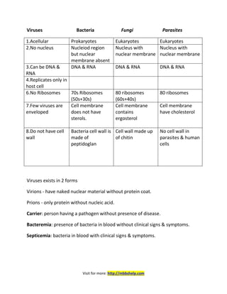
  • 1. Visit for more: http://mbbshelp.com Viruses Bacteria Fungi Parasites 1.Acellular Prokaryotes Eukaryotes Eukaryotes 2.No nucleus Nucleiod region but nuclear membrane absent Nucleus with nuclear membrane Nucleus with nuclear membrane 3.Can be DNA & RNA DNA & RNA DNA & RNA DNA & RNA 4.Replicates only in host cell 6.No Ribosomes 70s Ribosomes (50s+30s) 80 ribosomes (60s+40s) 80 ribosomes 7.Few viruses are enveloped Cell membrane does not have sterols. Cell membrane contains ergosterol Cell membrane have cholesterol 8.Do not have cell wall Bacteria cell wall is made of peptidoglan Cell wall made up of chitin No cell wall in parasites & human cells Viruses exists in 2 forms Virions - have naked nuclear material without protein coat. Prions - only protein without nucleic acid. Carrier: person having a pathogen without presence of disease. Bacteremia: presence of bacteria in blood without clinical signs & symptoms. Septicemia: bacteria in blood with clinical signs & symptoms.
  • 2. Visit for more: http://mbbshelp.com Normal flora in body  On Skin - Staph epidermidis  In Nose – Staph Aureus  Oropharynx -Strep Viridens like strep mutans  In Stomach – No bacteria  In Blood – No bacteria  Colon – In babies - Bifido bacterium o In adult - Bacteroids, Ecoli  Vagina: Lacto bacillus Major pathogenic mechanisms of bacteria:  Adherence to cell surface o Pili /Fimbriae are seen in most Gram –ve o Techoic acid is seen in most Gram +ve bacteria  Strep Pneumonia, Neisseria, and H.Influenza produce IgA proteases  Biofilm produced by Strep mutans, Staph epidermidis Anti-phagocytic Nature of bacteria:  Capsule / Slime layer inhibits phagocytic uptake  Strep pyogens has M-proteins that act as anti phagocyte component  Pili of Neisseria gonorrhea inhibits phagocytosis  Protein A of Staph Aureus inhibits phagocytosis by binding to FC fragment of Ig prevents phagocytosis.  Intracellular bacteria like Mycobacterium Tuberculosis inhibit phagosome & lysosome fusion.  Listeria escapes from phagosome before phagosome & lysosome fusion. Capsulated Organisms: Strep pneumonia Klebsiella pneumonia Haemophilus influenza
  • 3. Visit for more: http://mbbshelp.com Pseudomonas aeroginosa Pasturella Neisseria meningitides Cryptococcus neoformans Bordetella pertussis  Most capsules are made of polysaccharides.  Capsule of Bacillus anthracis is made up of d-glutamate. Toxins: Exotoxin Endotoxin 1.Exotoxin are protein toxin Lipopolysaccharides (LPS) 2. Secreted by gram –ve & +ve Secreted by gram –ve bacteria only [Gram +ve bacteria- Listeria] 3. Secreted outside cell wall 3. Endotoxin are structural components of outermember of cell wall, released only when cell is dead or during lysis. [Neisseria Meningitidis overproduces endotoxin and can secrete it] 4. Heat labile 4. Heat stable 5. Immunogenic 5. Not Immunogenic 6. Toxic component are A- active component B- Component helps to bind to receptors 6. Active component is lipid A Mechanism of Action of Toxins: Protein synthesis inhibitors  Diptheria Toxin has ADP ribosyl transferase activity and inactivates elongation factor ii Major targets heart, nerves, epithelium  Pseudomonas Aeroginosa also acts by ADP ribosyl transferase activity and inhibits EF ii o Primary target is Liver.
  • 4. Visit for more: http://mbbshelp.com  Shiga toxin produced by shigella dysentery interferes with 60s ribosomal subunit and inhibits protein synthesis.  EHEC [Entero haemorrhagic E.coli] produce verotoxin called as “ shiga like toxin” & interferes with 60s ribosome. Neurotoxins  Clostridium botulinum produces Botulinum toxin that inhibit release of Ach.  Clostridium tetani produce tetanus toxin that block or inhibit the release of inhibitory neurotransmitters. C-AMP inducers  C - vibrio cholera produces cholera toxin, acts by stimulating Gs, increases activity of adenylate cyclase that stimulate cyclic AMP  A - Bacillus Anthracis produces anthrax toxic that makes adenylate cyclase & increases C-AMP causes Edema  M – ETEC [Entero Toxic E.coli] causes traveller’s diarrhoea, its heat labile toxin stimulate Gs, increases activity of adenylate cyclase that stimulate cyclic AMP  P- Pertussis toxin acts by inhibiting Gi prtn & C-AMP Cyto toxins  Clostridium Perfringens produces (alpha) toxins, which has lecithinase activity that causes myonecrosis.  Staph Aureus produces (alpha) toxins, which damages the cells Super antigens  Toxic shock syndrome toxin of Staph Aureus (TSST1)  Erythrogenic toxin Strep pyogenes  They act as superantigens producing prolonged stimulation of T cells & decrease endotoxin clearance by the liver & cause profound endotoxin shock so also called as Endotoxin enhancers. Gram Staining 1.Crystal violet is reagent used that stains gram+ve and gram –ve into purple or blue colour.
  • 5. Visit for more: http://mbbshelp.com 2.Grams iodine acts as a fixative giving purple or blue colour to both gram +ve and gram –ve. 3. De-colourising agent used is alcohol or acetone. Gram +ve remains same, gram –ve becomes colourless. 4. Countor stain used is safranin. Gram +ve remain same purple or blue colour but gram –ve becomes or turns red/pink colour. Acid fast staining 1.Regent used is carbol fuchsin 2. Heat as a fixative 3.Decolourising agent used is acid alcohol. 4. Counter stain used is methylene blue. Acid fast stains red or pink Non-acid fast stains blue or purple. At the end of gram staining gram +ve is purple or blue, gram –ve is red or pink. At the end of acid fast staining acid fast pink, non-acid fast blue. Acid-fast bacilli: 1. Mycobacterium tuberculosis 2. Mycobacterium leprae 3. Legionella 4. Oocyst of cryptosporidium 5. Oocyst of isospora Nocardia is partially acid fast. Organism not staining with gram stain: 1. Treponema pallidum 2. Rickettsia 3. Chlamydia
  • 6. Visit for more: http://mbbshelp.com 4. Mycobacterium 5. Mycoplasma Special Stains: 1. Giemsa stain for Chlamydia, Plasmodium, Borellia 2. Zeihl neelsen staining for Mycobacteria tuberculosis 3. India ink staining for Cryptococcus neoformans. 4. Silver staining used for Legionella, Pneumocystis jerovosi Gram +ve Gram –ve Cocci  Staphylococcus  Streptococcus Cocci Neisseria meningitidis/gonorrhoea Moraxella Gram +ve Bacilli Gram -ve Bacilli  Actinomyces  Bacillus  Corynebacterium  Clostridium  Listeria  Mycobacterium  Nocardia E.coil Shigella Haemophilus Salmonella Chlamydia Klebsiella Spirochetes Rickettsia Bacterial growth curve: Log phase: there is high metabolic activity without cell division
  • 7. Visit for more: http://mbbshelp.com No of cells before log phase = no of cells after end of log phase Log phase/Exponential phase: rapid cell growth & rapid cell division. Stationary phase: No of new cells= no of dying cells Nutrients start getting used up & spore formation begins in stationary phase. Death phase: its due to depletion of nutrients & build up of waste products. Organisms Culture medium Haemophilus influenza Chocolate agar with factor V (NAD) factor X (Haematin) Neisseria Gonorrhoea Thayermartin medium or VPN medium Nesseria from sterile site Chocolate agar Corynebacterium diptheria Tellurite agar & loffler’s serum Mycoplasma pneumonia Eatons agar (requires cholestrol) Legionella Charcoal yeast extract & cystein Fungus Sabouroud’s dextrose agar with 10% KOH Enteric bacteria Eosin methyleve blue and mac conkeys agar Enteric pathogens Xylose lysine deoxychocolate agar Vibrio cholera Mycobacterium tuberculosis Clostridium TCBS Lowenstein Jenson medium, Bactec medium. Robertson’s cooked meat broth  Mac-conkeys differentiates lactose fermenters from lactose non- fermenters Lactose fermenters converts mac-conkeys agar into pink. Lactose non-fermenters converts mac-conkeys agar colourless.
  • 8. Visit for more: http://mbbshelp.com Cholesterol used in the culture of Mycoplasma. Cystein is used in culture of Legionella, Brucella, Pasturella, Francisella Protoporphyrin is used in culture of Haemophillus Obligate aerobes:  Bacillus Anthracis  Mycobacterium Tuberculosis  Pseudomonas Aeroginosa Obligate anaerobes:  Actinomyces  Bacteroides  Clostridium Obligate Intracellular bacteria: Rickettsia, Chlamydia Micro Aerophilic Bacteria:  Helicobacter Pylori  Campylobacter jejuni Requires low O2 tension Quellung Reaction: Swelling up of capsule when specific anti serum is added, seen with all capsulated bacteria. Urease +ve bacteria:  Proteus  Ureoplasma  Klebsiella  H.pylori
  • 9. Visit for more: http://mbbshelp.com  Cryptococus neoformans  Nocardia Spore forming bacteria:  Bacillus Anthracis  Clostridium  Coxiella burnetii Pigment Producing Bacteria: 1. Staph Aureus produces golden yellowish pigment. 2. Pseudomouas Aerugiuosa produces pyocyanin & fluorescin. 3. Serratia prod red pigment. 4. Actinomyces Israeli produces yellow sulphur granules. Types of Molility:  Tumbling motility: Listeria [culture medium]  Actinogenic motility: Listeria in the cells  Swaming motility: Proteus  Dartiling motility: Vibrio cholera, Campylobacter jejuni  Shooting star motility: Vibrio cholera  Cork- screw motility: Treponema pallidum  Falling leaf motility: Giardia Catalase +ve organisms: Produces infection in patients with chronic granulomatous disease (CGD)  Staph Aureus  Pseudomonas aeroginosa  Aspergillus  Candida  E.coli Phage coded toxins:  ShigA like toxin  Botulinum  Cholera
  • 10. Visit for more: http://mbbshelp.com  Diphtheria  Erythrogenic toxin of Strep Pyogenes Bacteriology Gram +ve cocci. Catalase (+) Catalase (–) Steaphylococus Streptococcus Coagulase (+) Coagulase (-) Staph Aureus Novobiocin test Novobiocin Novobiocin Sensitive Resistance Staph Epidermidis Staph Saprophyticus Staphylococcus Aureus:  Catalase (+), coagulase (+)  It ferments mannitol, produces Beta-hemolysis.  It produced yellow color colonies in blood agar.  Its normal flora in the nasal mucosa.  M/C route of infection is case or carrier.  M/C route of infection is skin.  It is the M/C cause of acute endocarditis  It is the M/C cause of acute osteomyelitis  It is the M/C cause of Nosocomial pneumonia  It is the M/C cause of Surgical wound infection  It is the M/C cause of Paronychia  It is the M/C cause of Spiral epidural abscess It also causes Botryomycosis and Mastitis.
  • 11. Visit for more: http://mbbshelp.com Transmission of Staph Aureus: 1. By hands 2. Sneezing 3. Surgical wounds 4. Canned foods Virulence: Protein A of Staph Aureus binds to Fc component of IgG and inhibits phagocytosis. Extracellular enzymes: 1. Coagulase converts to fibrinogen to fibrin 2. Hyaluronidase 3. Heat stable Nuclease or DNAase Toxins: 1. Cytotoxins: S.Aureus produces alpha Toxins which damages cells 2. Exterotoxin: Produced by them are heat stable and fast acting. Causes Gastroenteritis within 2-5 hrs after consuming salted canned foods and custards. 3. Toxic shock syndrome toxin (TSST1) 1. Super antigen 2. Common in female using tampons presents with  Hypotension  Fever  Erythematous rash  Multi organ failure  TSST1 enchances the endotoxin shock by decreasing the clearance of lipopolysacchasides. 4.Exfoliative toxin: responsible for staphylococcal-scalded skin Syndrome [SSSS] Neonates: Ritter’s Disease Elders: Toxic Epidermal necrosis [TEN] Milder forms: Pemphigus neonatorum Bullous impetigo
  • 12. Visit for more: http://mbbshelp.com Botryomycosis is a skin infection of staph aurcus: Rx of Staph Aureus:  Methicillin  Naphicillin  Oxacillin Methicillin resistance Staph Aureus [MRSA]  Vancomycin  Teicoplanin Vancomycin Resistance Staph Aureus [VRSA]  Linezolid  Quinpristine  Dalphopristine Staph Aureus diseases:  Skin: Surgical wound infection, Spiral epidural abscess, Paronychia, Bullous impetigo, Botryomycosis, Folliculitis, Furuncle, Cellulitis, Hordeolum.  Musculoskeletal: Osteomyelitis  Respiratory: Nosocominal Pneumonia  CNS: Meningitis  Endovascular: Endorcarditis [Acute] Staphylococcus Epidermidis:  Normal Flora: skin  Coagulase –ve, Novobiocin Sensitive.  It has predilection on growth and implanted foreign bodies like skin grafts, cathelers, and prosthetic valves.
  • 13. Visit for more: http://mbbshelp.com  It produces a biofilm and is M/C pathogen producing endocarditis, complicating patients of i.v catheters and exogenous implants.  It produces stitch abcess Staphylococcus Saprophyticus:  It novobiocin resistance, Coagulase –ve  It produces honeymoon cystitis [infection of urinary blader] in sexually active young women Streptococcus [catalase –Ve] on blood agar: Alpha α -hemolysis [partial] (green color due to biliverdin): Optochin sensitive Optochin resistant Strep Pneumonia/pneumococcus Strep viridans Beta β -hemolysis (complete): Bacitracin sensitive Bacitracin resistant Strep pyogenes (Group A Streptococcus) Strep Agalactiae (group B Step) Gamma γ-hemolysis (no hemolysis): 6.5% Nacl sensitive 6.5% Nacl resistant Strep bovis Enterococcus
  • 14. Visit for more: http://mbbshelp.com Streptococcus Pneumoniae/Pneumococcus:  Diplococci [they occur in pairs] lancet shaped, bile soluble.  Alpha hemolytic, Optochin sensitive.  Transmitted by resp droplets and is responsible for community acquired pneumonia & produces typical Pneumonia with rusty brown sputum.  M/C cause of adult meningitis.  M/C cause of otitis media & sinusitis in children.  Predisposing factors asplenia [Splenectomy]  Influenza  Measles  Alcoholism  Sickle cell anemia  COPD  CHF  It’s a capsulated organism, produces IgA proteases  Quellung’s reaction +ve  Produces lobar Pneumonia with productive cough.
  • 15. Visit for more: http://mbbshelp.com  M/C complication of pneumococcal pneumonia is Empyema. Rx: Macrolides for bacteria pneumonia Ceftriaxone for adult meningitis Amoxicillin in adults – otitis media, sinusitis Erythromycin in children Prevention:  Done by antibodies in capsule.  Paediatric vaccine consists of seven serotypes.  Adult pneumococcal vaccine consists of 23 serotypes and is recommended for all adults > 65 years and high risk individuals like Asplenia, Sickle cell anemia, DM, HIV, Chronic lung, renal, heart diseases.  Latex agglutination particle test produces pneumococcal capsule antigen in CSF. Streptococcus Viridens:  Catalase –ve, alpha hemolytic, optochin resistant, bile insoluble Eg: Strep mutans, Strep sanguis.  They are nomal flora of oropharynx.  Strep mutans is responsible for dental plague, dental caries & produces biofilm.  Strep viridens grows on damaged heart valves & is the m/c cause of sub– acute bacterial endocarditis after dental extraction/procedures.  Infective endocarditis presents with malaise, weight loss, splinter h’ages, night sweats, fever, heart murmurs, and Janeway’s lesion. Rx: Pencillin G with aminoglycosides for endocarditis Always give prophylactic antibiotic before dental produces for individuals with damaged heat valves. B-hemolytics:
  • 16. Visit for more: http://mbbshelp.com Streptococcus Pyogenes:  Also called as group-A streptococci.  It is Beta-hemolytic, bacitrian sensitive, PYR+ve.  They are divided into groups based on carbohydrate antigen of cell wall  It is divided into 20 lance field groups A-V (w/o I-J)  Most pathogenic streptococci is group A streptococci  Group A-strep are further divided into different types based on M-proteins [80 griffithe types] Pathogenesis:  Produces hyaluronidase, which is non-immunogenic. M-protein is antiphagocytic.  Streptolysin O: o Immunogenic o Hemolysis/cytolysin o O2 labile o Cardiotoxic o Poor prognostic factor  Streptolysin S: o Non-immunogenic o O2 stable o Causes hemolysis on surface only  Streptokinase is produced that breaks down fibrin clots.  They produce DNA are NADase  Strep pyogenes also produces exotoxin [A-C] also called pyrogenic toxin or erythrogenic toxin that acts as super antigen, which is responsible for scarlet fever.  They inhibit clearance of endotoxin by liver, thus acting as endotoxin enchancers. Diseases caused:
  • 17. Visit for more: http://mbbshelp.com Scarlet fever:  It is caused by erythrogenic/pyrogenic toxin  Type A is most common causative agent.  M/c infection caused is sore throat.  Streptococcal pharygitis accompanied by sandpaper rash, strawberry tongue, circum-oral pallor. Skin involvement in the form of: o Impetigo o Cellulititis o Erysipelas o Necrotising fasciitis Acute Rheumatic fever:  It develops only after throat infection by Strep pyogenes.  ASO titres are always raised.  Antibody produces against heart tissue.  It is type II hypersensitivity.  Jones criteria is used for diagnosis  Penicillin prophylaxis is indicated. Acute glomerulo nephritis [PSGN]:  Occurs after streptococcal pharygitis & streptococcal skin infection.  M/c its due M12 proteins.  ASO titre may or may not be raised.  Pencillin prophylaxis is not indicated.  Immune complex deposition producing smoky cocoa coloured urine.  Type III hypersensitivity. Dx:
  • 18. Visit for more: http://mbbshelp.com  Throat swab culture, Pike’s medium [transport medium for strep pyogenes]  ASO titre >200 indicate prior streptococcal infection mainly useful for rheumatic fever.  Aso titre may be low after skin infection. Rx:  Pharyngitis, Impetigo, Cellulitis - Penicillin  Necrotising fasciitis - surgical debridement, penicillin, clindamycin  Endocarditis - Pencillin G+ Aminoglycosides. Clindamycin reduces toxin synthesis & is used in toxic shock syndrome & Scarlet fever. Streptococcus Agalactiae:  It is group B, Beta hemolytic, Bacitracin resistant, CAMP +ve.  Hydrolyes hippurate.  Reservoir is vagina.  Risk factor is preterm rupture of membranes.  Its M/C cause of neonatal septecemia worldwide.  Neonatal GBS infection is acquired in utero.  Rx:  Pencillin  Treat the mother prophylactically intrapartum if vaginal cultures are +ve for GBS. γ -Hemolytic streptococci: Group D Streptococci: 1) Enterococcus 2) Non-entercoccal Group D Streptococci Enterococcus  Important species are enterococcus faecalis, Enterococcus faecium.
  • 19. Visit for more: http://mbbshelp.com  Enterococcus grows in 40% bile, hydrolyzes aesculin in bile, grows in 6.5% Nacl, PYR +ve.  Reservoir: human intestines  Usually survive in bowel & gall bladder.  It causes urinary & biliary tract infection and also associated with endocarditis. Rx:  Enterococci are resistance to Penicillin, so treated by Vancomycin.  In vancomycin Resistance Linezolid is used against all entercocci.  Quinupristin, Dalfopristin are effective against Vancomycin resistant enterococcus faecium.  E-Faecium all 3 drugs are used 1. Linezolid, Quinupristin, Dalfopristin  E.Faecalis only linezolid Mechanism of resistance:  Vancomycin resistance is due to change of D-ala, D-ala of cell wall NAM (N- acetyl muramic acid) with D-ala, D-lactate. Non Enterococcal Group D Streptococcus  Grow in bile, do not grow in 6.5% Nacl, PYR –ve Eg: Streptococcus Bovis  Strep Bovis associated c endocarditis after bowel surgeries/GI/GU surgeries. GRAM +ve Bacilli  Most bacillus are motile except Bacillus Anthracis  Branching bacilli are Actinomyces & Nocardia  Spore producing bacilli are Bacillus & Clostridium  Nocardia is partially Acid fast  Mycobacterium is acid fast
  • 20. Visit for more: http://mbbshelp.com  Gram +ve anaerobes are Actinomyces & Clostridium. Bacillus Anthracis:  It’s a large gram +ve, spore forming aerobic bacillus.  Capsule is made of polypeptide [D-glutamate].  It’s a potential warfare agent.  On microscopy it has Medusa head appearance on agar plates.  On gelatin stab it produces inverted fur tree appearance.  Bacillus Anthracis shows “ string of Pearls” appearance when grow on solid medium which differentiates it form B cereus.  On staining with polychrome methyline blue it produces Mac Fadyean‘s reaction  Toxin of Bacillus Anthracis has 3 factors: o Edema factor that acts by cyclic-AMP o Lethal factor causes cell death o Protective antigen Clinical features: Anthrax is a zoontic disease effecting herbivores. Humans became infected, when spores are inhaled from infected animals or with contact with infected animals. Anthrax types: 1. Cutaneous Anthrax 2. Pulmonary Anthrax 3. Gastrointestinal Anthrax 1.Cutaneous Anthrax:  It is also called as “ Hide porter’s disease”  M/c type of anthrax. There is a central necrotic lesion, which is covered by a black eschar. This lesion is called a “Malignant Pustule”  Resolves spontaneously. Fatal septicemia occurs in 10-20% of patients
  • 21. Visit for more: http://mbbshelp.com  Patient may have painful regional lymphadenopathy. 2. Pulmonary Anthrax:  It is also called as “ wool sorter’s disease”  Its life threatening pneumonia with cough, fever, facial edema & cyanosis.  There is characteristic mediastinal haemorrhagic lymphadenitis & mediastinal widening on X-ray.  Most of the times it’s fatal. 3. Gastointestinal Anthrax:  It is the rare form with primary lesion in tonsils causing edema, bloody diarrhoea, & high mortality. Rx:  Doc is penicillin  In penicillin allergy, Erythromycin is used.  Vaccine for B.Anthracis is Acellular vaccine Adsorbed [AVA] Bacillus Cereus:  It’s similar to bacillus anthracis, but does not show “ String of Pearls” appearance & is motile, non-capsulated.  Its usually food borne.  1st type is associated with fried rice [reheat foods] in Chinese restaurants.  2nd type is associated with improper cooked meat & vegetables.  Food poisoning due to B.cereus is also fast onset i.e within 6-8 hrs. 2 types of toxins:  Diarrhoeal type resemble heat labile toxin of ETEC  Emetic type resembles heat stable toxin of Staph Aureus. Clostridium:  It’s a Gram +ve, anaerobic, spore forming bacill.
  • 22. Visit for more: http://mbbshelp.com  Culture medium: Robertson’s cooked meat broth  C.perfringen’s & C.tetani are non-motile. Clostridum tetani:  Produces Tennis racket/drumsticks shaped terminal spores.  Common Reservoir is mud.  Transmission is by puncture wounds and trauma.  They require low O2 tension, strict anaerobes. Toxins: 2 types: Tetanolysin, Tetanospasmin Tetanospasmin:  It’s the toxin responsible for tetanus.  It’s transported retrograde in the axon.  It inhibits the release of inhibitory neurotransmitters causing muscle spasms and rigidity.  Tetanospasmin affects spinal cord mainly.  Strychnine poisoning also show similar muscle spasms appearance. Clinical features:  Average time of onset after injury is 7 days.  Trismus/Lock jaw.  Risus sardonicus  Opisthotonos  In tetanus deep tendon reflexes are exaggerated (UMN)  C.Tetani doesn’t invade tissues but the toxin is transported retrograde in the axons.  Tetanus patient are not infectious & there is no person to person transmission Rx:
  • 23. Visit for more: http://mbbshelp.com  By giving antitoxin i.e, Human tetanus immunoglobulin.  Antibiotics used are Penicillin & Metronidazole Alternative is Clindamycin.  Spasmolytic drugs like Diazepam.  Proper wound debridement.  Delayed wound closure.  Hyperbarric oxygen.  Prevention is done by active immunization using active toxoid. It’s a formaldehyde-inactivated toxin. Usually 2 doses of TT are given 1 month apart.  1st booster dose after 1 year, 2nd booster dose after 5 years.  Passive immunization is done by given human hyper immunoglobin.  Combined active & passive immunization with TT in one arm & Tetanus 23mmunoglobulin in other arm is usually given in patients of dirty wound, with no history of vaccination.  Tetanus spores are resistant to disinfectants. Clostridium Botulinum:  It’s an anaerobic, Gram +ve, spore forming bacilli.  Botulinum toxin is most potent bacterial toxic known.  Spores survive in soil.  Transmission is food borne or traumatic.  Botulinum toxin acts by inhibiting the release of Ach.  Botulinum toxin is coded by a prophage [viral DNA gets incorporated into bacterial chromosome in lysogenic cycle]  Botulinum toxin is heat labile i.e.boiling food removes the bacteria. Type of Botulism: Adult food borne botulism:  It’s due to the preformed toxin present in canned food, green beans, and peas.
  • 24. Visit for more: http://mbbshelp.com  Symptoms usually arise 1-2 days later with weakness, blurred vision, symmetric descending flaccid paralysis, severe constipation, nausea , vomiting ,diplopia,dysphagia & death is due to respiratory failure commonly.  Rx:  Resp support & trivalent antitoxin.  Prevention: Is by proper canning of foods & heating all canned foods.  No antibiotics are required. Wound botulism:  It is due to traumatic implantation of spores.  Resemble food poisoning without GI symptoms.  Diplopia, Paralysis & respiratory weakness. Rx:  Wound debridement.  Delayed closure.  Amoxicillin  Antitoxin & respiratory support. Infant botulism:  It’s the most common type.  It’s due to ingestion of botulimum spores, usually spores seen in honey.  The toxin is produced in the gut. There is constipation flaccid paralysis, diplopia weak crying, and respiratory arrest.  Toxin can be demonstrated in stool & serum. Rx:  Respiratory support.  Hyperimmune serum [Botulinum immunoglobulin]  Antibiotics generally not used  Prevention- avoid honey in the 1st year of life
  • 25. Visit for more: http://mbbshelp.com  Botox injections [toxins] are used to treat strabismus [crossed eyes], blepherospasms (eye twitching) and cervical dystonia/spasmodic torticollis. Clostridum Perfringens:  It’s a Gram +ve, spore forming, and anaerobic bacilli.  It causes gas gangrene, myonecrosis.  It shows stormy fernentation in milk media.  It shows double zone of B-hemolysis.  It produces Alpha-toxin, which has lecithinase activity, which disrupts the membrane of RBC’s platelets, endothetial cells produces massive hemolysis.  It also shows Nagler’s reaction on egg yolk agar. A zone of opacity is formed are the egg yolk agar plate with no antitoxin.  Enterotoxin product can cause food poisoning, watery diarrhoea similar to E.coli. Gas Gangrene:  Its myonecrosis due to contamination of wound with soil & feaces especially battle field wounds.  There is edema, gas production, and dense tissue pain.  C. Perfringens doesn’t produce spores in artificial medium.  Crepitus is present  Systemic symptoms: fever, disproportionate tachycardia.  There is rapid & high mortality due to circulatory failure Rx:  Surgery in main stray of treatment.  Penicillin & Clindamycin  Hyperbarric oxygen.
  • 26. Visit for more: http://mbbshelp.com Clostridium difficile:  Its responsible for antibiotic associated diarrhoea also called “pseudomembranous colitis “.  M/c diarroheal infection acquired in hospital.  Clindamycin, Ampicillin, Cephalosporin, Amoxicillin are associated antibiotics  Formation of whitish yellow plaques over colon is called as pseudomembrane  It can lead to toxic megacolon / sepsis  C.difficile is normal flora is Gut, so culture is not diagnostic  Demostration of toxin in the feaces is the gold standard  Rx: Doc is Metronidazole alternate is Vancomycin Corynebacterium diptheriae:  Its gram +ve rod that produces black colored colonies or Tellurite agar & Loeffler’s serum slope.  It produces metachromatin granules called Volutin granules/Babes-Ernst granules on Albert’s stain.  Bacteria are club shaped arranged in Chinese letter pattern.  Diptheria toxin is coded by a prophage and acts by inhibiting protein synthesis by inhibiting EF–II Pathogenesis:  It spreads via respiratory droplets.  It is not invasive it colonizes on pharynx producing a dirty pseudomembrane that can extend onto the trachea & larynx causing mechanical obstruction.  Systemic effects are due to the toxin mainly damage heart, nerves & epithelium. Respiratory Diptheria:
  • 27. Visit for more: http://mbbshelp.com Clinical features:  M/C type is Tonsilo pharyngeal [Faucial type]  Fever  Sore throat  Weakness  Bull neck appearance  Asphyxia  Myocarditis  Cardiac dysfunction  Recurrent laryngeal nerve palsy  Peripheral polyneuropathies  Dysphagia  The 1st muscle effected is palato-pharyngeus  Cause of death is circulatory failure. Dx:  It is usually clinical.  Demonstration of org by gram staining of throat swab & culture in specific media are useful investigations. Cutaneous Diphtheria:  It causes punched out ulcers in the skin & is caused by non-toxogenic strains of C.diptheriae.  Elek’s test: it’s done to demonstrate toxin production in vitro. Toxin produced by toxin producing strains reacts with the anti-toxin diffusing from the filter producing a precipitation line.  Schick test: a test used to determine whether or not a person is susceptible to diphtheria. Rx:  Penicillin G, alternative is erythromycin.
  • 28. Visit for more: http://mbbshelp.com  Prevention: by active immunization by diphtheria toxoid combined DPT is used. Pertussis increases the potency of diphtheria toxoid. Listeria:  It’s a gram +ve bacillus that shows tumbling motility at 20-25o C & non motile at 37o C [body temp] of 98o F  It’s a facultative intracellular non spore-forming bacillus.  Listeria monocytogenes is Beta-hemolytic. It shows tumbling motility in Broth & Actin jet motility in the cells.  Its characteristically shows cold growth.  Reservoir: Unpasteurized milk products  Stored cheese, cabbages  GI, GU, tracts of animals.  Transmission: food borne, also spreads vertically across placenta.  It produces listeriolysion O or LLO that that disrupts the phagosome membrane & allow it to escape before phagosome lysosome fusion with actin jet motility. Diseases caused: Listeriosis:  It is seen in pregnant women in 3rd trimester.  It causes fever, myalgia, choreoamnionitis, abortions, and stillbirths.  Healthy adults & children have mild symptoms of diarrhoea. Neonatal listeriosis:  It due to in-utero transfusion.  There is profound sepsis, disseminated granulomas with central necrosis & high mortality.  Late onset disease can be associated with meningitis, septicemia  Neonatal listeriosis is also called as “Granulomatosis infantisepticum”  Listeria is the m/c cause of meningits in kidney transplant patient.
  • 29. Visit for more: http://mbbshelp.com  Listeriosis not associated with pregnant presents, as meninigitsis & septicemia in immunocompromised patients like renal transplant patient, chronic gluco-corticoid therapy, malignancy & AIDS.  Prevention: Pregnant women should not eat cold stored foods. Actinomyces:  It’s an anaerobic, gram +ve, and filamentous branching bacilli.  Normal flora: gingival cervices, female genital tract.  Transmission: endogenous due to tooth pick injuries.  M/c Actinomyces Israeli prod indurated swelling & draining sinus tract that contains yellow sulphur granules the drains out on skin  M/c site is lower jaw  M/c type of actinomycosis is Cervico-facial produces “Lumpy-jaw” that occurs due to dental trauma & poor oral hygiene.  Pelvic actionomycosis is associated with use of IUD’s.  In CNS, it produces solitary brain abcess.  It also causes actinomycetoma.  On microscopy the Gram +ve filamentous bacteria have sunray appearance.  Culture on solid media prod colonies resembles molar tooth Rx:  Actinomycetoma by Streptomycin + Dapsone.  Doc is penicillin. Nocardia:  Its aerobic, gram +ve, partially acid fast, branching filmentous by bacteria.  Transmission is air borne & traumatic transplantation [mainly by inhalation]  It usually produces pulmonary infection in immune compromised or cancer patient.  Norcardiosis is mostly caused by Nocardia asteroides that produces bronchopulmonary pneumonia with cavitations causing cough, fever dyspnoea, and cavitations in Lungs.
  • 30. Visit for more: http://mbbshelp.com  Haematogenous spread to brain produces multiple brain abscesses. Cutaneous nocardiosis:  It is caused by Nocardia Brasiliensis.  It presents with draining granules & cellulitis. Rx:  Doc for Nocardia is Cotrimoxazole. Actinomyces Nocardia  Anaerobic Aerobic.  Non-acid fast Partially acid fast  Normal flora of Gingiva In soil dust /vegetative matter.  Produces solitary brain abcess. Produces multiple brain abcesses  Infection is endogenous Exogenous  Doc penicillin Doc- cotrimoxazole Mycobacterium tuberculosis:  Acid fast, obligate aerobe.  Cell wall of mycobacteria has concentration of lipid especially mycolic acid which is responsible for its acid fastness.  Mycobacterium is highly sensitive to disinfectants like NaOH & dessication.  M.tuberculosis produces niacin & heat sensitive catalase. Virulence factors:  KAT-G gene encodes for Oxidase & Catalase Mutations in KAT-G gene leads to resistance to Isoniazid.  Sulfatides produced inhibits phagosome, lysosome fusion & allows intracellular survival
  • 31. Visit for more: http://mbbshelp.com  CORD factor: main virulence factor [Trihelose Dimycolate] that causes serpentine growth in vitro & inhibits leukocyte migration & also disrupts mitochondrial phosphorylation.  Tuberculin protein: Responsible for delayed hypersensitivity reaction. Pathogenesis: Primary pulmonary Tuberculosis:  Localised in the sub pleural regiow, near the obique fissure in the middle lobe and upper part of lower lobes.  Ghon’s focus is the lesion in the lung that is due to inflammation & consolidation.  Ghon’s complex consists of a calcified focus of infection & an associated lymph node.  Depending on host immunity the primary Ghon’s complex can undergo healing, fibrosis or calcification (Ranke’s complex) or could progress to Secondary Tuberculosis in the form of cavitation, consolidation or miliary T.B. Secondary T.B or Reactivational T.B:  Usually develops in apical & posterior segment of upper lobe due to high oxygen concentration in these areas. C/F:  Productive Cough  Hemoptysis  Cavitations  Pleural effusion  Low grade fever especial during the evenings  Weight loss  Night sweats  Anorexia It can spread by hematogenous route & become miliary TB with wide spread organ involvement.
  • 32. Visit for more: http://mbbshelp.com Extra pulmonary Tuberculosis:  M/c site is lymph nodes mainly in cervical & supra clavicular region k/a Scrofula.  Skeleton T.B m/c involves the spine called as Potts spine.  M/c site of GI T.B is ileocecal junction.  It can involve the pericardium producing constrictive pericarditis and also involve meninges and Genito urinary tract. Dx:  TB is stained by Zeihl-Neelsen staining or by Auramine-rhodamine fluorescent dye.  Auramine-Rhodamine fluorescent dye is highly sensitive & rapid procedure is used for screenings of sputum but is not specific.  Sputum is best collected in the mornings before any meal.  3 sputum samples are taken.  Positive samples for auramine-rhodamine are confirmed by acid fast staining. Culture:  Lowenstein Jenson medium- it is a standard solid medium used but slow growing & -ve reports is given only after 8-12 weeks. There is production of niacin +ve acid-fast bacilli in the LJ medium from +ve sputum.  Bactec- it’s a liquid medium and gives results within 2 weeks.  Broth with palmatic acis  PCR & ligase chain reaction are fastest. Mantoux’s/tuberculin test:  It is done to demonstrate hypersensitivity to tuberculin protein.  It’s an intra dermal test using purified proteinn derivative PPD  The test is read after 72 hrs.
  • 33. Visit for more: http://mbbshelp.com  The area of in duration is looked at and is said to be +ve if its >10mm & -ve if <5mm  Its considered doubtful b/w 5-10mm  This test indicates only exposure to bacilli with or without clinical illness. Its not a confirmatory test for active infection  False –ve mantoux is seen in Sarcoidosis, Malnutrition, Miliary T.B, and Immune suppression, impaired cell mediated immunity.  False +ve mantoux is seen in infection by atypical mycobacterium infection, BCG vaccine  Catalase produced by mycobacterium is heat sensitive, so under standard catalase testing [68o C] its catalase –ve  Rx - Multiple drug treatment is given as ATT. Minimum 6 months of treatment is required for uncomplicated patients.  HRZE is given for 2 months followed by HR for next 4 months  H- Isoniaxid [INH]  R- Rifampician  Z- Pyranxinamide  E- Ethambutol Streptomycin is given in relapse/failure cases or defaulters BCG (Bacillus calmette Guerin) vaccine:  It is live attenuated vaccine, derived from attenuated Bovine strains. Its used to prevent disseminated disease & given by a intradermal injection.  It’s also used to treat Bladder cancer by folle’s catheter. Mycobacterium Leprae:  They are acid fast bacilli, obligate intracellular rods, that grows in macrophages in the form of cigar shaped bundles appearance called “Globi”  Reservior: human mucosa, skin, nerves.
  • 34. Visit for more: http://mbbshelp.com  Transmission: nasal discharge from an affected untreated patient or close contact with infected part for long period of time is required for infection.  Skin to skin contact is not an important route of infection.  Grows in cooler parts of body like ear lobes, skin, mucous membrane & peripheral nerves.  The disease caused by leprosy is also called as Hansen’s disease  Morphological index is total no of live bacilli in tissues that stains uniformly.  Bacteriological index is total no of live & dead bacilli in tissue.  Incubation period for leprosy is 5-7 yrs. Leprosy is divided into 5 clinical types: 1. Tuberculoid 2. Border line tuberculoid 3. Border line 4. Border line lepromatous 5. Lepromatous  Symptoms of leprosy are confined to skin & nerves.  M/c nerve involved is Ulnar nerve & Posterior auricular nerve.  Medial popliteal nerve is never involved in leprosy  Invasion & destruction of nerve by TH, cells is pathognomic of “tuberculoid reprosy”  Presence of bacilli in blood & other organs is a feature of “ lepromatous leprosy”  Lung & CNS are not involved in leprosy. Diagnosis:  Punch biopsy from advancing edge of lesion or nasar scrapings shows acid-fast bacilli Rx:
  • 35. Visit for more: http://mbbshelp.com Dapsone & Rifampicin Clofazamine is added for Rx of lepromatous leprosy. Tuberculoid Leprosy Lepromatous leprosy Strong cell mediated immunity Weak cell mediated immunity No of bacilli in tissue are low No of bacilli in tissue are medium Nerve & tissue damage is due to immune sys Due to bacteria. Few lesions present multiple lesions Lepramine test +ve Lepramine test –ve Skin lesion upto 3 in no, sharply defined. Nodular,multiple, poor margins. Hypopigmented, asymmetric. Symmetric, leonine faces. Langerhan Giant cells, macrophages, T cells Langerhan Giant cells & T cells are more are reduced. Granulomas are seen Granulomas are absent Type I lepra reaction (or) reversal reaction:  It’s a type IV hyper sensitivity reaction  Seen in border line leprosy  There is tissue edema, pain after starting treatment.  Rx- Glucocorticoids + Clofazamine  M/c nerve involument in type I real is Ulnar Nerve Type II Lepra Reaction:  Also called Erythema Nodosum Leprosum (ENL).  It’s a type III hypersensitivity reaction.  Seen in borderline lepromalous & lepromatous leprosy.  Usually follows dapsone therapy for lepromatous leprosy patient  Numerous painful, erythematous papules are seen.
  • 36. Visit for more: http://mbbshelp.com  M/c feature is TNF plays central role in this type II lepra reaction  Rx- Anti-pyretics & NSAID’s in mild cases.  Gluco corticoids are used in moderate to severe cases.  Doc for moderate to severe in type II lepra reaction is “ Thalidomide” o Single lesion pauci bacillary leprosy can be treated by giving ROM therapy  Rifampicin  Oflaxacin  Minocyclin Lepramine Test:  Its type IV hypersensitivity  Early reaction of Fernandez is similar to tuberculin reaction & is read in 24- 48 hrs.  Late reaction of Mitsuda is more meaningful reaction read after 4 weeks. o Mycobacterium leprae can grow in “armadillos”.  It can’t be grown in artificial medium, but can multiply in footpad of mice. Bacterial generation time is 12-13 days. Atypical Mycobacterium:  Further divided into 4 groups, based on pigment product  Group I photochromogen: They produce pigment only in the presence of light. Eg: M.kansasii causes chronic pulmonary disease in old people. M.marinum causes warty skin lesions called swimming pool granuloma/fish tank granulomas.  Group II is scotochromogen: Can produce pigment even in presence or absence of light. Eg: M.scrofulacium causes scrofula i.e. cervical lymphadenitis is produced  Group III/Non-photo chromogens: do not produce pigment Eg: M.Avium complex (MAC) disseminated pulmonary disease especially AIDS patients
  • 37. Visit for more: http://mbbshelp.com Rx-clarithromycin for patients MAC infection Mycobacterium intraellularae  Group IV is rapid growers. Their colonies appear within 7 days. Ex: - M.chelonie alos k/a title tubercle bacilli M.fortitium also k/a frog tubercle bacilli M.ulcerens prod buruli ulcer. GRAM –ve Cocci:  Neisseria meningitides.  Neisseria gonorrhoea  Moraxella Neisseria: Grow –ve diplocci that are oxidase +ve They grow on chocolate agar Neisseria Meningitidis Neisseria Gonorrhoea  Lens shaped Kidney shaped  Capsulated Non-capsulated  Resp droplets STD  Ferments both glucose & maltose Only glucose  Have a vaccine YWCA No vaccine  B-laclamase prod not seen B-laclamase production is seen Neisseria Meningitidis:  Gram –ve capsulated, diplococcic grows on chocolate agar with 5% CO2  It ferments mannitol & is oxidase +ve Virulence factors:  Capsular polysaccharide
  • 38. Visit for more: http://mbbshelp.com  Outer membrane protein endotoxin  Produces IgA proteases. Its divided into 5 groups based on capsular polysaccharide into Group A, B, C, W, Y.  Reservoir: Human Nasopharynx  Transmission: Respiratory droplets.  Neisseria meningitids, M/C cause Adult Meningitis.  It has over prod of endotoxin & it secretes endotoxin  Complement deficiency esp in C5-C9 factors [MAC deficiencies leads to infections with Neisseria]. Diseases Caused: Meningitis:  Its due to blood borne dissemination to meninges & presents with abrupt onset of o Fevers o Chills o Gen petechial rash with haemorrhagic lesion.  Rash is m/c distinctive feature  In fulminate cases, there is o Rapid septic shock, DIC , Ecchymosis, Bilateral adrenal haemorrhages (Waterhouse friedrichsen syndrome),Death Lab diagnosis: Done by gram staining of CSF  PCR amplification of DNA of CSF is most sensitive method  Latex agglutination test identifies capsuals of N.meningitidis.  Growth med is muiler-Hinton or chocolate agar with 5% CO2.
  • 39. Visit for more: http://mbbshelp.com  Thayer Martin med is a sclective media for culture.  Nasopharyngeal & throat specimen. Rx: Ceftriaxone is Doc Rifampicin is Doc for meningo-coccal prophylaxis. Prevention: is done by YWCA vaccine. There is no vaccine against group B Neisseria Gonorrhoea:  Gram –ve, kidney shaped, non-capsulated, B-lactamase producing diplococci  Reservoir: human genital tract  Transmission: is by sexual contact & during birth Pathogenesis:  Pili are important for all attachment to mucosal surface. They inhibit phagocytic uptake & is responsible for antigenic variation.  It also produces IgA proteases.  Most abundant gonococcal surface protein is Sporin. Disease Caused:  Urethritis  Proctitis  Endocervicitis [female]  Pelvic inflammatory disease  Its m/c cause of Septic arthritis  Its infants it causes Opthalmia neonatorum. Dx:  Gram staining of urethral exudates with presence of Gram –ve diplococci in neutrophils is suggestive of Gonorrhoea.  Selective medium is Thayer martin medium.
  • 40. Visit for more: http://mbbshelp.com  Diagnosis is common done by genetic probes in western countries.  Gonorrhoea is B-lactamase producing, so most are penicillin Resistant. Rx:  Cefriaxone is Doc for Gonorrhoea  Ciprofloxacin 500mg  Ofloxacin 400mg  Doxycycline (or) erythromycin Prevention:  No vaccine for Gonorrhoea.  Use of condoms is best for prevention.  Incubation period for N. Gonorhea is 7 days  Giving erythromycin ointment in eyes after birth prevents neonatal Opthamia neonatorum. Moraxella:  Gram –ve diplococci similar to Neisseria that causes otitis media, sinusitis, Bronchopneumonia in elderly patient with COPD.  Causes Angular conjunctivitis. Rx: Zinc sulphate GRAM –ve Bacilli Pseudomonas Aeroginosa:  Gram –ve aerobic, oxidase +ve rod shaped bacillus.  It produces a blue green pigment called as “pyocyanine” and ‘ fluroscein’.  It has a capsule that produce slime layer.  It produces non-lactose fermenting colonies on Mac-conkey’s agar.  It’s present in fresh waters, water aerosols and also in raw vegetables and flowers.
  • 41. Visit for more: http://mbbshelp.com  It produces an endotoxin that causes septicemic shock.  Pseudomonas exotoxin A acts by inhibiting elongation factor EF II & inhibition of protein synthesis. The primary organ involved is liver. Diseases Caused:  It causes Gastroenteritis, hot tub folliculitis, and corneal ulcers in contact lens users.  It causes cellulitis with production of blue green pus & causes septicemia in ‘burn patients’  It’s an important source of pneumonia & septicemias in neutropenic patient.  Its catalse +ve & can cause infection in chronic Granulomatous disease (CGD).  Skin lesion is in form of Ecthyma Gongrenosum with black necrotic area associated with fever & shock.  It’s also causes UTI’s in catheterised patient & is an important source of pneumonia in patients of cystic fibrosis. Rx- Piperacillin, carbenicillin, Ticarcillin & Aminoglycoside. Legionella:  Its gram –ve fastidious bacteria, cultured in charcoal agar & requires cysteine & iron for its growth.  It’s stained by silver. Reservoir: water tanks, air conditioners, streams.  Causes “pneumonia in smokers” with a high alcohol intake and in immunosupressed patient, like kidney transplant patient.  It’s a facultative intra cellular pathogen.  There is no human-to-human transmission. Mode of transmission: is an aerosol from contaminated Air conditioners.
  • 42. Visit for more: http://mbbshelp.com Diseased Caused: Legionaires disease: it’s an atypical pneumonia associated with Diarrhoea, mental confusion, hyponatremia. Pontiac fever: its acute febrile self-limiting disease with malaise & myalgia. Legionella haemophilli: m/c legionella affecting human. Legionella micdadei is partially acid fast like nocardia. Diagnosis: it’s done bye direct fluorescent antibody testing. Rx: Azithromycin, fluroquinolones. Francisella tularensis:  Its gram –ve, intra cellular facultative pathogen.  Reserviors: Rabbits, Deers, rodents.  Spread: through Tick Bite.  Its important zoonosis.  Spreads through tick bite [dermacentor] produces “ Ulcero glandular disease” Characterized by ulcer at bite site with lymphadenitis, fever during skinning of rabbits.  Aerosols during skinning can produce pneumonia (commonly seen in U.S.A).  “Tularemia” is a disease caused by francisella. o “Tularemia’ is also called as ulceroglandular disease.  Usually it is diagnosed by serological methods. Rx- DOC is Streptomycin. Ingestion of under cooked rabbit meat can prod gastro intestinal tularemia. Bordetella Pertussis:  It produces whooping cough with inspiratory whoop.  It’s an encapsulated organism.
  • 43. Visit for more: http://mbbshelp.com  Reservoir: are vaccinated human beings.  Transmission: Respiratory droplets, it attaches to nasal mucosa by its heam agglutinins.  It produces a pertussis toxin that acts by CAMP by causing inhibition (Gi) of –ve regulator of adenylate cyclase. [ADP ribosylase Gi protein]  Characteristic feature are lymphocytosis, hypoglycemia & increased histamine sensitivity. Whooping Cough:  Its highly contagious disease with incubation period of 7-10 days.  Initially there is rhinorrhoea  Sneezing  Fever  Malaise followed by peroxims of cough and whoof.  The frenulum is swollen & there could be capillary ruptures due to intense cough.  Complications like Pneumonia, Seizures & Encephalopathy can occur.  Pertussis vaccine is “ acellular Pertussis toxoid given as DTaP.  The immunogens used for vaccine are pertussis toxoid, haem agglutinin & outer membrane protein like pertactin.  Cough with sub conjunctival haemorrhages. Rx:  Hospitalization & Erythromycin is DOC.  Erythromycin should be given to all household contacts.  Maximum infectivity of bordetella is during catarrhal stage.  Culture on Bordet-genjou or potato agar produces aluminium paint appearance.  Culture films have thumbprint appearance.  Best specimen for diagnosis is naso-pharyngeal aspiration.  The immunization due to pertussis vaccine that’s wanes of in 5-10 years.  The immunity is mainly of IgA.
  • 44. Visit for more: http://mbbshelp.com Brucella:  It’s zoonoses.  Reservoir is domestic live cattle  Spread by unpasteurised dairy products & by direct contact by animal or animal placenta as in slaughterhouses.  It produces undulent fever called as brucellosis that presents as: Acute septicemia High-grade fever with myalgia Arthralgias Profuse sweating & hepatomegaly.  Diagnosis is usually made by serological examination.  Culture is hazardious. Rx:  Rifampacin + Doxycyclin  Prevention: is by pasteurisation of milk, vaccination of cattle & vaccination of high-risk humans.  Brucella produces granulomatous response with the parasite multiplying in reticulo endothelial cells & causing septiciemia. Campylobacter Jejuni:  It’s a curved rod with polar flagella in the form of gull’s wings.  Its micro aerophilic.  Transmission: poultry.  It can cause infection even after low infectious dose producing inflammatory diarrhoea with blood & pus in stools.  Its an important cause of infectious diarrhoea that is self limiting  Ten or more bloody stools per day.  Imp complication is Guillain Barre Syndrome & Reactive arthritis . Culture: done on schirro agar at 42 degrees.
  • 45. Visit for more: http://mbbshelp.com Rx: mostly supportive. Helicobacter Pylori:  It gram –ve spiral bacilli.  It’s micro aerophilic.  It grows in campy or schirro agar.  Its urease +ve, oxidase +ve.  Transmitted by feco – oral route.  It’s motile with flagella.  It produces mucinase that destroys mucous layer of stomach.  Its imp cause for chronic gastritis & duodenal ulcers.  It’s a type 1 carcinogen associated with gastric adenocarcinoma and maltoma [B cell lymphoma].  Diagnosis is done biopsy and culture.  Stained by giemsa staining.  Urea breath test is usually used in diagnosis of H.pylori.  Its oxidase +ve, urease +ve. Rx: Triple regimens like  Omeprazole +Amoxicillin + Clarithromycin. Entero bacteraciae  They are gram –ve oxidase -ve, reduce nitrate to nitrites.  Catalase +ve. Entero Bactericae Lactose fermenters [Pink colories on mac conkey’s agar] Non- Lactose fermenters [Colorless colonies on mac conkey’s agar] Ex: Citrobacter Non-motile Motile H2s
  • 46. Visit for more: http://mbbshelp.com Enterobacter Escherichia Klebsiella [CEEK] [Non H2s producing] producing Ex: Shigella Yersenia [ShY] Ex:Proteus Salmonella [PS] E. Coli:  They are gram –ve capsulated facultative anerobes  Oxidase –ve, lactose fermenters.  Biochemical test of E.coli IMVC ++-- [indole +, mythelene –ve, vogs roskou – ve, citrate –ve]  Reservoir: human colon.  Transmission: Endogenous.  They prod pink colour colonies on eosin methylene blue. They prodouce greenish shine.  Enterohaemorrhagic E.coli usually prodouces colourless colonies.  Spread through bovine fecal contamination. Antigenic structure of E.coli:  Capsular antigen ‘K’ is heat labile & is associated with virulence.  Somatic antigen ‘O ‘ is heat stable.  Flageller antigen ‘H’.  Normal colorn stains belong to group ‘o’. Different types of E.coli: 1. Entero pathogenic E coli [EPEC]. 2. Entero invasive E.coli [EIEC] 3. Enterotoxigenic E.coli [ETEC] 4. Enterohaemorrhagic E.coli [EHEC] 5. Entero aggrevative E.coli [EAEC] EPEC:
  • 47. Visit for more: http://mbbshelp.com  Enteropathogenic e.coli causes prolonged water diarrhoea in infants & children [paediatric group].  There is effacement of brush border microvilli.  Its non-toxigenic & non invasive. EIEC:  It invades large bowel, similar to shigellosis produces watery diarrhoea with excess of leukocytes, fever and abdominal pain. It causes inflammatory diarrhoea. Rx: Fluroquinolones  It penetrates cells. Penetration of HELA /HEP-2 cell culture is diagnostic.  Sereny test is used in diagnosis of EIEC. ETEC:  Its m/c cause of traveller’s diarrhea.  ETEC labile toxin acts by inducing C-AMP.  It produces dianhoea.  ETEC stable toxin acts rapidly by stimulating guanylate cyclase.  Capsule impedes phagocytosis. Rx: Rehydration. EHEC:  Also called as verotoxigenic E.coli.  O157 H7 is MC serotype.  Transmitted through contaminated bovine feces.  It prodouces diarrhea, haemorrhagic colitis & haemolytic uremic syndrome [HUS]  Virotoxin is shiga like toxin. It acts by inhibiting protein synthesis by binding to 60s ribosome.
  • 48. Visit for more: http://mbbshelp.com  It does not ferment sorbitol. Its non-invasive no inflammatory response & no fever.  Bloody diarrhoea in absence of fecal leukocytes is an indicator of EHEC.  It produces colourless colories on mac conkey’s agar.  Antibiotics are not given as they increase risk of hemolytic uremic syndrome [HUS] EAEC:  Imp cause of persistant diarrhea.  Produces stalk brick appearance,  Produces yeast toxin Entero aggregative heat stable toxin [EAST]. Diseases caused by E.coli.  M/c cause of UTI’s by endogenous contamination.  Pyelonephritis associated pili allows adherens to urothelium.  Gram –ve bacilli > 105 /ml of urine in asymptomatic patient in a urine sample is said to be having UTI.  Any bacteria in supra pubic aspirate indicated UTI.  E. Coli is the m/c cause of Neonatal meningitis in India.Its mainly due to maternal fecal contamination during parturition.  E.Coli is the M/C cause of intra abdominal abscess, peritonitis, and cholangitis.  E.Coli is the 2nd M/C cause of Nosocomial pneumonia. Rx: Fluroquinolones are DOC for UTI. Klebsiella:  Klebsiella pneumonia is gram –ve, enterobactereciae.  It’s a Lactose fermenters, has a large polysaccharide capsule producing mucoid lactose fermenting colonies on Mac Conkey’s agar.  Klebsiella is also called as Friedlander’s bacillus. Diseases Caused:
  • 49. Visit for more: http://mbbshelp.com  It’s an imp cause of pneumonia in alcoholics, diabetics and patient of chronic lung disease.  It produces frequent abcesses in the lung.  Sputum is thick and bloody called current jelly sputum.  Klebsiella pneumoniae produces bulging fissure sign of the lung lobes.  Its an imp source of catheter associated UTI’s. Rx: Cabipenem is the most active antibiotic against klebsiella. Shigella:  They are gram –ve, oxidase –ve rods.  They are non-lactose fermenters, non-motile, non-H2S producing.  Classified on basis of somatic O antigen.  M/C Shigellosis worldwide is by Shigella sonnei.  M/C Shigellosis in India is Shigella Flexneri.  Most Severe Shigellosis is caused by Shigella Dysentery type 1.  Selective media for shigellosis is Deoxycholate citrate agar.  Hektoen enteric agar is used for growth of Shigella and Salmonella.  Triple sugar Iron agar differentiates Salmonella and Shigella from other gram –ve rods in the stool.  Reservoir of shigella is colon. No animal carriers.  Transmission is by feco oral route. Pathogenesis:  Endotoxin triggers inflammation and irritation of bowel.  It has no H antigen (flagellar antigen).  It invades M cells, polymerises acting jet trails. Produces very shallow ulcers.  Shiga toxin is produced by Shigela Dysentery type 1. It acts by proten synthersis inhibition by binding to 60s ribosome.  Shiga toxin is neurotoxic, cytotoxic, and enterotoxic. Clinical Features:
  • 50. Visit for more: http://mbbshelp.com  It causes bacillary dysentery.  A very low infective dose is enough to start infection.  It’s highy acid resistant with an incubation period of 1-4 days.  It produces bloody diarrhea, abdominal cramps, fever, tenismus, and progresses to septicemia. Complications:  Haemolytic Uremic Syndrome (HUS), toxic dilatation and colonic perforations.  Majority of lesions are in the distal colon. Rx:  It’s a self-limiting disease requiring electrolytic fluid and electrolyte replacement.  Antibiotics are used only in severe infections. Yersinia:  It causes plague.  Its gram –ve enterbactereciae.  It has bipolar staining.  Coagulase +ve, zoonosis.  It’s a potential biowarfare agent.  It has a safety pin appearance.  It shows stalactile growth in ghee broth.  Reservoir: Rodents, squirrels.  Spread by fleabite Xenopsylla Cheopis.  Transmission: human – human by respiratory droplets.  The plaque spread by wild rodents, fleabites are called sylvatic plague. Bubonic Plaque:  Characterized by regional bubos, high-grade fever, conjunctivitis, after fleabite.
  • 51. Visit for more: http://mbbshelp.com  It leads to septicemia & death if untreated.  It’s most common plague.  Bubos are commonly seen in femoral axillary, cervical, inguinal region. Pneumonic Plaque:  It is by septic embolic from a bubos or by inhalation of organism from infected person.  Its highly contagious & most fatal type of plague.  The bacteria produce evelope antigen F1 that inhibit phagocytosis & has type III secretion system that suppresses cytotoxine production. Rx:  Doc for plaque streptomycin.  Prophylaxis of choice – tetracyclin.  Culture: hazardious.  Diagnosis: sero diagnosis & direct immunofluroroscence. Type III secretion system:  Protein appendage produced by gram –ve bacteria. It has needle like structure & help the bacteria to secrete protein into eukaryotic cells or the host cells.  Bacteria showing type III secretion system: o Shigella o Pseudomonas o Salmonella o E.coli o Yersenia o Chlamydia. Salmonella:  Its gram –ve, non lactose fermenters, motile, H2S producers.  Its sensitive to gastric acid, high infective dose required.
  • 52. Visit for more: http://mbbshelp.com  Culture characterstics: salmonella typhi produces jet black colonies on Wilson Blair media due to production of H2S.  The enrichment media is Selenite F Broth.  Salmonella is classified on the basis of somatic antigen ‘o’, flagellar antigen H & Virulence antigen Vi.  H- antigen is responsible for immunogenecity producing fluffy clumps with anti sera.  O antigen produces chalky granular clumps when mixed with anti sera.  Vi antigen is associated with virulence. Pathogenicity:  Transmission is by feco oral route by human carriers through contaminated food and water.  Stomach acid kills Salmonella bacilli. Decrease in gastric acid production and sickle cell disease predisposes the patient to Salmonella infection. Thyphoid fever [enteric fever]  Typical feature: step – ladder pyrexia with relative bradycardia.  Rose spots located on chest usually appear in 1st week of infections.  A large infective dose is required if gastric acid secretion is normal.  Infection usually begins in ileo – coecal region.  Salmonella reaches mesenteric lymph nodes & then the blood producing bacteremia.  The infection is mainly caused S typhi [Typhoid] and S.paratyphi A, B, C (milder infection).  Blood reports are +ve in 80% of patients by 1st week.  Liver & spleen get infected with additional release of bacteria into the blood.  Patient complains of constipation & abdominal pain.
  • 53. Visit for more: http://mbbshelp.com  S.paratyphi survives intracellularly being resistant to macrophage killing with reduced fusion of lysosome and phagosome.  Gall bladder & biliary tract is infected & excrete the bacilli in bile.  Stool cultures become +ve by 3rd week. Complications:  Necrosis of peyer’s patches can lead to perforation.  Cholecystitis [biliary tract].  Thromboflebitis.  Abscess formation.  3% of patients become carriers.  Those who excrete bacilli for more than a year is k/a chronic carrier.  Fecal carriers are more common. Diagnosis:  Blood culture is diagnostic -Gold standard, +ve in 1st week.  Widal test is +ve only after 1st week, peaks at 3rd week and declines.  Stool cultures are +ve from 3rd week & are useful in a patient on antibiotics.  Diagnosis is made by isolation of organism from blood, bone marrow, urine & tissue biopsy.  Widal test has no value in detection of carriers. Demonstration of Vi agglutinin indicates carrier state and is used for screening of cariers.  Widal test measures H & O agglutinins for typhoid & para typhoid bacilli.  ‘O’ agglutinin has a better diagnostic value and is common in Typhi & para typhi.  ‘H’ agglutinins are different for Typhi & para typhi & can differentiate the infection.  Widal test is never confirmatory. False +ve widal is seen in cases of prior infection or immunization.  Widal test +ve: if titre > 1:60
  • 54. Visit for more: http://mbbshelp.com Rx: DOC for typhi ciprofloxalin. DOC for typhi in children is Ceftriaxone. Prevention: against typhoid is done by proper sanitation.  Ty21A is a live attenuated oral vaccine available for typhoid.  Other vaccines are VIR-EPA. Note:  Salmonella enteridis, salmonella Typhimurium are responsible for gastroenteritiswhich presents with nausea, vomiting & diarrhea usually after ingestion poultry products.  They can lead to septicemia, osteomyelitis especially in sickle cell anemia patient.  Widal could be +ve but gastroenteritis is never caused by S.typhi.  Gastroenteritis is self-limiting and antibiotics are avoided. Proteus vulgaris:  They are gram –ve, enterobacteracia, and non-lactose fermenters, H2S producing.  Has peritrichous flagella and shows swarming motility.  Its urease +ve.  Proteus vulgaris also called Proteus Mirablis.  Proteus differs from other enterobacteracia by presence of enzyme phenyalanine- D- aminase.  Urease production makes the urine alkaline [↑ Urinary PH ] and ↑risk of triple stones [NH4,Mg.Phosphate] also called as struvite or staghorn calculi.  Proteus is a common cause of UTI with alkaline urine.  Ox strain of P.Vulgaris cross react with rickettsial organanisms & is the basis of Weil-Felix reaction.
  • 55. Visit for more: http://mbbshelp.com Gardnarella:  It causes bacterial vaginosis.  Its Gram –ve, pleomorphic rod.  Its oxidase –ve, similar to Entero bactercia.  It’s responsible for vaginal discharge in a female patient, post antibiotic therapy or menses.  The number of lacto bacillus is reduced which increases the vaginal PH due to antibiotic use and predisposes to gardnerella infection. Pathogenesis:  Bacterial vaginosis is thin grey fluid discharge from the vagina with vaginal odour.  Diagnosis is made when vaginal PH is >4.5 with the presence of clue cells that are epithelial cells covered by bacteria on vaginal smear or sample.  Whiff Test: Is addition of KOH to vaginal sample producing fishy amine odour.  Gardnerella is gram variable [i.e sometimes +ve and -ve] Vibrio Cholera:  It’s a gram –ve curved rod with polar flagella.  It is oxidase +ve and grows well on alkaline medium.  Culture medium: TCBS [Thiosulphate Citrate Bile salt Sucrose].  Reservoir: Human colon.  Vibrio cholera O1 is divided into 2 sub types: ELTOR and Classical cholera.  V.Cholera has fish in stream appearance and shows darting motility.  It catalase +ve, oxidase +ve indole +ve, sucrose fermentor & converts nitrates to nitrites.  Ogawa serotype of ELTOR is M/C strain causing cholera.  O139 strain is called as Bengal vibrio cholera.
  • 56. Visit for more: http://mbbshelp.com  It differs from ELTOR in production of O139 lipopolysaccharide (LPS) toxin and a capsule. Cholera:  Spreads by feco oral route.  Bacilli are sensitive to gastric acid and high infective dose of atleast 10*7 bacilli is required.  Incubation period is of 1-2 days .  Patient presents with painless rice watery stools, a non –inflammatory diarrhea.  Achlorhydria (absence of gastric Hcl) predisposes to cholera & salmonella infections.  ‘O’ blood group individuals are more susceptible for cholera.  Cholera enterotoxin acts by ADP ribosylating Gs protein & increasing Cyclic AMP causing efflux of chloride & water.  Vibrio cholera is non-invasive. Major problem is tremendous fluid loss and hypovolemia. Diagnosis:  Stool examination and culture on TCBS.  Stool collected from rectum is best sample.  It shows darting motility or shooting star motility, which is inhibited by adding antisera under dark field microscopy. Rx:  TOC is rehydration with proper fluid and electrolyte replacement.  ORS for moderate to mild dehydration.  For severe dehydration ringer- lactate i.v fluid is best.  Doc for adults Tetracyclin (doxycyclin).  Doc for children <8yrs is Furazolidone. Halophilic vibrio:
  • 57. Visit for more: http://mbbshelp.com 1. Vibrio para hemolyticus: is a capsulated vibrio showing bi polar staining .It exhibits kanagawa phenomen causing gastroenteritis after consumption of under cooked sea food(shell fish). 2. Vibrio vulnificus: is highly salt tolerant causing infection of eye, ear & wounds producing cellulitis after exposure to sea water.It also causes gastroenteritis after consumption of sea food. Pasturella:  P.multocida causes` cat bite fever’.  Rservoir is mouths of cats and dogs.  Transmission is by cat bite.  It produces cellulitis, lymphadenitis after cat bite.  It’s capsulated and produces endotoxin. Rx: most bites are treated with Amoxicillin + Cluvulanic acid (augmentin) and proper wound cleaning. Eikennella Corrodens:  Prod cellulitis after human bites and fist fight injuries.  It corrodes agar and has bleach like odour. Rx: Amoxilcillin+Clavulanic acid. Bartonella Henslea:  It produces `cat scratch fever’ and bacillary angiomatosis in AIDS patients. Hacek Group:  They are gram –ve organism resp for 10% cases of infective endocarditis. H -Haemophillus A – Actino bacillus C- Cardio bacterium
  • 58. Visit for more: http://mbbshelp.com E- Eikennella K –Kingella. Haemophilus:  Gram –ve bacilli requiring factor V and factor X-hematin for its growth on blood agar.  Its shows satellite phenomenon i. e growth near staph aureus on blood agar.  Chocolate agar contains both factor V and X.  Spread: respiratory droplets.  Its capsulated organism, which is most important virulence factor.  It’s a polysaccharide capsule.  Haemphillus type B has polyribitol phosphate capsule.  It produces IgA protease & helps in colonizing in the mucosa.  Quelleng reaction is +ve.  Latex particle agglutination test is used to detect capsule antigens in the CSF. Disease caused:  It’s an important cause of meningitis in children between three months to 2 yrs.  It produces pneumonia, bronchitis and otitis media.  It’s an imp cause of bronchitis in COPD patient.  It’s an imp cause of epidemic epiglottitis. Rx:  Ceftriaxone.  Nasal and oropharyngeal carriers are also treated.  Rifampacin for meningococcal prophylaxis given to close contacts. Prevention:
  • 59. Visit for more: http://mbbshelp.com  Capsular polysaccharides protein vaccine. The polyrubitol capsule is bound to protein to make it‘T’ cell dependent.  Polysaccharides moiety is combined with outermembrane protein to make it more immunogenic.  Vacination also prevents Haemophillus Influenza Type B also. H. Ducreyi:  It produces chancroids, which is a soft genital ulcer that is extremely painful transmitted by STD. Rx: Ceftriaxone. Bacteroides:  They are gram –ve anerobic, commonly present in colon.[mostly]  Its an endogenous infection occurring due to bowel defects, trauma, surgery, cancer.  It produces septicemia, mixed peritonitis & abominal abscess after abdominal surgeries. Rx: Metronidazole is Doc [for all anerobes]  Abscesses should be drained surgically.  Antibiotic resistance is common with bacteroids.  Giving prophylactic antibiotics before GI and biliary tract surgeries does prevention.  Bacteroids Melanogenica is melanin producing also k/a Prevotella.  It produces gingival abscess & can grow in mouth.  Fuso bacterium is fusiform oral flora. Grows with spirochetes in the mouth & is responsible for Vincent’s angina. Treponema Pallidum:
  • 60. Visit for more: http://mbbshelp.com  It’s a spirochete, that can’t be grown in artificial media, grown in rabitts testis. It has an endoflagellum called axial flament.  Its thin spirochete not properly stained by gram staining.  It’s transmitted by sexual contact or across the placenta. Primary syphilis:  It has an incubation period upto 10 to 90 days.  It produces non-tender indurated chancer that is highly contagious.  It heals spontaneously in 3-6 weeks.  Organism can be visualized by dark field microscopy or fluroscent microscopy of the lesion.  1-3 months later the patient develop secondary syphilis. Secondary syphilis:  There is a maculo papular rash [copper colored rash] with involvement of palms & soles.  There is patchy aloplecia, condylomata lata, which are multiple warty lesions in genital & perianal region that are highly infectious. Tertiary Syphilis:  It occurs years later in an untreated patient.  It’s characterized by syphilitic granulomas called `gummas’.  CNS & CVS involved, causes aortitis. Congenital syphilis:  It can cause still births, keratitis, 8th nerve damage [auditory N] & notched teeth [ Hutchinson’s teeth].  Most babies are asymptomatic with major complaint being Rhinitis [Snuftles] at birth.  Later on there is saddle nose & a desquamating rash.
  • 61. Visit for more: http://mbbshelp.com Diagnosis: The organisms are visualized by inmuno fluroscence or dark field microscopy. Serological diagnosis:  Done by non-treponemal antibody screening test. The antibody here binds to cardiolipin, which is an antigen, found in cow’s heart.  Cardiolipin antigen is used in screening tests like VDRL, rapid plasma reagent test [RPR].  These test are very sensitive to diagnose primary and secondary syphilis but are not specific.  The diagnosis is usually confirmed by FTA-ABS. Specific tests for treponemal antibody are:  These bind to spirochetes and are more specific but are more expensive. 1. Fluroscent treponemal antibody absorption test [FTA-ABS] 2. Treponema pallidum heam agglutination test (MHATP).  These are confirmatory test. Rx:  Benzathine Penicillin. Long acting form of penicillin G for primary & secondary syphilis.  Penicillin G for neuro & congenital syphilis.  Jarish – Heximer reaction occurs 24 hrs after antibiotic Rx with increase in temperature, rigor & occurs due to death of spirochetes. Borellia:  It’s a spirochete.  Borellia burgdoferi causes Lyme’s disease.  Reservoir, are white footed mile & white tailed deers.  Ixodes Tick does transmission.  Borellia burgdoferi invades skin after tick bite & spreads via blood stram to involve heart, joints & CNS.
  • 62. Visit for more: http://mbbshelp.com  Arthritis is due to immune complex deposition. Lyme’s disease:  Initial symp is erythema migrans, which is annular bull’s eye skin lesion.  There is fever, headache, myalgias & lymphoadenopathy.  Several weeks later there could be involvement of facial nerve leading to Bells Palsy, meningitis.  Cardiac defects like arrythmias, myocarditis.  Very late in the disease patients develop arthralgias & migratory arthritis. Diagnosis:  Sero diagnosis by immunofluroscence & ELISA.  DNA probes & western blotting techniques are also used. Rx: Doxycyclin & Ceftriaxone. Leptospira:  Is a thin sphirochete with terminal hooks.  Can be seen on dark field microscopy.  Reservoir: wild & domestic animals [zoonosis].  Transmission: contact with animal urine, sewer workers, contaminated recreational water (contaminated with rat urine).  Leptospirosis is also k/a swineherd’s disease or swamp or mud fever.  It presents with influenza like disease with GI symptoms [weil’s disease]  There is hepatitis & rental failure [Hepatorenal syndrome] Rx: penicillin G or Doxycyclin. Characteristic Chlamydia Rickettsia Mycoplasma 1. Obligate intra cellularity Yes Yes No 2. Production of ATP No Limited Normal 3. Peptidoglycan in Modified Normal No
  • 63. Visit for more: http://mbbshelp.com cell wall Rickettsia:  It’s an aerobic gram –ve bacilli that are obligate intra cellular parasite. Bacteria Diseases Vector R. Rickettsia Rocky mountain spotted fever Ticks R. Atari Rickettsial pox Mite R. Prowazaki Epidemic typhus Human louse R. typhi Endemic typhus Fleas R.tsutsugamushi Scrub typhus Mites Coxiella burnetti Q-fever Air borne Rochalimara Trench fever Louse Ehrlichiar Ehrlichiosis Tick R. Conorri Indian tick typhus Ticks  R.ricketsii produces Rocky Mountain spotted fever.  R.akari produces Rickettsial pox.  R.ricketsii is spread by Dermacentor a hard tick.  It also shows transovarian transmission.  It damages endothelial lining of capillaries producing vasculitis in many organs incluin skin, lungs, brain and kidneys.  It causes Rocky Mountain spotted fever characterized by high-grade fever, headache, petechial rash on ankles and wrist then spreading to trunk and face (centripetal rash). Diagnosis:  Is done by clinical symptoms and history of tick bite.
  • 64. Visit for more: http://mbbshelp.com  Start Rx without laboratory confirmation.  Serological immunofluroscent assay is most widely used.  Weil-Felix reaction is a heterophile agglutination test using OX strains of Proteus.  Ox 19 strains are used in endemic & epidemic typhi.  Ox-k strain is used in scrub typhus.  Ox 19 & Ox -2 are used in Rocky Mountain spotted fever.  Weil-felix reaction is of no value or –ve in a fever Q-fever, trench fever & Rickettsial pox. Rx: Doxycyclin. Note:  Weils Disease- Leptospirosis.  Weil-Felix reaction- to diagnose Rickettsia. Coxiella Burnetti [Q-fever]:  It’s an obligate intracellular bacteria.  Reservoir: domestic stalk.  Pregnant animal have high titres.  Transmission: inhalation of aerosols from amniotic fluid or placental tissue.  It survives drying and producing spores.  Q fever: presents with fever, pneumonia & granulomatous hepatitis.  Diagnosis is by serology.  Weil– Felix test –ve. Rx: Doxycyclin [ no vector & no rash in Q fever] Chlamydia:  Its obligate intracellular bacteria, cannot be stained by gram stain.  Can’t produce its own ATP.  It has modified peptidog lycan layer lacking muramic acid.
  • 65. Visit for more: http://mbbshelp.com  Chlamydia occurs in 2 forms. Elementary body: Extracellular, infective form. Reticulate body: Intracellular, replicating form.  It’s seen in human genital tracts & eyes.  Its m/c bacterial STD.  STD’s are due to chlamydial serotypes D to K.  It causes non – gonococeal cervicitis, urethritis, pelvic inflammatory disease.  Chlamydia trachomatis:  Produces inclusion conjunctivitis & pneumonia in neonates & infants. Children have staccato cough.  Chlamydia serotypes L1, L2, L3 produce lymphogranuloma venerum [LGV] characterised by swollen lymph nodes & genital elephentiasis.  Chlamydia Trachomatis serotypes A, B; Ba & C is leading cause of preventable infectious blindness [Trachoma].  Tachoma is characterised by Follicular conjunctivitis, Entropion [inversion of eye lids], corneal scarring & blindness. Diagnosis:  It can’t be cultured on inert medium.  So requires tissue culture or embryonated eggs.  DNA probes are used and PCR is helpful in diagnosis.  Cytoplasmic inclusion bodies can be seen on Giemsa staining. Rx: Doxycyclin.  SAFE stratergy is used for trachoma. Chlamydia psittaci:  Produce atypical pneumonia on exposure to parrots.  Chlamydia pneumophylli also called Chlamydia pneumonia.  Has a potential association with Atherosclerosis. Mycoplasma:
  • 66. Visit for more: http://mbbshelp.com  It’s the smallest free-living bacteria.  It does not have cell wall.  Has sterol in its membrane.  It requires cholesterol for its growth.  Produces fried egg colonies on Eaton’s agar.  Mycoplasma pneumonia is m/c cause of atypical pneumonia. There is dry cough without production of sputum.  Transmission- Respiratory droplets. Diagnosis:  Primarily clinical diagnosis is done.  ELISA & immunofluorescence can be used.  Mulberry shaped colonies on stool containing media.  Mycoplasma shows cold agglutinins. Rx: Azithromycin, clarithromycin or Erythromycin. Zoonotic infections: 1. Bacillus Anthracis 2. Listeria 3. Francisella tularensis 4. Brucella 5. Campylobacter jejuni 6. Q-fever [Coxiella burnetti) 7. Chlamydia 8. Salmonella 9. Brucellosis 10. Yersina pestis [plague}
  • 67. Visit for more: http://mbbshelp.com Mycology  Study of fungi is k/a mycology.  All fungal spores are reproductive.  Fungi are eukaryotes with 80s ribosomes.  Cell wall consists of Chitin, mannan & other polysaccharides.  Cell membrane contains ergosterol.  Cytoplasmic membrane contains sterols.  Some fungi are useful to man such as edible mushrooms. Certain yeasts are used in fermentation and some fungi in elaborating antibiotics (Penicillium).  Imidozole, antifungal agents act by inhibiting the synthesis of ergosterol.  Polyene antifungal like Amphoteracin B acts by binding to ergosterol Classification of fungi: Based on morphology:  Yeast is single celled. Ex: Cryptococcus neoformans, Saccharomyces.  Yeast like contains pseudohyphae. Ex: Candida albicans.  Moulds have branching filaments called hyphae. Ex: Dermatophytes, Aspergillus, Rhizopus, Mucor, and Penicillium.  Dimorphic fungi exist in 2 forms. Yeast like in tissues [i.e at 37® C] & Filamentous forms in soil & culture [i.e at 25®C] Ex: Histoplasma capsulatum, Sporothrix schenckii, Blastomyces dermatitidis, Coccidiodis immitis, Paracoccidiodis brasiliensis. Based on sex spores:  Zygomycetes- zygospores & oospores.  Ascomycetes- ascospores.  Basidiomycetes- basidiospores.  Deuteromycetes/ Fungi imperfecti. Asexual spores can be vegetative spores or aerial spores.
  • 68. Visit for more: http://mbbshelp.com  Vegetative spores- Blastospores, Arthrospores, Chlamydospores  Aerial spores- Conidiospores, Microconidia, Macroconidia, Sporangiospores. Dx:  Direct examination:  10% KOH wet mount.  KOH with Calcofluor white  Gram stain  India Ink preparation  Wood’s lamp exam  Culture:  Sabouraud’s dextrose Agar (SDA)  Brain heart infusion (BHI)  Tissue sections:  Methamine silver stain  Periodic acid Schiff (PAS) stain. Fungal Infections Superficial Fungal Infections Pityriasis versicolor/Tinea Versicolor:  Caused by Malassezia furfur (Pityrosporum oribiculare)  Presents with hypogigmented spots on back, chest, abdomen & neck.  KOH staining shows characteristic sphegetti & meat balls/ banana & grape appearance.  Diagnosis is done by fluroscence under wood lamp  It involves only dead layers of skin (keratinized layer) and is an endogenous infection.
  • 69. Visit for more: http://mbbshelp.com Rx: Topical Selinum sulphide  In pre-mature infants on IV lipid supplement, it can produce fungiemia. Cutaneous fungal infections  It can be in the form of dermatophytes or cutaneous candidiasis. Dermatophytes:  Dermatophytoses are also k/a Tinea or Ring Worm.  Itching is the m/c symptom of Tinea infection.  They are of 3 types –Trichophyton, Microsporum, Epidermophyton. Epidermophyton Trichophyton Microsporum Tissue inv: Skin & Nails. Skin, Hair, Nails. Skin & Hair. On SDA: No microconidia Micro > Macro Micro < Macro Shape (macroconidia): Pear shaped Pencil shape Spindle shape Tinea capitis is the ringworm infection of scalp. Most serious form of infection is called Favus [causes permanent hair loss] Tinea barbae/barber’s itch is seen in beard area. Tinea corporis is the infection of the non-hairy skin of the body Tinea cruis/Jock itch involves perineum or inguinal region Tinea pedis is infection around foot. Tinea unguam is infection around nail.  Transmission occurs from infected person by towels, combs, public places etc.
  • 70. Visit for more: http://mbbshelp.com  Tinea produces arthrospores in the lesion. Ectothrix- arthrospores found on surface of hair shaft Endothrix- arthrospores found within the hair shaft. Dermathophytid reaction is allergic reaction to circulating fungal antigen. Dx:  Wood lamp examination with UV light of infected hair shows fluorescence.  KOH mounting shows hyphae & arthroconidia.  Culture is by Sabouraud’s Dextrose Agar Rx: Topical Terbinfine Ointment Oral Griseofluvin is the DOC for dermatophytes. Sub-Cutaneous Infections Sporotrichosis:  Sporothrix schenckii is the only dimorphic fungus causing subcutaneous infection.  Sporotrichosis is also called as Rose Garden’s Disease.  Usually transmitted by thorn prick of rose and other parts of trees.  Produces subcutaneous swelling with lymph nodes enlargement.  There is a painless papule at the site of infection, which often ulcerates.  M/c site of infection is extremities. Dx:  Yeast phase appears as Cigar shaped cells (in tissues)  Filamentous form is seen on SDA.  Asteroid bodies can be seen with in the tissue, it is due to antigen antibody reaction. Rx:  Intraconazole [cutaneous sporotrichosis]
  • 71. Visit for more: http://mbbshelp.com  i.v Amphoteracin B [extra cutaneous ]  KI Mycetoma:  It is a chronic granulomatous infection of the subcutaneous tissue, usually affects foot and rarely the other parts of body.  It is of 2 types- Bacterial & Fungal.  This disease begins as a subcutaneous swelling usually of foot that produces characteristic abscess. The abscess bursts forming multiple sinuses discharge pus that contains granules.  A condition resembling mycetoma is k/a botryomycosis. Dx:  Diagnosis is made from examination of granules.  Granules are washed & examined with naked eye & under microscope.  Under naked eye they can be yellow or black/brown in nature.  However, staining and microscopy do confirmation.  If fungal, then septate hyphae are seen.  If bacterial, gram +ve bacteria are seen. Rx:  Actinomycotic mycetoma- Streptomycin & Dapsone  Mycotic mycetoma- may require amputation. Chromoblastomycosis:  It is the disease confined to the subcutaneous tissue of the feet and lower legs.  It’s caused by black mould that belong to the family Dematiaceae.  It causes warty nodules/ cauliflower lesions on skin. Dx:
  • 72. Visit for more: http://mbbshelp.com  On H&E staining, yeast like bodies appears that shows copper penny appearance. They are also known as muriform cells/ sclerotic bodies. Rx:  Itraconazole with or w/o 5-fluorocytosine.  Amphoteracin B. Rhinosporidiosis:  It is caused by Rhinosporidium seeberi.  It is a chronic granulomatous disease characterized by formation of friable polpys on the nose k/a strawberry polyps.  These polyps are also seen on mouth and eye and are rich in blood supply that bleeds on touching.  The infection is seen mainly seen on coastal areas of India & Sri Lanka and is acquired by swimming or frequent contact with stagnant water or aquatic life. Dx:  The fungus has not been cultivated.  Diagnosis depends on the demonstration of sporangia.  Biopsy sample typically shows Sporangium filled with endospores. Rx:  Surgical removal of polyps.  Deep Fungal Infections Produces systemic pulmonary disseminated disease. Histoplasmosis:  It is caused by Histoplasma capsulatum.
  • 73. Visit for more: http://mbbshelp.com  It is a dimorphic fungus.  Primarily it involves reticuloendothelial system and also produces acute pulmonary symptoms.  Infection is acquired from inhalation of spores coming from soil/dust contaminated with birds/bats droppings.  Disseminated histoplasmosis infection in immunocompromised patient presents with fever & hepatosplenomegaly. Dx:  Tissue samples shows yeast forms that typically shows figure of 8 appearance.  When cultured on SDA shows thick walled spherical spores with tubercles.  Histoplasmin skin test Rx:  Intraconazole  Amphoteracin B Coccidiomycosis:  It is caused by Coccidiodes immitis.  It is a dimorphic fungus.  It occurs as primary infection of the lung in endemic areas of South-West USA & Nothern Mexico.  It’s highly pathogenic & infection is acquired by inhalation of barrel shaped arthro-spores from the environment.  Disseminated disease presents as Respiratory infection, Arthritis, Rheumatism.  It’s also called as Valley fever or Desert dump. Dx:  Biopsy or culture on SDA shows yeast form, which is a thick double walled spherule, filled with endospores.
  • 74. Visit for more: http://mbbshelp.com  Coccidiodin skin test. Rx: Amphoteracin B Blastomycosis:  It is caused by Blastomyces dermatitidis.  It is a dimorphic fungus, with septate hyphae & single globose conidia.  Source of infection is inhalation of conidia from soil & wet rotten wood.  Produces pulmonary symptoms that can be chronic and spread to skin, bone & GU tract.  It’s also known as North American Blastomycosis. Dx:  In tissues it appears as budding yeast.  10% KOH shows broad base, budding yeast with thick double contoured walls. Rx: Amphoteracin B Paracoccidiodomycosis:  It is caused by Paracoccidiodes brasiliensis.  It is a dimorphic fungus.  It produces pulmonary infection that spreads by haematogenous route to mucous membranes of mouth, nose, lymph nodes & adjacent skin, producing chronic granulomatous reaction.  It is also k/a South American Blastomycosis. Dx:  It produces budding yeast with capitain’s wheel formation. Rx: Amphoteracin B.
  • 75. Visit for more: http://mbbshelp.com  Oppurtunistic infections: Candidiasis:  M/c agent responsible is candida albicans.  Candida albicans forms pseudo-hyphae both in culture & tissues.  Its normal inhabitant of skin, GI tract, oral & vaginal cavities.  Predisposing factors: Diabetes, AIDS, Pregnancy, OCP’s, Infants & elderly, prolonged administration of antibiotics, patients on immunosuppressive drugs & i.v catheters. Mucocutaneous lesions: Oral thrush, Vulvovaginitis, Balanitis, Conjunctivitis, Keratitis.  Oral thrush is adherent white plaque seen on oral mucosa common in AIDS patient.  Vulvo-vaginal candidiasis is seen in 3rd trimester of preg, common in diabetics, with intense use of antibiotics.  Esophageal candidiasis is seen in AIDS & immunocompromised patient. Systemic candidiasis: UTI, Intestinal candidiasis, pulmonary candidiasis, endocarditis, meningitits, septicaemia.  Candida can cause endocarditis, septicemia, disseminated infection can occur in neutropenic & immunocompromised patient. Dx:  On KOH mount shows pseudohyphae & budding yeast.  Gram staining- shows oval cells with pseudohyphae.  Tests to differentiate C.albicans from other species are: o Germ tube test/Reynold’s Braude phenomenon: formation of germ tubes when incubated in human serum [at 37®C] o Chlamydospore formation. o Carbohydrate assimilation tests. Rx:
  • 76. Visit for more: http://mbbshelp.com  Amphoteracin B with 5-fluorocytosine in systemic infections.  Nystatin for superficial infection. Cryptococcosis:  It is caused by Cryptococcus neoformans.  It is a capsulated fungus.  It is urease +ve yeast.  Infection is acquired by inhalation of dust from pigeon droppings.  Produces meningitis commonly in AIDS patients, Hodgkin’s lymphoma.  It produces soap bubble lesion in brain. Dx:  India ink staining stains the capsule.  Gram staining.  Niger seed agar is the selective agar.  Latex particle agglutination test detects polysaccharide capsular antigen in CSF  Cryptococcus are best seen in tissues with methenamine silver & periodic and shift [PAS] Rx: Amphoteracin B + Flucytosin Pneumocystis jirovesi [Pneumocystis carinii pneumonia]:  It was previously classified as protozoa.  Molecular studies (ribotyping) proved it as a fungus, an ascomycete.  It lacks ergosterol. Imp Risk Factor HIV infections Immunosuppressive therapy like glucocorticoids Cancer therapy Pathogenesis  It is an air borne disease.  Transmission: Person to Person
  • 77. Visit for more: http://mbbshelp.com  Most infections are asymptomatic, but damage type1 pneumocytes & causes hypertrophy of type 2 pneumocytes.  It produces Interstitial Pneumonia. Symptoms: o Dyspnoea o Fever o Non-productive cough o Tachypnea o Tachycardia o Cyanosis Dx:  Mainstray of diagnosis is Broncho-alveolar lavage using fiber optic bronchoscopy.  Lung biopsy is done, when diagnosis can’t be made by Broncho alveolar lavage.  Most sensitive method of diagnosis is DNA amplification by PCR Stains used are - methenamine silver & toluidine blue, giemsa stain. Rx: DOC for prevention & treatment is Cotrimoxazole. Start prophylaxis for PCP with CD4 count =/> 200 in HIV patient. Aspergillosis:  Aspergillus fumigatus is the main opportunistic pathogen.  It is caused by inhalation of Aspergillus conidia present in soil/air.  It is an opportunistic fungus that is filamentous with dichotomous branching.  The branching is usually at 45® angle (acute angle)  Aspergillus is a septate fungus.
  • 78. Visit for more: http://mbbshelp.com Types of Aspergillus  M/c cause of otomycosis – A.niger.  M/c cause of aflatoxin production – A.flavus (produces hepatoma)  M/c cause of fungal keratitis – A.fumigatus.  Resp disease like Allergic Broncho pulmonary aspergillosis [ABPA] is seen in Asthma patients.  Fungus ball/Aspergilloma is seen in cavities of TB or other lung cavities presenting with cough & hemoptysis.  Sometimes, pulmonary aspergillosis is angioinvasive.  Invasive Aspergillosis occurs only in immunocompromised patients with granulocyte count < 500.  Patients of acute Leukemia & tissue transplant recipients (especially near construction site) are at  risk.  It can also lead to meningitis, pneumonia & cellulitis.  It is also m/c cause for fungal corneal ulcer after injury with vegetative matter and animal tail. Dx:  Earliest CT findings include pulmonary nodules with halo sign.  Sputum examination is done & on direct microsopy septate hyphae are seen.  Selective media used for A.niger is Czapek dox agar. Rx:  Lobe-ectomy for fungus ball.  Gluco corticoids for ABPA  Amphoteracin B & Itraconazole for Invasive Aspergillosis. Zygomycosis:  They are non-septate, filamentous fungi.  Zygomycetes, Rhizopus, Mucor, and Absidia cause it.
  • 79. Visit for more: http://mbbshelp.com  Zygomycosis includes mucormycosis & entomophthoramycosis.  Predisposing conditions are Diabetic ketoacidosis [DKA] & Leukemia.  Rhizopus causes zygomycosis & otomycosis  M/c presentation is Rhinocerebral zygomycosis.  Pulmonary zygomycosis is angioinvasive. Dx:  On SDA medium Rhizopus has rhizoids, & sporangiophores arise in groups directly above the rhizoids.  Absidia also has rhizoids but sporangiphores arise from the aerial mycelium in between the rhizoids.  In Mucor, sporangiophores are present but rhizoids are absent.
  • 80. Visit for more: http://mbbshelp.com
  • 81. Visit for more: http://mbbshelp.com