1. FORM 4 INTEGRATED SCIENCE NOTES 2010/2011
1
TRANSPOSRT SYSTEMS
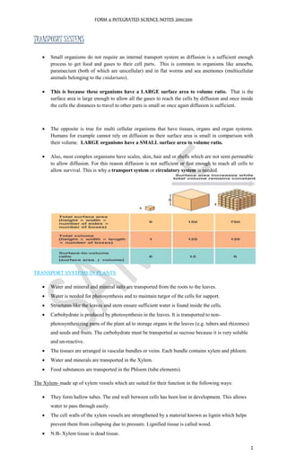
Small organisms do not require an internal transport system as diffusion is a sufficient enough
process to get food and gases to their cell parts. This is common in organisms like amoeba,
paramecium (both of which are unicellular) and in flat worms and sea anemones (multicellular
animals belonging to the cnidarians).
This is because these organisms have a LARGE surface area to volume ratio. That is the
surface area is large enough to allow all the gases to reach the cells by diffusion and once inside
the cells the distances to travel to other parts is small so once again diffusion is sufficient.
The opposite is true for multi cellular organisms that have tissues, organs and organ systems.
Humans for example cannot rely on diffusion as their surface area is small in comparison with
their volume. LARGE organisms have a SMALL surface area to volume ratio.
Also, most complex organisms have scales, skin, hair and or shells which are not semi permeable
to allow diffusion. For this reason diffusion is not sufficient or fast enough to reach all cells to
allow survival. This is why a transport system or circulatory system is needed.
TRANSPORT SYSTEMS IN PLANTS
Water and mineral and mineral salts are transported from the roots to the leaves.
Water is needed for photosynthesis and to maintain turgor of the cells for support.
Structures like the leaves and stem ensure sufficient water is found inside the cells.
Carbohydrate is produced by photosynthesis in the leaves. It is transported to non-
photosynthesizing parts of the plant ad to storage organs in the leaves (e.g. tubers and rhizomes)
and seeds and fruits. The carbohydrate must be transported as sucrose because it is very soluble
and un-reactive.
The tissues are arranged in vascular bundles or veins. Each bundle contains xylem and phloem.
Water and minerals are transported in the Xylem.
Food substances are transported in the Phloem (tube elements).
The Xylem- made up of xylem vessels which are suited for their function in the following ways:
They form hallow tubes. The end wall between cells has been lost in development. This allows
water to pass through easily.
The cell walls of the xylem vessels are strengthened by a material known as lignin which helps
prevent them from collapsing due to pressure. Lignified tissue is called wood.
N.B- Xylem tissue is dead tissue.
2. FORM 4 INTEGRATED SCIENCE NOTES 2010/2011
2
The Phloem- made up of sieve tube elements which are living cells. If they die they cannot perform their
function (not transport food).
Sieve tube elements have no nucleus.
They have a reduced cytoplasm which is controlled by the nucleus of a companion cell.
The end plate is full of holes and is called a sieve plate, this allows fluid to pass easily between
the sieve tube elements.
UPTAKE OF WATER BY ROOTS
1. Soil Grains- are covered with a tiny film of water as a result of surface tension. If the root hair
cell makes contact with a soil grain, it too becomes covered with a film of water due to surface
tension.
2. Water enters the root hair cell by osmosis. The root hair cell is adapted for its function because its
projection touches soil grains and becomes covered with water. The projection gives it a large
surface area.
3. Water moves across the cortex cells by osmosis and over the surface of the cells.
4. Water enters the xylem in the centre of the root.
NB- for mineral salts on the other hand the concentration is greater in the root hairs than in the soil,
therefore to absorb them, they are pumped into the root by active transport against their diffusion
gradient.
HOW DOES TRANSPORT OOCUR?
1. Capillarity- this is the rising of liquid in very narrow tubes. In plants the xylem is extremely
narrow therefore, water and dissolved minerals only rise up to a certain height.
2. Root Pressure- this is the force exerted by the root which pushes water into the xylem and up the
plant. This is caused by the absorption and active transport of minerals into the xylem vessels of
the root. This increases the concentration of solutes therefore, water moves in from the
surrounding soil naturally. As water enters the root, the water which was already there is pushed
upwards.
3. Cohesion Theory- the evaporation of water from leaves drives the movement of water from the
roots. When the water is lost from the top of the xylem a tension is created which lifts the water
up the xylem. The columns of water in the xylem are held together by cohesion and adhesion.
The cohesion pulls allowing for transport of water from root to leaf of the tallest trees.
4. Transpiration- Loss of water through the leaf. Dependent upon temperature, humidity, light
intensity, wind and water supply and Translocation- Movement of food substances in plants,
usually by mass flow.
3. FORM 4 INTEGRATED SCIENCE NOTES 2010/2011
3
TRANSPORT SYSTEMS IN HUMAN BEINGS
The transport medium – blood
A system of tubes to carry the blood- veins, arteries
A pump to create pressure and move the blood around- heart
Location where substances are exchanged- capillaries
Parts of the Blood
1. Red blood cells-They are called erythrocytes. They have a characteristic shape called biconcave
disc. Their biconcave shape gives them a large surface area for the diffusion of oxygen, it also
gives it a smooth shape which allow red blood cells to flow easily through narrow blood vessels.
The main function of the red blood cells is to transport oxygen from the lungs to the tissues.
The cytoplasm of the red blood cell is totally filled with a red pigment called haemoglobin.
The RBCs differ from other cells because they have no nucleus and because of this they
cannot repair themselves when damaged so they die very often (2-3million/second) and have
a life span of about 120 days
When oxygen concentration is high haemoglobin combines with oxygen to form
oxyhaemoglobin.This occurs as blood passes the lungs.
Haemoglobin is dark, brownish red while oxyhaemoglobin is a bright scarlet red.
In conditions of low oxygen concentration (the tissues), the oxygen separates and the
oxyhaemoglobin turns back into haemoglobin
Haemoglobin molecules contain iron, this is why iron is necessary in the diet.
Red blood cells assist in the transport of carbon dioxide from the tissues to the lungs, because
they contain an enzyme which assists carbon dioxide to combine with water for return to the
lungs.
2. White Blood Cells- They form the immune system. They defend the body from diseases. There
are different types:
Phagocytes: -move between tissue cells to sites of infection.
-They destroy bacteria by engulfing and digesting them.
-Many phagocytes are also killed in the process
-The mixture of dead phagocytes and bacteria is called pus.
4. FORM 4 INTEGRATED SCIENCE NOTES 2010/2011
4
Lymphocytes: -Germs(bacteria and viruses) have a unique protein coat on their outside
-This outer protein coat is called antigen
-When lymphocytes recognize a foreign antigen, they produce antibodies
in response
-Antibodies are chemicals which wrap around the germs causing them to
burst and clump together
-The clumps of germs are then engulfed and destroyed by phagocytes
-germs produce poisons called toxins. Some antibodies make these
harmless (antitoxins)
Haemorrhage- Is a loss of blood. A severe haemorrhage is life threatening because too many red blood
cells are lost so not enough oxygen can be carried to the tissues and also because blood pressure is
reduced and so is the rate of flow.
3. Platelets-also called thrombocytes are protein fragments which help the blood to clot preventing
excess blood loss.
Platelets are sticky, irregularly shaped and colourless
Their sticky surfaces along with other substances form clots to stop bleeding
A clot begins to form when blood is exposed to air
A clot can be external (scab) or internal (bruise , black and blue mark)
Calcium and Vitamin K must be present for clots to form
5. FORM 4 INTEGRATED SCIENCE NOTES 2010/2011
5
4. Plasma- The fluid in which blood cells are surrounded.
It is straw colours and consists of 90% water and 10% dissolved substances and plasma
proteins.
Main function is to carry the blood cells around the body.
It also carries dissolved nutrients, hormones, carbon dioxide and urea.
It also distributes heat around the body
HUMAN BLOOD GROUPS
GROUP
ANTIGEN
PRESENT
ANTI-BODY
PRESENT
CAN RECEIVE
FROM
CAN DONATE
TO
A A Anti-B A and O A and AB
B B Anti-A B and O B and AB
AB (Universal
Recipient)
A and B None A,B,AB and O AB
O (Universal
Donor)
None Anti- A and Anti-B O A,B,AB and O
Red blood cells have chemicals known as Antigens in their surface. Blood plasma(serum)
contains specials proteins known as Antibodies.
Blood Transfusion
A blood group must be matched before a transfusion is given.
If incompatible blood is given AGGLUTINATION will take place. This is where the blood
would clump together, possibly killing the person.
This happens because Antibodies are produced to fight off the foreign Antigens.
Blood is refrigerated in blood banks. Sodium Citrate is added as an anticoagulant, which stops the
blood from clotting.
Blood can only be stored for about three weeks, after this time too many blood cells are damaged.
The blood must also be tested for the Rhesus factor. People are either positive or negative.
This is also an Antigen. People who have it are termed as Rh+
, those who don’t are termed as
Rh –
6. FORM 4 INTEGRATED SCIENCE NOTES 2010/2011
6
Blood Vessels - means of transporting blood.
1. Arteries – Transports blood away from the heart.
Features:
Blood pressure in arteries is high so that blood retains enough pressure to go all the way
around the circulatory system.
They have a small (narrow) diameter (lumen), this helps maintain the blood pressure.
They have a thick muscular layer and thick elastic fibres that line their walls contracting
against the blood.
They do not have a valve as blood is pumped in one direction.
All arteries contain oxygenated blood EXCEPT the Pulmonary Artery.
2. Veins- Transport blood to the heart.
Features:
They have a thinner layer of muscle and elastic fibres as blood is under low pressure.
They have valves to stop the back flow of blood.
They have a relatively large diameter (lumen), which allows for easy passage of blood.
They are squeezed as they pass between muscles, which help to return blood to the heart.
The veins usually carry deoxygenated blood EXCEPT for the Pulmonary Vein.
3. Capillaries-are where exchange of substances between blood and tissues takes place. The blood
moves slowly to allow time for diffusion.
Features:
They have a permeable wall which is one cell thick, this allows for rapid diffusion of
food, water and gases.
Their diameter (lumen) is just wide enough for one red blood cell to pass through.
They form an extensive network around vital organs, providing an exchange surface for
the transport of materials.
DIAGRAMS OF VEINS ARTERIES AND CAPILLARIES
7. FORM 4 INTEGRATED SCIENCE NOTES 2010/2011
7
The human circulatory system is a double system because it has two loops. One loop carries
blood from the heart to the lungs and back to the heart. The other loop carries blood from the
heart to the body and back to the heart.
It is also a closed system because the blood is contained inside blood vessels.
Table 1: Substances transported in blood
Substance Transported from Transported to Why it needs to be
transported
Oxygen Lungs Body cells Respiration
Digested foods
(fats, glucose, amino
acids)
Digestive organs
(intestinal villi) and liver
Body cells Growth and cell
metabolism
Urea and other
nitrogenous waste
Liver and body cells Kidneys Excretion
Hormones Ductless endocrine glands Various organs as
needed
Regulation of body
functions
Heat Muscles, liver All tissues Regulation of body
temperature
Carbon dioxide Body cells and tissues Lungs Excretion
THE STRUCTURE OF THE HEART
8. FORM 4 INTEGRATED SCIENCE NOTES 2010/2011
8
The heart is a four-chambered muscular structure. It is used to pump blood around the body. It is made of
cardiac muscle which is a special muscle that does not get fatigued.
PARTS OF THE HEART:
1. Atrium- receives blood from the veins
2. Ventricles- have a thick muscular wall, when these contract the blood pressure in the ventricles
becomes very high.
3. Semi lunar valves- are found in the pulmonary artery and the aorta. These prevent blood from
flowing backwards into the ventricles.
4. Atria-ventricular valves (tricuspid and mitral) - prevent blood from flowing backwards into the
atria. Tendons call heart strings prevent these valves from failing under pressure.
5. The right side of the heart pumps deoxygenated blood from heart to lungs to heart.
6. The left side of the heart pumps oxygenated blood from heart to body to heart.
THE HEARTBEAT:
1. Blood carries little oxygen, and large amounts of carbon dioxide (deoxygenated blood), enters the
right atrium from the head and body through the Vena Cava.
2. Then the deoxygenated blood is pumped through the Tricuspid Valve into the right ventricle.
3. From the Right Ventricle blood is pumped into the Pulmonary Artery to be carried into the lungs.
In the Lungs, the carbon dioxide is exchanged for a new supply of oxygen.
4. The now oxygenated blood is returned to the Left Atrium via the Pulmonary Vein.
5. The blood is pumped through the Bi-cuspid Valve to the Left Ventricle. From there it is carried to
the various organs of the body. And then the process repeats itself.
6. The heart has two phases. During the SYSTOLE phase the ventricles contract and force blood
into the arteries and during the DIASTOLE phase the ventricles relax drawing blood into the
atrium.
NB: Active and Passive immunity and vaccines, immune deficiency virus.
Blood transfusion in terms of pregnancy, Rhesus factor, transmitted diseases.
Physiological effects of exercise, performances enhancing drugs. (Blood doping
increases the number of red blood cells). Steroids, diet and training programs.