2. Terminologies
Glands are basically divided into
Endocrine glands – are glands that secrete their products
through the basal lamina and lack aduct system.
Exocrine glands _ secrete their products through aduct.
The glands in this group can be divided into three group:-
3. Merocrine glands – cells secrete their substances by exocytosis
(mucous and serous glands).
Apocrine glands – aportion of the secreting cells body is lost
during secretion.
Holocrine glands – the entire cell disintegrates to secrete its
substances (sebaceous gland).
4.
5. The type of secretory product of an exocrine gland may
also be one of three categories:
Serous glands – secrete awatery , often protein rich
product.
Mucous glands – secrete aviscous product , rich in
carbohydrates .
Sebaceous glands – secrete alipid product.
6. Definition:- salivary galnds are compound , tubuloacinar, merocrine , exocrine glands the
ducts of which open in the oral cavity.
7. Classification of salivary glands
According to location
According to size
According to the nature of secretion
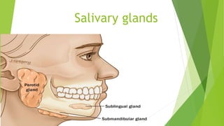
8. According to location
Glands of oral vestibule
1- labial glands (upper and lower)
2- buccal glands
3- parotid glands
Gland of oral cavity proper
1- palatine glands (hard ,soft palate and uvula)
2- glands of the floor of the mouth (sublingual,submandibular,glossopalatine)
3-glands of tongue (Weber , Von Ebner , Balandin Nuhn)
9. According to size
Major salivary glands
1- parotid Gs
2- submandibular Gs
3- sublingual (major) Gs
Minor salivary glands
1-labial & buccal Gs 2-palatine Gs
3- Glossopalatine Gs 4- minor sublingual glands
5- Weber Gs 6-Von Ebner Gs
7- Balandin Nuhn Gs
10. According to the nature of secretion
Pure serous Gs
1- Parotid gland (of adult)
2- Von Ebner Gs
Pure Mucous Gs
1-palatine Gs 2- Glossopalatine Gs
3- minor sublingual glands 4- Weber Gs
Mixed Gs
1-labial & buccal Gs 2- submandibular Gs
3- sublingual (major) Gs 4- Balandin Nuhn Gs
5- parotid (infant & old age)
11. Development and growth
The individual salivary glands arise as aproliferations of oral epithelial
cells , forming afocal thickening that grows into the underlying
ectomesenchyme.
Continued grows results in the formation of small bud connected to the
surface by acord of epithelial cells with mesenchymal cells condensed
around the bud.
Clefts develop in the bud forming two or more new buds , continuation of
this process called branching morphogenesis.
Signaling molecules including members of the fibroblast growth factor
family , transforming growth factor B and their receptors play amajor
role in the development of branches.
12. Salivary gland branching morphogenesis.
Transmitted light microscopy of a living
submandibular salivary gland dissected
out of a mouse embryo after 12 days of
gestation (panel A ), placed into explant
culture, and photographed after being
cultured for an additional 12 hrs (B), 24
hrs (C), 48 hrs (D), and 72 hrs (E). The
gland starts as a single bud, which is
subdivided by clefts (arrowheads), which
progressively deepen. The clefts
eventually widen to define secondary
ducts (arrow) connected to the main duct.
This process of branching morphogenesis
progresses rapidly from a single bud to a
complex branched structure in 3 days,
and it continues to branch as the embryo
develops. Scale bar = 100 μm.
13. The development of alumen within abranched epithelium
generally occurs in this order 1- in the distal end of the main cord
and in branch cords 2-in the proximal end of the main cord 3- in
the central portion of the main cord .
The lumen form within the ducts befor they develop within the
terminal buds.
Some studies suggested that lumen formation may involve
apoptosis of centrally located cells in the cell cords.
14. Following development of the lumen in the terminal buds , the
epithelium consists of two layers of cells .
The cells of the inner layer differentiate into the secretory cells of
the mature gland , mucous or serous.
Some cells of the outer layer forms the contractile myoepithelial
cells that are present around the secretory end pieces and
intercalated ducts .
As the epithelial components increase in size and number the
associated mesenchyme is dimenshed , although athin layer of C.T
remains surrounding each secretory end piece and duct of the adult
gland.
15. Thicker partitions of connective tissue(septa) continuous with
capsule and within which run the nerves and blood vessels
supplying the gland , and divide the gland into lobes and lobules.
16. The Parotid glands begin to develop at 4 to 6th week of
intrauterine life , the submandibular glands at 6 week , and
the Sublingual and Minor salivary glands at 8 to 12 weeks.
The cells of the secretory end pieces and ducts attain
maturity during the last 2 months of gestation.
17. Histological structure of salivary gland
Parenchymal elements
1- Terminal secretory portion
2- Non secretory cells
3- Duct system
connective tissue stroma
1- Cells , nerves, blood vessels
2- Ground substances
18.
19. Terminal secretory portion
The terminal portions are concerned mainly with
the production of saliva.
There are three types
1- serous acinus
2- Mucous acinus
3- Mixed
20. Serous cells
Morphology :- pyramidal with broad base in the
basement membrane and apex towards narrow lumen.
Each cell has arounded nucleus situated in the basal
third of the cell.
Apical part contains eosinophilic secretory granules
(zymogen granules) that clearly seen by toluidine blue
stain.
21.
22. The lumen usually has finger like
extensions located between
adjacent cells called intercellular
canaliculi that increase the size of
luminal surface of the cells
23. Large amount of RER arranged basaly
and laterally to the nucleus.
Golgi complex 4-6 situated apical or
lateral to the nucleus .
Numerous mitochondria situated basally
and laterally to the nucleus.
Cytoplasmic organells include lysosomes,
free ribosomes , microtubules and
microfilaments.
24. Inter cellular junction between serous cells
Tight junction
The adhering junctions and desmosomes – hold adjacent
cell together and also help in cell signaling.
Gap junctions - allow passage of ions and small
molecules between the cells.
25.
26. Transmission EM of serous cell of
rat parotid gland.
N nucleus
Lu lumen
SG secretory granules
Arrow head inter cellular spaces
Ly lysosomes
rER rough endoplasmic reticulum
27. Mucous cells
The mucous cell is high cuboidal in shape and larger than the
serous cells .
It has broader apex bordering awide lumen .
The nuclei of mucous cell are flattened and basally located.
Mucous cell lack intercellular canaliculi , expect for those
covered by demilunes.
28. Mucous cells have more golgi apparatus (10-12) saccules because
of the greater amount of carbohydrate that is added to the secretory
protein.
The mucous cells exhibits large , irregular , electron – lucent
mucigen granules which are readily distinguished from those of
serous cells by their pale appearance.
The cells appear pale due to their mucinous granules of high
carbohydrate content which gets dissolved during preparation of
sections giving an empty appearance , the mucinous granules are
clearly seen by PAS stain.
29.
30.
31. Mucous secretion differ from serous secretion in two ways :-
1- They have little or no enzymatic activity and mainly
serves for lubrication and protection of oral tissues.
2-The ratio of carbohydrate to protein is greater.
32. Secretion of zymogen and mucigen granules
Serous cell
Secretion of zymogen
granules by exocytosis
without loss of cytoplasm.
Mucous cell
Secretion of mucigen
granules is different from
zymogen granules.
The membrane may
fragment and being lost with
the discharge of mucous.
33. Transmission electron
micrograph (TEM) showing
several secretory granules in
the apical surface of the cell.
A granule has just secreted its
contents by exocytosis into
the lumen.
34. Salivary gland. Coloured
scanning electron micrograph
(SEM) of a section through a
parotid salivary gland, showing
numerous serous secretory
granules (small, spherical),
within the serous glands.
Serous glands contain serous
acini.
Endoplasmic reticulum (brown
stacks at right and upper left),
35. Mixed secretory portion
In mixed glands the proportion of serous and mucous cell vary
from predominantly serous as in human submandibular gland , to
predominantly mucous as in human major sublingual gland .
Separate serous and mucous units may exist , in addation to
secretory units composed of both cell types .
In the later arrangement the mucous cells form atypical tubular
portion that’s capped at the blind end by crescents of several
serous cells known as crescent og giannuzzi or demilune of von
Ebner.
39. Myoepithelial cells
Myoepithelial are contractile cells
located around the terminal secretory
portions and the intercalated duct.
They are situated between the
basement membrane and the basal
plasma membrane of the
parenchymal cells.
40. It is stellate in shape and has small flattened body with elongated
nucleus and numerous long branching processes that embrace the
secretory and duct cells.
The plasma membrane of myoepithelial cells joins the basal
membrane of parenchymal cells by desmosomes.
In ordinary H&E stain only their nuclei are visible.
Immunofluorescent studies indicate presence of actin and myosin.
43. Function of Myoepithelial cell:-
1- Myoepithelial cells have contractile function as it
help to expel saliva from the terminal portion and
intercalated duct.
2-It may provide support for the parenchymal cells
during active secretion.
3- Contraction of the myoepithelial cells of the
intercalated ducts may shorten and widen the duct ,
helping to maintain their patency.
44. Recent studies showed that myoepithelial cells are involved
in signaling the secretory cells and protecting the salivary
gland tissue.
The myoepithelial cells also produce anumber of protein
that has tumor suppressor activity such as proteinase
inhibitors and angiogenic factors which act as barriers
against invasive epithelial neoplasm.
45. Oncocytes
This is aspecial large eosinophilic cell with small ,
centrally pyknotic nucleus and loaded with
mitochondria.
It tends to be found in the ducts particularly in parotid
and submandibular glands.
This cell may represent an age change it may form
tumor called oncocytoma.
48. Intercalated duct
The primary saliva produced by the secretory end pieces
passes first through the intercalated duct ducts .
The ducts are lined by asimple cuboidal epithelium.
49. The intercalated duct cells have centrally placed nuclei which
appear prominent with scanty cytoplasm contain some RER and a
small golgi complex.
The apical cell surface has afew short microvilli projecting into
the lumen , the lateral surfaces are joined by apical junctional
complexes , scattered desmosomes and gap junctions .
50. Function of intercalated duct:-
They convey the secretion from the terminal secretory units to the
striated ducts.
Lactoferrin (aprotein having an antibacterial function) has been
localized in the cells of the intercalated ducts.
They are capable of reabsorbing protein from the lumen (modifaction).
They may represent areserve of UMC providing adegree of
regenerative capability in the event of injury of the gland.
51. Striated ducts
The striated ducts are lined by simple high columnar cells .
The cells of the striated ducts have alarge amount of eosinophilic
cytoplasm and alarge , spherical , centrally positioned nucleus .
Ultra-structurally Basal cytoplasm of striated duct cells show deep
infolding of the plasma membrane and many large mitochondria are
packed between the infolding , this large surface area supplied with
high level of energy is involved in active transport.
Adjacent cells are joined by well developed tight junctions and
junctional complex and lack of gap junction.
52.
53.
54. Functions of the striated duct
Convey secretion from inter calated duct to excretory duct.
They actively reabsorb sodium and chloride ions and secrete
potassium and bicarbonate in the primary secretion thus the
secretion is changed from an isotonic or hypertonic to
hypotonic secretion with low sodium and chloride and high
potassium and bicarbonate.
Amylase enzyme is added from the serum to the saliva by
the cells of striated duct.
55. Excretory duct(Interlobular)
The excretory ducts are located in the connective tissue septa between
the lobules of the gland.
The basal striation of striated duct become less prominent as the
excretory duct increase in size.
Near the striated duct the excretory duct are lined by
apseudostriatified epithelium of tall columnar cells.
In larger excretory duct the epithelial lining may contain goblet cell.
56.
57. Function of excretory duct
They convey the salivary secretion toward the oral cavity.
They have the ability to reabsorb sodium and secrete
potassium and bicarbonate.
58. Main duct
As the excretory duct approach the oral cavity ,
epithelium changes to non keratinized stratified
squamous epithelium merging with that of the oral
cavity at the ductal orifice.
59. Connective tissue
The C.T elements of the salivary glands forms the capsule which
serrounds the parenchymal elements of major salivary glands.
It forms septa that divide the gland into lobes and lobules.
The cells : fibroblast , plasma cells , mast cells, macrophage and fat
cells.
Collagen , reticulr fibers and ground substances(glycoprotein and
proteoglycans).