1. Development of the MSD-based assay for determining the PD-1 Antibody concentration in mouse serum and tumor
Khushbu Bhatt, Tatiana Tolstykh, Lakshmi Srinivasan, Michael Lampa, Yu-an Zhang, Mikhail Levit, Andrew Hebert, Dmitri Wiederschain, Timothy Wagenaar
Sanofi Oncology, 640 Memorial Drive, Cambridge, MA 02138
Abstract
Introduction
PD-1 (Programmed cell death protein 1/ CD279) is an immune-inhibitory
cell-surface receptor expressed on T-cells. Its ligands PD-L1 and PD-L2
(Programmed death ligand 1/2) (CD274) are present on the tumor cells
and various antigen presenting cells (APCs) such as dendritic cells and
macrophages. Upon binding to its ligands PD-1 initiates a signaling
cascade within the T cell to attenuate the T cell response which contributes
to immune evasion of the tumor cells. PDL-1 is highly expressed in
melanoma, renal cell carcinoma (RCC) and non-small cell lung cancer
(NSCLC). Thus expression of PD-1 on TILs (tumor infiltrating
lymphocytes) and its interaction with PDL-1 and PDL-2 on tumor and
other immune cells suppresses the recognition of tumor antigen by the T-
cells. Therein lies the therapeutic rationale that by blocking the
interaction of PD-1 with its ligands the anti-tumor immune response can
stimulated.
Results
Conclusion
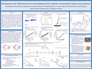
MSD based assay with high specificity and sensitivity to analyze the PD-1
antibody concentration as low as 0.5 ng, with minimal background and
matrix effect was developed and validated. This assay was used to
determine the antibody levels at various time points in serum and tumor.
Acknowledgement
I would like to thank my mentor Tatiana Tolstykh and my team members
Lakshmi Srinivasan, Timothy Wagenaar, Michael Lampa, Yu-an Zhang for
training me in the lab and helping me with the project. Thanks to Mikhail Levit
and Andrew Hebert for helping me out with the execution of MSD assay. I would
also like to thank the Section Head Dmitri Wiederschain for his continued
motivation, guidance, support and patience during my time in Sanofi.
Materials and Methods
MSD (Meso Scale Discovery)
Meso Scale Discovery’s MULTI-ARRAY®
technology is an unique immunoassay platform
which enables detection of protein markers in
single and multiplex format utilizing the electro-
chemiluminescence technique.
MSD Assay’s improved sensitivity, expandable
dynamic range, multiplexing ability and no
matrix effect gives an edge over the other
traditional techniques like ELISA, RIA.
Simple WES : Protein Simple
Simple Western is an automated size-based
Western analysis which separates and detects
proteins as large as 440 kD in as little as 3 hours.
Total Protein Assay from Protein Simple was
used to analyze the total protein content in the
Pd-1 antibody in which target proteins are
separated by size, labelled with biotin reagent
and are then detected by chemiluminescence
using Streptavidin-HRP
Purity/Total Protein Concentration of PD-1 (RMP1-14) antibody
Evaluation of binding of PD-1 antibody to PD-1
MSD Assay Development
-Optimizing variables:
1. PD-1 antigen capture level
2. Detection antibody concentration
-Standard curve generation
-Effect of serum on the assay
-In-vivo validation of the assay
PK-PD Study to evaluate PD-1 antibody in mouse
Determining the kinetics of PD-1 antibody in serum and tumor
Target Engagement using FACS
Assay Development Outline
MSD Assay Development
Low background, high signal to noise ratios, 3 log dynamic range (0.5 ng-10,000 ng), high sensitivity, low matrix (serum) effect
Optimized Variables: PD-1 coating concentration-2 ug/ml , Detection antibody concentration- 0.5 ug/ml
Minimum PD-1 antibody detection level: 0.5 ng/ml
MSD Kit
Results
PK/PD study to evaluate mouse PD1 antibody
Cancer Immunology: PD-1 and Beyond. (n.d.). Retrieved from https://www.smartpatients.com/pathways/pd-1
The PD-1 receptor and its ligands PDL-1 and PDL-2 are important
inhibitory molecules on T cells that contribute to tumor immune evasion.
Checkpoint blocking antibodies targeting PD-1 and PD-L1 have shown
significant anti-tumor activity in a range of human cancer types. To
investigate the effect of checkpoint blockade in murine tumor models
several surrogate antibodies targeting PD-1 have been described in the
literature. To identify an optimal dosing regimen of the PD-1 antibody
RMP1-14 a more thorough understanding of the antibody
pharmacokinetics was required. To this end, a Meso Scale Discovery
(MSD) assay was developed to determine the concentration of PD-1
antibody in mice serum and tumor. Different variables like PD-1 plate
coating concentration and detection antibody concentration were
optimized to yield an assay with three-log dynamic range, low
background, minimal matrix effect and high signal to noise ratio. The
optimized MSD assay was used to evaluate RMP1-14 pharmacokinetics
after a single IV injection in tumor bearing mice. The MSD assay
indicated dose proportional expose of RMP1-14 with a strong correlation
of serum and tumor antibody levels.
Protein Simple WES
PD-1
ISOTYPE
Typical dose-response obtained when RMP1-14 was
serially diluted
Normalized with BSA as standard
Purity/Total Protein Concentration of
RMP1-14 (PD-1 antibody)
Binding of PD-1 antibody to PD-1
confirmed using FACS
EL4 cell line which constitutively express
high levels of PD-1 was used to confirm
antibody binding
PD-1 antigen capture level Detection antibody concentration Standard curve generation and
effect of serum (33% ) on the assay
Dose proportional increase in serum concentration
PK supports dosing every 3 to 4 day intervals
Correlation in tumor and serum PK of PD-1 antibody
Significant decrease in MFI of PD-1 on intratumoral CD8 T cells at
336 hrs.
Serum and Tumor PK of PD-1 antibody
Serum
Tumor
- Time points when mice were taken down
T u m o r P K D a ta
T im e (h rs )
RMP1-14,ng/mgoftotalprotein
6 1 6 8 3 3 6
0
5 0
1 0 0
1 5 0
2 0 0
P B S
5 m g/kg
1 0 m g/kg
2 0 m g/kg
S e ru m P K D ata
T im e, h rs
RMP1-14,ug/ml
6 168 336
0
500
1000
1500
P B S
5 m g/kg
10 m g/kg
20 m g/kg
Serum and Tumor PK of PD-1 antibody
TumorSerum
Target Engagement confirmed
by FACS