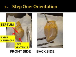
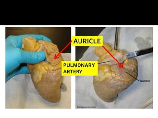
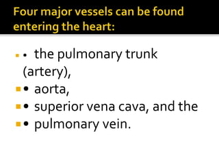
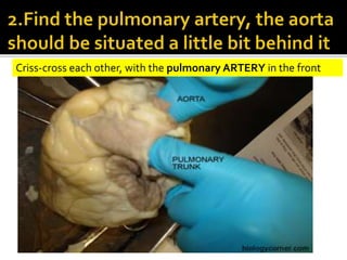
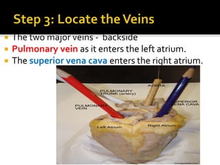
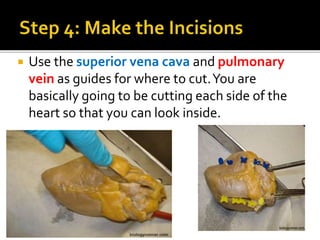
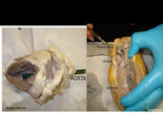
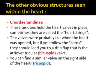
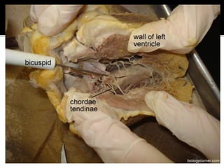
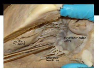
LS2BFET
UNIT 1
PRACTICAL 1
FRONT SIDE BACK SIDE
SEPTUM
RIGHT
VENTRICLE
LEFT
VENTRICLE
AURICLE
PULMONARY
ARTERY
 • the pulmonary trunk
(artery),
• aorta,
• superior vena cava, and the
• pulmonary vein.
Criss-cross each other, with the pulmonary ARTERY in the front
 The two major veins - backside
 Pulmonary vein as it enters the left atrium.
 The superior vena cava enters the right atrium.
 Use the superior vena cava and pulmonary
vein as guides for where to cut.You are
basically going to be cutting each side of the
heart so that you can look inside.
 Your colour pencils you used to mark the
heart in step 2 can also now be used to see
where those vessels connect within the heart.
 For instance, the aorta pencil can now be
seen ending in the left ventricle.
 You can also now see how much thicker the
walls of the left ventricle are compared to
the right ventricle. ???
 Chordae tendinae
 These tendons hold the heart valves in place,
sometimes they are called the "heartstrings".
 The valves were probably cut when the heart
was opened, but if you follow the "cords"
they should lead you to a thin flap that is the
atrioventricular (bicuspid) valve.
 You can find a similar valve on the right side
of the heart (tricuspid).
ENJOY!

Practical 1 pig heart dissection

  • 1.
  • 2.
    FRONT SIDE BACKSIDE SEPTUM RIGHT VENTRICLE LEFT VENTRICLE
  • 3.
  • 4.
     • thepulmonary trunk (artery), • aorta, • superior vena cava, and the • pulmonary vein.
  • 5.
    Criss-cross each other,with the pulmonary ARTERY in the front
  • 6.
     The twomajor veins - backside  Pulmonary vein as it enters the left atrium.  The superior vena cava enters the right atrium.
  • 7.
     Use thesuperior vena cava and pulmonary vein as guides for where to cut.You are basically going to be cutting each side of the heart so that you can look inside.
  • 8.
     Your colourpencils you used to mark the heart in step 2 can also now be used to see where those vessels connect within the heart.  For instance, the aorta pencil can now be seen ending in the left ventricle.  You can also now see how much thicker the walls of the left ventricle are compared to the right ventricle. ???
  • 10.
     Chordae tendinae These tendons hold the heart valves in place, sometimes they are called the "heartstrings".  The valves were probably cut when the heart was opened, but if you follow the "cords" they should lead you to a thin flap that is the atrioventricular (bicuspid) valve.  You can find a similar valve on the right side of the heart (tricuspid).
  • 13.