More Related Content
Similar to In Situ Formation of Metal Oxide Nanoparticles in Nafion Membranes
Similar to In Situ Formation of Metal Oxide Nanoparticles in Nafion Membranes (20)
In Situ Formation of Metal Oxide Nanoparticles in Nafion Membranes
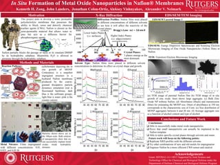
- 1. X-Ray Diffraction
0
2000
4000
6000
8000
0 20 40 60 80 100
In Situ Formation of Metal Oxide Nanoparticles in Nafion® Membranes
Kenneth H. Zong, John Landers, Jonathan Colon-Ortiz, Aleksey Vishnyakov, Alexander V. Neimark
Motivation
Metal Nitrates: Films impregnated
with different concentrations: 0.01,
0.025, and 0.05 M.
Nafion partially blocks the passage of Sarin or its simulant DMMP
by its hydrophobic subphase. Meanwhile H2O is allowed to
permeate through the hydrophilic subphase.
Solvent Type: Nafion films were placed in different solvent
concentrations to determine its effect on crystal shape and growth.
Grant: HDTRA1-14-1-0015 Supported by Joint Science and
Technology Office for Chemical and Biological Defense under the
Department of Defense Chemical and Biological Defense Program
Higher Index Planes
(i.e. edges/corners)
Acknowledgements
Methods and Materials
Reaction Pathway:
0.05 M
0.025 M
Zn Ni Mg Fe Co
0.01 M
Conclusions and Future Work
EDS/SEM/TEM Imaging
Diffraction Profiles: Nafion films were placed
in different concentrations of different solvents
to see how it will affect the reactivity of the
crystal planes.
𝐁𝐫𝐚𝐠𝐠′
𝐬 𝐋𝐚𝐰: 𝒏𝝀 = 𝟐𝒅 𝐬𝐢𝐧 𝜽
Landers ©
Crystal Planes:
3 planes emerge as the
most dominant.
100 002 101
Lower Index Planes
(i.e. surfaces)
K.H. Zong ©
Conclusions:
Able to successfully make metal oxide nanoparticles.
Prove that small nanoparticles can actually be implanted in the
Nafion template.
Able to tailor specific crystal planes through solvents and strain.
Future work will focus on the following points:
Replicate XRD profiles with the different solvents.
Try other combinations of new and old metals for impregnation.
Engineer Nafion be a more efficient CWA sensor and catalyst.
Co
Ni Zn
Fe
Co Ni Fe Zn
10 μm
100 μm
EDS/SEM Layered Image 1
EDS/SEM: Energy Dispersive Spectroscopy and Scanning Electron
Microscopy Imaging of Zinc Oxide Nanoparticles (Yellow Dots) in
Nafion.
Particles shown above are to
the 100μm scale. Bulk solution
of nanoparticles show that we
can successfully implant metal
oxides inside hydrophilic
domains.
TEM: Transition Electron Microscopy Imaging
0.5in
100 μm 100 μm
Intensity
This project aims to develop a water permeable
polyelectrolyte membrane that possesses the
ability to block, sense and detoxify chemical
warfare agents (CWA). Nafion is chosen as the
semi-permeable material that allows water to
pass but acts as a diffusion barrier for
phosphororganic agents.
2θ
(a) TEM image of parental Nafion film (b) TEM image of in situ
growth of zinc oxide. Inset shows large aggregate. (c) Bulk Zinc
Oxide NP without Nafion. (d) Absorbance (black) and transmission
(blue) for estimating the MONP size. Onset of absorbance at 350 nm
corresponds to the characteristic size of 4 nm. (e) Size distribution of
MONP aggregates between 6 and 20 nm. (f) Average aggregate size
as a function of alcohol content and type of alcohol.
(a)Schematic depicting the in
situ growth of MONP.
Centerpiece is a snapshot
segregated structure in a
hydrated Nafion membrane
produced by the coarse-
grained dissipative particle
dynamics simulation (red is
fluorinated backbone, dark
blue is the sulfonate groups,
light blue is water, pink is the
counter cation).
c
500 nm