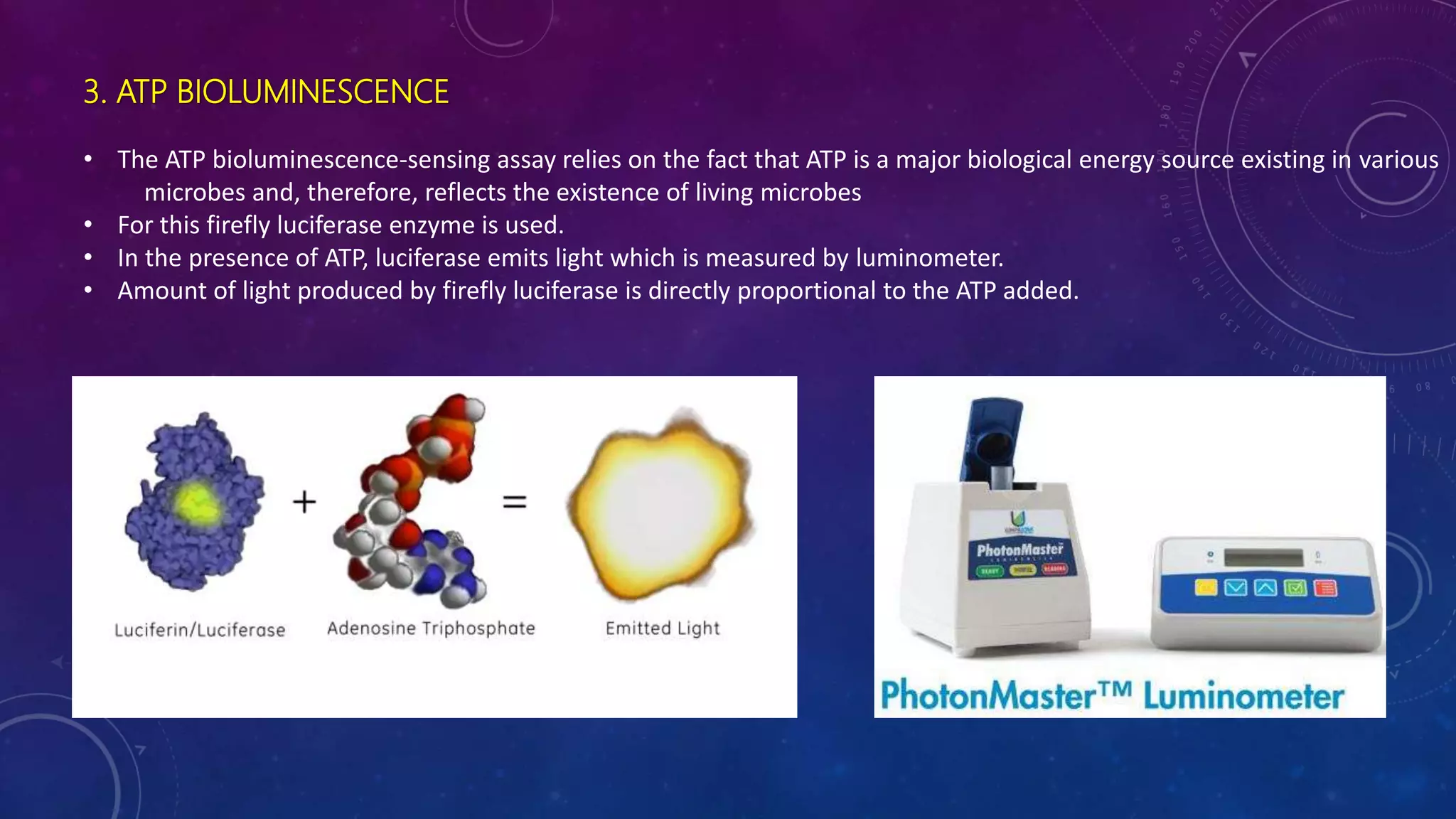
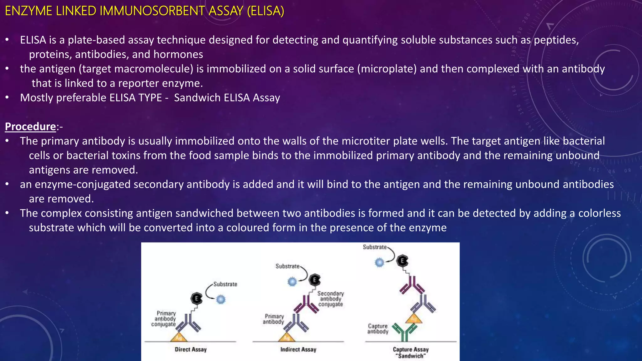
RAPID METHODS FOR
DETECTION OF
PATHOGENS IN INDIA
JUBY MATHEW
Introdution
Foodborne pathogens cause a lot of diseases
Few characteristic pathogens are :-
o Pathogenic Bacteria
• Bacillus cereus
• Campylobacter jejuni
• Clostridium botulinum
• Esherichia coli
• Salmonella sp.
o Pathogenic Viruses
• Hepatitis A
• Noroviruses
o Pathogenic Parasites
• Cylospora cayetanensis
• Toxoplasma gondii
• Trichinella spiralis
 Nucleic Acid – Base Method
 Biosensors
 Immunological Methods
 Chemical Method
Rapid Detection of Pathogens
NUCELIE ACID –BASE METHODS
It operates by detecting specific DNA or RNA sequences in the target pathogen.
Done by hybridizing the target nucleic acid sequence to a synthetic oligonucleotide (probes or primers) which is
complementary to the target sequence.
It includes
• Polymerase chain reaction (PCR),
• Multiplex polymerase chain reaction (mPCR),
• Real-time/quantitative polymerase chain reaction (qPCR),
• Loop-mediated isothermal amplification (LAMP)
.
1. POLYMERASE CHAIN REATION (PCR)
It allows the detection of a single bacterial pathogen that present in food by detecting a specific target DNA sequence
It amplifies a specific target DNA sequence in a cyclic three steps process
Procedure :-
• the target double-stranded DNA is denatured into single-stranded DNA at high temperature. (94-98⁰C)
• two single-stranded synthetic oligonucleotides or specific primers which are the forward and reverse primer will anneal
to the DNA strands. (48-72⁰C)
• primers complementary to the single-stranded DNA are extended with the presence of deoxyribonucleotides and a
thermostable DNA polymerase. (68-72⁰C)
The PCR amplification products are visualized on electrophoresis gel as bands by staining with ethidium bromide
PCR have been used in the detection of numerous foodborne pathogens like Listeria monocytogenes, Escherichia coli,
Staphylococcus aureus, Campylobacter jejuni, Salmonella spp. and Shigella spp.
2. MULTIPLEX PCR (mPCR)
• Several sets of specific primers are used in mPCR assay
• Primer design is very important for the development of mPCR, as the primer sets should have similar annealing
temperature in order to produce a successful mPCR assay.
• Can detect 5 or more pathogens
• Used for detection of Salmonella Enteritidis, Staphylococcus aureus, Shigella flexneri, Listeria monocytogenes, and
Escherichia coli O157:H7
3. REAL TIME QUANTITATIVE PCR (qPCR)
• This method is able to monitor the PCR products formation continuously in the entire reaction by measuring the fluorescent
signal produced by specific dual- labelled probes or intercalating dyes.
• The fluorescence intensity is proportional to the amount of PCR amplicons.
mPCR machine qPCR machine
4. LOOP MEDIATED ISOTHERMAL AMPLIFICATION (LAMP)
• LAMP is a novel nucleic acid amplification method developed by Notomi.
• It provides a rapid, sensitivity and specific detection of foodborne pathogens.
• LAMP is based on auto-cycling strand displacement DNA synthesis carried out by Bst DNA polymerase large
fragment under isothermal conditions between 59°C and 65°C for 60 min.
• In LAMP, four primers comprising two inner primers and two outer primers are used to target six specific regions
of target DNA.
• Large amount of amplicons can be produced by LAMP within 60 min which is usually 103-fold or higher as
compared to simple PCR.
• The amplicons can be detected by agarose gel electrophoresis or SYBR Green I dye .
BIOSENSOR METHODS
Biosensor is an analytical device that consists of two main elements: a bioreceptor and a transducer.
The bioreceptor responsible for recognizing the target can either be a:
(1) Biological material: enzymes, antibodies, nucleic acids and cell receptors, or
(2) Biologically derived material: recombinant antibodies, or
(3) Biomimic: imprinted polymers and synthetic catalysts.
Transducer converts biological interactions into measurable signals
1) Optical
2) Electrochemical
3) Mass- based
4) Thermometric
5) Micromechanical
6) Magnetic
The recent biosensors that commonly used for the detection of foodborne pathogens are optical, electrochemical and
mass-based biosensors
Types of Biosensors are:-
 Bacterial bioluminescence
 Electrical Impedance Biosensors
 ATP Bioluminescence
1. BATERIAL BIOLUMINESCENCE
• Gene responsible for bacterial bioluminescence (lux gene) has been identified and cloned
• The DNA carrying this gene can be introduced into the host specific phages
• These genes do not posses the intracellular biochemistry necessary to express this gene, remain dark.
• On transfer of lux gene to the host bacterium during infection results in light emission that can be easily
detected by luminators
Specificity of this assay depends on phage specificity
E.g.:- Bacteriophage p22 is specific for Salmonella typhimurium
2. ELECTRICAL IMPEDANCE BIOSENSORS
• Detects microbes directly due to the production of ions from metabolic end products or indirectly from liberation of CO₂.
• Method –
• Population of microbes provided with nutrients (non-electrolyte) like lactose
• Microbes utilizes it, and convert into lactic aid (ionic form).
• Impedance is measured over 20hrs after inoculation in specific media.
3. ATP BIOLUMINESCENCE
• The ATP bioluminescence-sensing assay relies on the fact that ATP is a major biological energy source existing in various
microbes and, therefore, reflects the existence of living microbes
• For this firefly luciferase enzyme is used.
• In the presence of ATP, luciferase emits light which is measured by luminometer.
• Amount of light produced by firefly luciferase is directly proportional to the ATP added.
IMMUNOLOGICAL METHODS
1. Enzyme Linked Immunosorbent Assay
2. Radioimmunoassay
ENZYME LINKED IMMUNOSORBENT ASSAY (ELISA)
• ELISA is a plate-based assay technique designed for detecting and quantifying soluble substances such as peptides,
proteins, antibodies, and hormones
• the antigen (target macromolecule) is immobilized on a solid surface (microplate) and then complexed with an antibody
that is linked to a reporter enzyme.
• Mostly preferable ELISA TYPE - Sandwich ELISA Assay
Procedure:-
• The primary antibody is usually immobilized onto the walls of the microtiter plate wells. The target antigen like bacterial
cells or bacterial toxins from the food sample binds to the immobilized primary antibody and the remaining unbound
antigens are removed.
• an enzyme-conjugated secondary antibody is added and it will bind to the antigen and the remaining unbound antibodies
are removed.
• The complex consisting antigen sandwiched between two antibodies is formed and it can be detected by adding a colorless
substrate which will be converted into a coloured form in the presence of the enzyme
2. RADIOIMMUNOASSAY
It consists of adding a radioactive label to an antigen, allowing the labelled antigen to react with its specific antibody and
measuring the antigen that combined with the antibody by the use of a counter to measure radioactivity
CHEMICAL METHODS
Limulus Lysate for Endotoxins
• Limulus Amoebocyte Lysate (LAL) test was introduced in 1970s by Fredrick Bang and Jack Levin
• This employs a lysate protein obtained from the blood cells of horseshoe crab (Limulus polyphemous)
• This protein is sensitive to endotoxins
• Presence of endotoxins will cause gel formation of lysate material.
Limulus polyphemousFrederick Bang
Detction of pathogens

Detction of pathogens

  • 1.
    RAPID METHODS FOR DETECTIONOF PATHOGENS IN INDIA JUBY MATHEW
  • 2.
    Introdution Foodborne pathogens causea lot of diseases Few characteristic pathogens are :- o Pathogenic Bacteria • Bacillus cereus • Campylobacter jejuni • Clostridium botulinum • Esherichia coli • Salmonella sp. o Pathogenic Viruses • Hepatitis A • Noroviruses o Pathogenic Parasites • Cylospora cayetanensis • Toxoplasma gondii • Trichinella spiralis
  • 3.
     Nucleic Acid– Base Method  Biosensors  Immunological Methods  Chemical Method Rapid Detection of Pathogens
  • 4.
    NUCELIE ACID –BASEMETHODS It operates by detecting specific DNA or RNA sequences in the target pathogen. Done by hybridizing the target nucleic acid sequence to a synthetic oligonucleotide (probes or primers) which is complementary to the target sequence. It includes • Polymerase chain reaction (PCR), • Multiplex polymerase chain reaction (mPCR), • Real-time/quantitative polymerase chain reaction (qPCR), • Loop-mediated isothermal amplification (LAMP) .
  • 5.
    1. POLYMERASE CHAINREATION (PCR) It allows the detection of a single bacterial pathogen that present in food by detecting a specific target DNA sequence It amplifies a specific target DNA sequence in a cyclic three steps process Procedure :- • the target double-stranded DNA is denatured into single-stranded DNA at high temperature. (94-98⁰C) • two single-stranded synthetic oligonucleotides or specific primers which are the forward and reverse primer will anneal to the DNA strands. (48-72⁰C) • primers complementary to the single-stranded DNA are extended with the presence of deoxyribonucleotides and a thermostable DNA polymerase. (68-72⁰C) The PCR amplification products are visualized on electrophoresis gel as bands by staining with ethidium bromide PCR have been used in the detection of numerous foodborne pathogens like Listeria monocytogenes, Escherichia coli, Staphylococcus aureus, Campylobacter jejuni, Salmonella spp. and Shigella spp.
  • 6.
    2. MULTIPLEX PCR(mPCR) • Several sets of specific primers are used in mPCR assay • Primer design is very important for the development of mPCR, as the primer sets should have similar annealing temperature in order to produce a successful mPCR assay. • Can detect 5 or more pathogens • Used for detection of Salmonella Enteritidis, Staphylococcus aureus, Shigella flexneri, Listeria monocytogenes, and Escherichia coli O157:H7 3. REAL TIME QUANTITATIVE PCR (qPCR) • This method is able to monitor the PCR products formation continuously in the entire reaction by measuring the fluorescent signal produced by specific dual- labelled probes or intercalating dyes. • The fluorescence intensity is proportional to the amount of PCR amplicons. mPCR machine qPCR machine
  • 7.
    4. LOOP MEDIATEDISOTHERMAL AMPLIFICATION (LAMP) • LAMP is a novel nucleic acid amplification method developed by Notomi. • It provides a rapid, sensitivity and specific detection of foodborne pathogens. • LAMP is based on auto-cycling strand displacement DNA synthesis carried out by Bst DNA polymerase large fragment under isothermal conditions between 59°C and 65°C for 60 min. • In LAMP, four primers comprising two inner primers and two outer primers are used to target six specific regions of target DNA. • Large amount of amplicons can be produced by LAMP within 60 min which is usually 103-fold or higher as compared to simple PCR. • The amplicons can be detected by agarose gel electrophoresis or SYBR Green I dye .
  • 8.
    BIOSENSOR METHODS Biosensor isan analytical device that consists of two main elements: a bioreceptor and a transducer. The bioreceptor responsible for recognizing the target can either be a: (1) Biological material: enzymes, antibodies, nucleic acids and cell receptors, or (2) Biologically derived material: recombinant antibodies, or (3) Biomimic: imprinted polymers and synthetic catalysts. Transducer converts biological interactions into measurable signals 1) Optical 2) Electrochemical 3) Mass- based 4) Thermometric 5) Micromechanical 6) Magnetic The recent biosensors that commonly used for the detection of foodborne pathogens are optical, electrochemical and mass-based biosensors
  • 9.
    Types of Biosensorsare:-  Bacterial bioluminescence  Electrical Impedance Biosensors  ATP Bioluminescence
  • 10.
    1. BATERIAL BIOLUMINESCENCE •Gene responsible for bacterial bioluminescence (lux gene) has been identified and cloned • The DNA carrying this gene can be introduced into the host specific phages • These genes do not posses the intracellular biochemistry necessary to express this gene, remain dark. • On transfer of lux gene to the host bacterium during infection results in light emission that can be easily detected by luminators Specificity of this assay depends on phage specificity E.g.:- Bacteriophage p22 is specific for Salmonella typhimurium
  • 12.
    2. ELECTRICAL IMPEDANCEBIOSENSORS • Detects microbes directly due to the production of ions from metabolic end products or indirectly from liberation of CO₂. • Method – • Population of microbes provided with nutrients (non-electrolyte) like lactose • Microbes utilizes it, and convert into lactic aid (ionic form). • Impedance is measured over 20hrs after inoculation in specific media.
  • 13.
    3. ATP BIOLUMINESCENCE •The ATP bioluminescence-sensing assay relies on the fact that ATP is a major biological energy source existing in various microbes and, therefore, reflects the existence of living microbes • For this firefly luciferase enzyme is used. • In the presence of ATP, luciferase emits light which is measured by luminometer. • Amount of light produced by firefly luciferase is directly proportional to the ATP added.
  • 14.
    IMMUNOLOGICAL METHODS 1. EnzymeLinked Immunosorbent Assay 2. Radioimmunoassay
  • 15.
    ENZYME LINKED IMMUNOSORBENTASSAY (ELISA) • ELISA is a plate-based assay technique designed for detecting and quantifying soluble substances such as peptides, proteins, antibodies, and hormones • the antigen (target macromolecule) is immobilized on a solid surface (microplate) and then complexed with an antibody that is linked to a reporter enzyme. • Mostly preferable ELISA TYPE - Sandwich ELISA Assay Procedure:- • The primary antibody is usually immobilized onto the walls of the microtiter plate wells. The target antigen like bacterial cells or bacterial toxins from the food sample binds to the immobilized primary antibody and the remaining unbound antigens are removed. • an enzyme-conjugated secondary antibody is added and it will bind to the antigen and the remaining unbound antibodies are removed. • The complex consisting antigen sandwiched between two antibodies is formed and it can be detected by adding a colorless substrate which will be converted into a coloured form in the presence of the enzyme
  • 16.
    2. RADIOIMMUNOASSAY It consistsof adding a radioactive label to an antigen, allowing the labelled antigen to react with its specific antibody and measuring the antigen that combined with the antibody by the use of a counter to measure radioactivity
  • 17.
    CHEMICAL METHODS Limulus Lysatefor Endotoxins • Limulus Amoebocyte Lysate (LAL) test was introduced in 1970s by Fredrick Bang and Jack Levin • This employs a lysate protein obtained from the blood cells of horseshoe crab (Limulus polyphemous) • This protein is sensitive to endotoxins • Presence of endotoxins will cause gel formation of lysate material. Limulus polyphemousFrederick Bang