
Recommended
Recommended
More Related Content
What's hot
What's hot (20)
Similar to Right-sided diverticulitis differential diagnosis of complicated appendicitis
Similar to Right-sided diverticulitis differential diagnosis of complicated appendicitis (17)
Recently uploaded
Recently uploaded (20)
Right-sided diverticulitis differential diagnosis of complicated appendicitis
- 1. Cirujano General 2019; 41 (3): 226-229 www.medigraphic.com/cirujanogeneral www.medigraphic.org.mx Clinical case ABSTRACT The case of a 23 year-old female patient who had emergency surgery for presumed appendicitis is presented. During surgery, after appendicitis was discarded, diagnosis of cecal diverticulitis with a perforated phlegmon was made, and an ileocecal resection done. Histopathological analysis confirmed a diverticulum with chronic inflammation and acute exacerbation. The objective of this paper is to present a case that could mimic a common diagnosis. RESUMEN Se presenta el caso de una paciente de 23 años, con un cuadro de dolor abdominal sugerente de apendicitis aguda. La paciente fue sometida a una intervención quirúrgica de urgencia encontrando una masa inflamatoria en el ciego, secundaria a un divertículo perforado localizado en el borde antimesentérico al mismo nivel de la válvula ileocecal. Se realizó la resección del área afectada. El estudio histopatológico de la pieza reveló un divertículo con inflamación crónica y en estado de agudización. El objetivo del presente caso clínico es referir que esta pato- logía es fácilmente confundida con diagnósticos comunes. General Cirujano July-September 2019 Vol. 41, no. 3 / p. 226-229 * General surgeon, M Sc in Health Sciences. Department of Health Sciences, Ciudad Juárez Autonomous University, Mexico. ** 1st year resident. Department of General Surgery Hospital General de Zona Núm. 6, Instituto Mexicano del Seguro Social. *** Pathologist. Department of Pathology. Hospital General de Zona Núm. 6, Instituto Mexicano del Seguro Social. Received: 27/08/2018 Accepted: 24/04/2019 Right side diverticulitis, differential diagnosis of complicated appendicitis. Clinical case Diverticulitis del lado derecho, diagnóstico diferencial de apendicitis complicada. Presentación de caso clínico Juan de Dios Díaz-Rosales,* Cecilio R Salva,** Irving Velázquez-Meraz*** Keywords: Diverticulum, diverticulitis, appendicitis, abdominal pain, acute abdomen. Palabras clave: Divertículo, diverticulitis, apendicitis, dolor abdominal, abdomen agudo. INTRODUCTION Right-sided diverticulitis is a rare entity in the West, accounting for 1 to 3.6% of all cases of diverticular disease.1 Approximately 80% of right-sided diverticula are located on the anterior face of the cecum, near the ileocecal valve, and are usually asymptomatic.2 Because of the low incidence and location, the diagnosis of this entity is complex. Despite the sensitivity and specificity of ultrasound and tomography, two out of three patients with right-sided diverticulitis are operated on (sometimes unnecessarily) under the presumptive diagnosis of appendicitis.3 The objective of this article is to describe the case of a patient with a clinical picture suggestive of complicated appendicitis. The transoperative finding was complicated right- sided diverticulitis. How to cite: Díaz-Rosales JD, Salva CR, Velázquez-Meraz I. Right side diverticulitis, differential diagnosis of complicated appendicitis. Clinical case. Cir Gen. 2019; 41(3): 226-229. CLINICAL CASE A 23-year-old female patient with no significant personal or family history. She presented to the emergency department with abdominal pain of 20 hours’ evolution, initially in the epigastrium with subsequent irradiation to the right iliac fossa, with initial intensity (8/10, visual analog scale), progressive until becoming disabling (10/10), with irradiation to both lower quadrants and pelvis, exacerbated by changes in position and the Valsalva maneuver. She also presented chills, diaphoresis, nausea without vomiting, asthenia, adynamia, and anorexia. On physical examination, her vitals were BP of 115/70 mmHg, respiratory rate 21 per minute, heart rate of 110 per minute, and temperature of 38 oC. The abdomen was found to be flat, rigid, with decreased peristalsis,
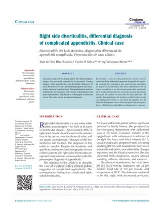
- 2. 227 Díaz-Rosales JD et al. Right side diverticulitis, differential diagnosis of complicated appendicitis Cirujano General 2019; 41 (3): 226-229 www.medigraphic.com/cirujanogeneral www.medigraphic.org.mx pain in the periumbilical area and lower right quadrant, with a palpable tumor, positive Rovsing’s and Dunphy’s signs, and pain upon percussion. Labs showed leukocytosis of 17,000 per mm3 with a 75% neutrophilia. She had 10 points on the Alvarado score. A clinical diagnosis of complicated appendicitis was made and the patient programmed for an appendectomy. A Mcburney’s incision was made, a slightly edematous cecal appendix was observed, with a small fecalith inside. An appendectomy was performed, with ligation of the mesoappendix and the appendix base, section, and extraction. On the junction of the cecum with the ascending colon at the antimesenteric border, a plastron was found. Upon pressure the affected area discharged pus. Resection of the affected segment (cecum and a segment of 5 cm of distal ileum) was decided, with the closure of the distal end with Hartmann’s technique (two planes of polyglactin 910) and a terminal ileostomy. The segment corresponded to a perforated cecum diverticulum classified as Hinchey II-III (Figure 1). The pathology report confirmed a 1 × 0.5 cm diverticulum with lymphocytic infiltration and polymorphonuclears, macrophages, and fibrin (diverticulitis) (Figures 2 and 3). The appendix report showed no evidence of pathology (lymphoid follicles, germinal nodes with macrophages, and submucosa with loosely vascularized connective tissue). The patient was in good post-surgical condition and was discharged on the third day, medicated only with analgesia (paracetamol 500 mg for three to five days). DISCUSSION Right-sideddiverticulitisisarareentity.However, it should be kept in mind as a differential diagnosis of complicated appendicitis.4 Other diagnoses in the course of an appendectomy can be diverticula of the appendix,5 ileocecal invaginations,6 epiploic appendicitis,7 and torsion of the omentum,8 among others. Both ultrasound (in expert hands),9 computerized tomography,10 and even magnetic resonance imaging (in pregnant women)11 can provide data to guide the diagnosis and avoid unnecessary surgery. (Hinchey I). However, most of the time the definitive diagnosis of cecal diverticulitis is made during the transoperative period, even with previous imaging studies.12 If the diagnosis of uncomplicated diverticulitis is made (Hinchey Ia and Ib stages), conservative management with antibiotics is effective13 and in the case of moderate inflammation (Hinchey II) a closed drain may be placed, while in a severe inflammatory case (Hinchey III and IV) hemicolectomy is Figure 1: Open cecum showing the entry site of the diverticulum and the puncture site on the antimesenteric border (clamp tip), as well as inflammation of peridiverticular fat. Figure 2: Surgical piece (cecum after formalin treatment), with sagittal section where the path from the intestinal lumen to the peridiverticular fat is observed.
- 3. Díaz-Rosales JD et al. Right side diverticulitis, differential diagnosis of complicated appendicitis 228 Cirujano General 2019; 41 (3): 226-229 www.medigraphic.com/cirujanogeneral www.medigraphic.org.mx recommended, as the presence of carcinoma in the cecum cannot be ruled out.14 A therapeutic dilemma ensues facing a moderate inflammatory process (as was the case), with perforation, collection of pus, and risk of fistula. In these cases, one could opt for ileocecal resection,15 a cecostomy,16 an omentum patch, or simple observation and antibiotic management with closed drainage. In this case, ileocecal resection was chosen, being a feasible procedure with lower morbidity than a right hemicolectomy,17 with immediate ileocolic anastomosis.18 Given the possibility of various surgical scenarios in the Emergency Room, good therapeutic judgment must be exercised to provide the best treatment. CONCLUSION Right diverticulitis should be considered in the differential diagnosis of appendicitis and its treatment should be applied based on the findings and hospital possibilities, which range from conservative management to diverticulectomy, to right hemicolectomy. REFERENCES 1. Yardimci E, Hasbahçeci M, Idiz UO, İdiz UO, Atay M, Akbulut H. Is there need for surgery to confirm diagnosis of right-sided diverticulitis in spite of relevant clinical and radiological findings? Turkish J Trauma Emerg Surg. 2016; 23: 61-65. 2. Kyziridis DS, Parpoudi SN, Antoniou ND, Konstantaras DCh, Moysidis MG, Christoforidis ECh, et al. Cecal diverticulitis is a challenging diagnosis: a report of 3 cases. Am J Case Rep. 2015; 16: 206-210. 3. Maya MZ, Padrón AG. Diverticulitis cecal. Presentación de un caso y revisión de la literatura. Cir Gen. 2010; 32: 125-127. 4. Nemeth K, Vaughan S. Cecal diverticulitis: a diagnostic conundrum. West J Emerg Med. 2015; 16 (2): 316- 317. 5. Escobar F, Valentín Vega N, Valbuena E, Barón M. Diverticulitis apendicular, revisión de la literatura científica y presentación de dos casos. Rev Colomb Cir. 2013; 28: 223-228. 6. Díaz-Rosales J de D, Enriquez-Dominguez L, Castillo-Moreno JR, Gutierrez-Ramirez PG. Adult intussusception by tumor in ileum: a diagnostic dilemma. Int J Students Res. 2012; 2: 18-20. 7. Molinares AB, Castrillón GA, Restrepo R. Apendicitis epiploica. Reporte de cuatro casos. Rev Colomb Cir. 2006; 21: 196-200. 8. Bizueto-Monroy JL, Montoya-Pérez BI, Saldaña-Torres F. Torsión de epiplón: causa rara de dolor abdominal recurrente. Cir Gen. 2017; 39: 171-174. 9. Chou YH, Chiou HJ, Tiu CM, Chen JD, Hsu CC, Lee CH, et al. Sonography of acute right side colonic diverticulitis. Am J Surg. 2001; 181: 122-127. 10. Shin S, Kim D, Kang UR, Yang C-S. Impact of CT imaging on predicting the surgical management of acute diverticulitis. Ann Surg Treat Res. 2018; 94: 322. 11. Cobben LPJ, Groot I, Blickman JG, Puylaert JBCM. Right colonic diverticulitis: MR appearance. Abdom Imaging. 2003; 28: 794-798. 12. Oter V, Oter S, Kafadar MT. Cecal diverticulitis mimicking acute appendicitis: Management and Figure 3: In the histological sections, a blind sac covered by mucosa protruding through the mucosal muscularis bundles is identified, communicating with the colonic lumen. The serosa presents an inflammatory infiltration corresponding to acute diverticulitis. Hematoxylin-eosin stain with 10x magnification.
- 4. 229 Díaz-Rosales JD et al. Right side diverticulitis, differential diagnosis of complicated appendicitis Cirujano General 2019; 41 (3): 226-229 www.medigraphic.com/cirujanogeneral www.medigraphic.org.mx short-term results of surgery. J Clin Anal Med. 2018; 9: 222-225. 13. Espinosa J, Sharma R, Lucerna A, Stranges D. Medical approach to right colon diverticulitis with perforation. Case Rep Emerg Med. 2017; 2017: 2563218. 14. Gilmore T, Jordan C, Edelstein E. Right-sided diverticulitis mimics appendicitis. J Emerg Med. 2013; 44: e29-e32. 15. Wyble EJ, Lee WC. Cecal diverticulitis: changing trends in management. South Med J. 1988; 81: 313-316. 16. Park HC, Kim BS, Lee BH. Management of right colonic uncomplicated diverticulitis: outpatient versus inpatient management. World J Surg. 2011; 35: 1118- 1122. 17. Poon RT, Chu KW. Inflammatory cecal masses in patients presenting with appendicitis. World J Surg. 1999; 23: 713-716. 18. Frías Espinosa JE, Iglesias MJ. Diverticulitis exclusiva de ciego. Rev Cuba Cir. 2012; 51: 332-337. Correspondence: Juan de Dios Díaz-Rosales Departamento de Ciencias Médicas, Instituto de Ciencias Biomédicas. Av. Benjamín Franklin No. 4650, Zona PRONAF. Ciudad Juárez, Chih. Tel: 52 656-375-1759 E-mail: jdedios.diaz@uacj.mx