1. Sensing Parainfluenza virus infection:
The role of RIG-I and MDA5
John A L Short, Rick Randall E-mail: jals4@st-andrews.ac.uk
Parainfluenza: The Essentials
What is a Parainfluenza virus?
Parainfluenza virus (PIV) is a single stranded, RNA negative sense virus
Infects the upper and lower respiratory tract
Who is at risk of infection?
All children are infected at some stage
Immunocompromised patients e.g. AIDS
What are the symptoms?
Severe fever, coughing, sneezing, croup
There are NO antiviral drugs or vaccines
1
RIG-I and MDA5: viral sensors
RIG-I detects short double stranded viral RNA and 5’ triphosphate capped
single stranded viral RNA
MDA5 detects viral long double stranded viral RNA
RIG-I MDA5
IFNCell
Antiviral
IFN
2
Virus Infects Cell
RIG-I and MDA5 sensors
recognise viral RNA
Sensors activate pathway for
Interferon (IFN) expression
IFN is secreted & absorbed by
neighbouring uninfected cell
Cell expresses antivirals,
generating antiviral state
Inhibition of viral replication
upon viral infection
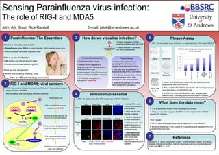
3 How do we visualise infection?
Plaque Assay
Plaques formed from virus
spreading from infected cell to
neighbouring cells over 10 days
Use cells that “knock down” or
decrease levels of RIG-I and MDA5
2o Antibody conjugated to alkaline
phosphatase that reacts with
substrate to stain plaques black
A549 NPro cells is a +ve control:
NPro inhibits IFN expression
Immunofluorescence
Cells infected for 2 days
Marker for IFN signalling to
neighbouring cells: cellular antiviral
MxA stained with Cy5
Cells express Green Fluorescent
Protein (GFP) when IFN is induced
2o Antibody conjugated to
fluorophore Texas Red
Plaque
2
MergeAnti-MxAAnti-PIV5GFP
Plaque
1
4
Mock
GFP +IFN-IFNAnti-PIV5
Immunofluorescence
- AIM: To determine the IFN response to PIV5 infection
Key: Green = GFP; Red = Viral NP;
Blue = Antiviral MxA
Adding IFN to cells
induces expression of
antiviral MxA
PIV5 infects cells,
forming a plaque
No IFN induction =
no MxA expression
IFN expressing cells
leads to induction of
MxA in neighbouring
uninfected cells
Heterogeneous
cellular response:
Only some cells
express IFN, the
majority respond to IFN
signaling
Infect monolayer of A549
alveolar epithelial cells with PIV5
NPro +ve
Control
Naïve MDA5 KD
RIG-I KD RIG-I/
MDA5 KD
0
0.5
1
1.5
2
2.5
3
Naïve
MDA5KD
RIG-IKD
RIG-
I/MDA5
KD
Npro+ve
Control
Relative sizes of largest plaques following
PIV5 infection
PlaquesizerelativetoNaive
5 Plaque Assay
- AIM: To visualise virus infection in cells lacking RIG-I and MDA5
Naïve and MDA5 knock down (KD) cell lines had similar
plaque sizes after viral infection
RIG-I and the RIG-I/MDA5 double KD cells had larger plaque
sizes than Naïve and MDA5 KD cells
In RIG-I KD and RIG-I/MDA5 KD cells, plaques were
different sizes, indicating asynchronous viral IFN evasion
6 What does the data mean?
IFN is expressed in some cells following virus infection
Plaque assay data suggests that RIG-I is more important than MDA5 in
sensing PIV infection
The Future:
Why is there a heterogeneous cellular response to virus infection?
Are there differences in the number IFN expressing cells between RIG-I KD
and MDA5 KD cell lines?
7 Reference
Randall, R. E. and S. Goodbourn (2008). "Interferons and viruses: an interplay
between induction, signalling, antiviral responses and virus countermeasures."
J Gen Virol 89(Pt 1): 1-47.
Stain cells with 1o antibody
for virus nucleoprotein