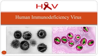
2. Introduction
Etiologic agent of Acquired Immunodeficiency Syndrome
(AIDS).
Discovered independently by Luc Montagnier of France and
Robert Gallo of the US in 1983.
Former names of the virus include:
Lymphadenopathy associated virus (LAV) (Luc Montagnier)
Human T cell lymphotrophic virus (HTLV-III) (Robert Gallo)
AIDS associated retrovirus (ARV)
1986, HIV name was given by International Committee on Virus
Nomenclature.
2
3. Introduction
HIV-2 discovered in 1986, antigenically distinct virus
endemic in West Africa.
30 million worldwide are infected. 2 million deaths
every year & 2.5 million new cases every year.
Leading cause of death of men aged 25-44 and 4th
leading cause of death of women in this age group.
HIV-1 in humans was believed to be acquired from
chimpanzee (Pan troglodytes troglodytes) by cross species
infections (simian immunodeficiency virus or SIVcpz).
HIV-2 through sooty mangabeys.
3
4. Characteristics of the virus
Icosahedral (20 sided), enveloped virus of
the lentivirus subfamily of retroviruses.
Retroviruses transcribe RNA to DNA.
Two viral strands of RNA found in core
surrounded by protein outer coat.
Outer envelope contains a lipid matrix within
which specific viral glycoproteins are
imbedded.
These knob-like structures responsible for
binding to target cell.
4
5. HIV
The outer shell of the virus is known as the
Viral enevlope. Embedded in the viral
envelope is a complex protein known as env
which consists of an outer protruding cap
glycoprotein (gp) 120, and gp 41.
Within the viral envelope is an HIV protein
called p17(matrix), and within this is the
viral core or capsid, which is made of
another viral protein p24(core antigen).
5
7. Group Specific Antigen (Gag)
It encodes for core and shell proteins.
Expressed as a precursor protein, p55.
Cleaved into p15, p17 and p24.
p 24 can be detected in serum during early
stages of infection till the appearance of
antibodies.
The decline of anti-p24 antibody from
circulation indicates progression of illness and
is an indication of antiviral treatment
7
8. Envelope (Env)
Envelope (Env) gene codes for envelope
protein gp160; gp120 and gp41.
gp160 cleaved to form gp120 and gp41.
gp120 forms the 72 knobs which protrude from
outer envelope.
gp41 is a transmembrane glycoprotein
antigen that spans the inner and outer
membranes and attaches to gp120.
gp120 and gp 41 both involved with fusion and
attachment of HIV to CD4 antigen on host cells.
8
9. Polymerase (Pol)
Polymerase (Pol) codes for viral enzymes
such as reverse transcriptase.
Expressed as precursor protein p100.
Cleaved into p 31, p 51 and p 64.
Located in the core, close to nucleic acids.
Responsible for conversion of viral RNA
into DNA, integration of DNA into host cell
DNA and cleavage of protein precursors.
9
10. Sexual transmission, presence of
STD increases likelihood of
transmission.
Blood transfusion.
Parenteral transmission.
Exposure to infected blood or blood
products.
Transplantation of infected tissues
or organs.
Mother to fetus, perinatal
transmission variable.
10
Modes of transmission
12. Types of Exposure and Relative Risk
S.N. Types of Exposure Relative risk per
exposure (%)
1. Sexual intercourse: anal, vaginal, oral 0.1-1.0
2. Transfusion of blood and blood products >90
3. Tissue and organ donations 50-90
4. Injection and injuries 0.5-1.0
5. Mother to baby 30
12
13. Viral Replication
First step, HIV attaches to susceptible
host cell.
Site of attachment is the CD4 antigen found
on a variety of cells
helper T cells
macrophages
monocytes
B cells
microglial brain cells
T cells infected later on.
13
14. Early Phase HIV Infection
In early phase HIV infection, initial viruses
are M-tropic. Their envelope glycoprotein
gp120 is able to bind to CD4 molecules and
chemokine receptors called CCR5 found on
macrophages.
Mutation of CCR5 in some Europeans are
Completely resistant to HIV infection if the
mutation is homozygous or are susceptible
but progress of AIDS is delayed if the
mutation is heterozygous.
14
Maraviroc
15. In late phase HIV infection, most of the
viruses are T-tropic, having gp120
capable of binding to CD4 and CXCR4
found on T-lymphocytes.
15 HIV (arrows) Infecting a T-lymphocyte
16. Life Cycle
HIV attaches to two cell-
surface receptors (the CD4
antigen and a specific
chemokine receptor).
The virus and cell membrane
fuse, and the virion core enters
the cell.
The viral RNA and core
proteins are released from the
virion core and are then
actively transported to the
nucleus.
16
17. The viral RNA genome is
converted into double-
stranded DNA through an
enzyme unique to viruses,
reverse transcriptase.
The double-stranded viral
DNA moves into the cell
nucleus.
Using a unique viral
enzyme called integrase,
the viral DNA is
integrated into the cellular
DNA. The integrated
virus is called provirus.
17
18. 18
Viral RNA is synthesized by
the cellular enzyme RNA
polymerase using
integrated viral DNA as a
template.
Two types of RNA
transcripts shorter spliced
RNA and full-length
genomic RNA are produced.
Shorter spliced RNAs are
transported to the cytoplasm
and used for the production
of several viral proteins that
are then modified in the
ribosomes of the cell.
19. Full-length
genomic RNAs are
transported to the
cytoplasm.
New virion is
assembled and then
buds off.
Mature virus is
released.
19
Enfuvirtide
Indinavir,
Ritonavir,
Darunavir
Raltigravir
Zidovudine
(NRTI)
20. Viral Replication
The gp120 protein on virus binds
specifically to CD4 receptor on host cell
with high affinity.
gp41 causes fusion of the virus to the cell
membrane.
After fusion virus particle enters cell.
Viral genome exposed by uncoating
particle.
Reverse transcriptase produces viral
DNA from RNA.
Becomes a provirus which integrates into
host DNA.
Period of latency occurs.
20
21. Viral Replication
After a period of latency lasting up to
10 years viral replication is triggered and
occurs at high rate.
CD4 cell may be destroyed in the
process, body attempts to replace lost
CD4 cells, but over the course of many
years body is unable to keep the count at
a safe level.
Destruction of large numbers of CD4
cause symptoms of HIV to appear with
increased susceptibility to opportunistic
infections, disease and malignancy.
21
22. Clinical Features: According to CDC, clinical course of
HIV infection
Group I- Acute HIV infection: Acute onset of fever, malaise, sore throat, myalgia,
arthralgia, skin rash and lymphadenopathy. Viral nucleic acid or viral p24 antigen
may be detected. Antibodies to HIV usually negative. (3 to 6 months)
Group II- Asymptomatic infection: Show positive HIV antibody tests and are
infectious. Person usually well.
Group III- Persistent generalised lymphadenopathy: Enlarged nodes at two or
more extragenital sites for at least 3 months.
Group IV- Symptomatic HIV infection: CD4 T lymphocyte count falls below
400 per cu. mm. Symptoms like diarrhea, fever, weight loss, night sweats and
opportunistic infection develops. Some patients develops AIDS related complex or
conditions.
22
23. Primary HIV Syndrome
Cold or flu-like symptoms may occur 6 to 12 weeks after infection.
Symptoms are relatively nonspecific.
HIV antibody test often negative but becomes positive within 3 to 6
months (window period), this process is known as seroconversion.
Large amount of HIV in the peripheral blood.
Primary HIV syndrome resolves itself and HIV infected person
remains asymptomatic for a prolonged period of time, often years
(Clinical Latency).
23
24. Clinical Latency Period
HIV continues to reproduce, CD4 count
gradually declines from its normal value
of 500-1200.
Once CD4 count drops below 500, HIV
infected person at risk for opportunistic
infections.
The following diseases are predictive of
the progression to AIDS:
Persistent Herpes-zoster infection
Oral candidiasis (thrush)
Oral hairy leukoplakia (Epstein Barr virus)
Kaposi’s sarcoma (KS) (Herpes Virus)
24
Candidiasis
Oral Hairy Leukoplakia
25. Oral Hairy Leukoplakia (OHL)
Being that HIV reduces immunologic activity, the
intraoral environment is a prime target for chronic
secondary infections and inflammatory processes,
including OHL, which is due to the Epstein-Barr virus
under immunosuppressed conditions .
25
Kaposi’s sarcoma (KS)
Kaposi’s sarcoma is a rare cancer of the blood
vessels that is associated with HIV caused due to
Herpes virus. It manifests as bluish-red oval-shaped
patches that may eventually become thickened.
Lesions may appear singly or in clusters.
OHL
Kaposi’s sarcoma
26. AIDS
CD4 count drops below 200, person is considered to have advanced HIV disease
If preventative medications not started the HIV infected person is now at risk for:
a. Pneumocystis carinii pneumonia (PCP)
b. Cryptococcal meningitis
c. Toxoplasmosis
If CD4 count drops below 50:
a. Mycobacterium tuberculosis
b. Cytomegalovirus
c. Lymphoma
d. Dementia
e. Most deaths occur with CD4 counts below 50.
26
28. ‘Typical’ HIV-1 infection
symptoms
HIV-1 p24 antigen
0 1 2 3 4 5 6 / 2 4 6 8 10
weeks years
HIV antibodies
Time following infection
HIV viral load
HIV proviral DNA
symptoms
‘window’
period
1° infection
28
29. Immunologic Manifestations
Immune abnormalities associated with increased viral replication.
Decrease in CD4 cells due to virus budding from cells, fusion of uninfected cells
with virally infected cells and apoptosis.
B cells have decreased response to antigens possibly due to blockage of T cell/B
cell interaction by binding of viral proteins to CD4 site.
CD8 cells initially increase and may remain elevated.
As HIV infection progresses, CD4 T cells drop resulting in immunosuppression
and susceptibility of patient to opportunistic infections.
Death comes due to immuno-incompetence.
29
30. Laboratory Diagnosis of HIV Infection
Methods utilized to
detect:
Antibody
Antigen
Viral nucleic acid
Virus in culture
30
Screening tests
ELISA
Rapid test
HIV spot and comb tests
Supplemental tests:
Western blot test
Indirect immunofluorescence test
Radio Immuno assay
PCR
31. ELISA Testing
Antibodies detected in ELISA include those directed against: p24, gp120, gp160
and gp 41, detected first in infection and appear in most individuals.
31
Other Screening Tests
Agglutination tests using latex particles, gelatin particles or microbeads are
coated with HIV antigen and will agglutinate in the presence of antibody.
Dot-Blot Testing utilizes paper or nitrocellulose impregnated with antigen,
patient serum is filtered through, and anti-antibody is added with enzyme
label, color change is positive.
A rapid, cost-effective and may become an alternative to standard ELISA
and Western blot testing.
32. Western Blot
Most popular confirmatory test.
Antibodies to p24 and p55 appear
earliest but decrease or become
undetectable.
Antibodies to gp31, gp41, gp 120,
and gp160 appear later but are
present throughout all stages of the
disease.
32
Interpretation of results.
No bands, negative.
In order to be interpreted as positive a minimum of 3 bands directed against the
following antigens must be present: p24, p31, gp41 or gp120/160.
CDC criteria require 2 bands of the following: p24, gp41 or gp120/160.
33. Detection of p24 HIV antigen
Most useful for the following:
early infection suspected in seronegative patient
Newborns
Monitoring disease progress
33
Polymerase Chain Reaction (PCR)
Looks for HIV DNA in the WBCs of a person.
PCR amplifies tiny quantities of the HIV DNA present, each cycle of PCR
results in doubling of the DNA sequences present.
34. Virus isolation
Virus isolation can be used to definitively diagnose
HIV.
Best sample is peripheral blood, but can use CSF,
saliva, cervical secretions, semen, tears or material
from organ biopsy.
Cell {peripheral blood mononuclear cell (PBMC)}
growth in culture is stimulated, amplifies number of
cells releasing virus.
Cultures incubated one month, infection confirmed
by detecting reverse transcriptase or p24 antigen in
supernatant.
34
35. Resistance:
Temperature: Inactivated at 56o
C in 30 minutes and in seconds at 100o
C.
Disinfection:
35% Isopropyl alcohol: inactivation in 10 minutes.
70% ethanol
0.5% lysol
2% freshly prepared glutaraldehyde
0.5% sodium hypochlrite
3% hydrogen peroxide
Extremes of pH (pH 1.0, pH 13.0)
Resistant to Lyophilisation
35