1. Structural analysis of the root hair protein Villin4
Significance:
• Villin4 has relevance in agricultural science
• A firm understanding of Villin4 works also contributes
to an understanding of how plants interact with their
environment to uptake water
• The Smirnov lab group has spent the last year
characterizing the headpiece domain of this protein to
better understand its function
• We have chosen to begin with the headpiece because
much of the rest of the protein well conserved among
the Gelsolin/Dematin protein family
• The other unique region is the linker, which in Villin4
is much longer than other well studies homologues,
such as the model animal variant from chickens
Russell McFarland, Jessalyn Rogers, Heather Miears, Sergey Smirnov
• Recently Villin-like proteins have been
described in plants and have been termed
“plant villins”
• Villin-like proteins have the distinct property
of binding & bundling actin fibers
• We are most interested in Villin4 and its
functions contributing to root hair
morphology (Fig. 1, 3)
• Root hairs are single cell extensions from
plant roots roughly 0.5 mm long
• Root hairs increase plants’ ability to uptake
water and nutrients
A litt
• Villin has six conserved gelsolin-like domains
which generate the core structure (Figure 2)
• On the N-terminus there is a long linker region
(roughly 100 amino acids) which is much
larger than that of animal villins
• The C-terminal headpiece shows significant
differences from animal villins as well
• In animals, the headpiece domain separated
by a linker region serves to bundle multiple
actin fibers together
Pollard, T. (2002). Formins initiate new actin filaments. Nature Cell Biology 4, E191. doi:10.1038/ncb0802-e191a
Background A little more about Villin
FPLC size-exclusion:
Ni-NTA purification:
Methods:
• We used Ni-NTA purification and then size-exclusion FPLC to purify our protein. The differences
that each step makes can be seen on the blue gels.
• From there we are using two methods for structural determination: NMR and crystallography
• NMR determines indirect constraints which can be used to model the protein in the computer,
this gives much more data but also takes longer
• Crystallography is used to model the protein using electron density and gives a simple, static
structure
• A spin-down actin binding assay will assess binding activity
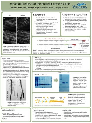
Figure 1. Comparison of wild type villin to atvln4-1, a
reduced functionality villin mutant. The left side shows
high levels of interconnectedness, while on the right
this is not the case. This shows that the actin filaments
are not bundled by villin.
Figure 2. comparison of plant and vertebrate (animal) villins by domain. The largest noticeable difference
between the two is the linker region, which in animals is 40 amino acids, in plants this is much longer; between
100 and 200 amino acids in length.
Figure 3. Comparison of root hairs of
wild-type Arabidopsis and a Villin-
defective mutant.
Figure 4. Gels from the two-
step purification process of
HP domain. These are made
to determine whether the
protein is present and pure.
This is done on
polyacrylamide gel and dyed
with a protein-binding dye.
Figure 5. Diagram of actin filament
formation. This can be done in a lab
setting and is the basis of the actin
binding assay we are carrying out on
Villin4. By running this with
individual domain or carefully
selected proteins, we can determine
how the protein binds.
Data Collected
• We have begun collecting NMR data
• The data collected has been of incredibly high
quality such that structural determination by NMR is
faster than by crystallography
• We have also begun to test crystallization conditions
Future Work
• Our intent is to finish collecting NMR/X-ray data
over the summer
• In parallel with the headpiece domain, the Smirnov
lab group intends to begin characterizing the linker
region
Acknowledgments
WWU Office of Research and
Sponsored Programs Pilot Grant
(2017).
Headpiece domain
we’re interested
in. This should
bind actin.