SlideShare a Scribd company logo
1 of 102
Cytoskeleton
&
Extracellular
Matrix
Pradeep Singh
M.Sc. Medical Biochemistry
HIMSR, JAMIA HAMDARD
Fig: A section of mouse intestine
stained for actin (red), the
extracellular matrix protein laminin
(green), and DNA (blue). Each blue
dot of DNA indicate the presence of a
cell.
CYTOSKELETON
Introduction
 Eukaryotic cells contain protein
filaments that are collectively called as
cytoskeleton.
 These protein filaments are involved in:
─ establishing cell shape
─ provide mechanical strength
─ help in locomotion of cell
─ chromosome separation
─ intracellular transport of organelles
 The three major cytoskeleton
filaments are:
1. Actin filaments
2. Microtubules
3. Intermediate filaments
 In addition, a large number of
accessory proteins, including the
motor proteins, are also required
for the properties associated with
each of these filaments.
1. ACTIN FILAMENTS
1. Actin
• Actin was first isolated in 1942, from muscle cells in which it constitutes
approximately 20% of the total cell protein.
• The three-dimensional structures of both individual actin molecules and
actin filaments were determined in 1990.
• Actin filaments (F-Actin) also known as microfilaments, made up of small
subunits called G-Actin.
• G-Actin are globular proteins consisting of 375 amino acids. G-Actin is
described as having a pointed end and a barbed end.
Pointed End: Minus end
Barbed End: Plus end
The structure of an actin monomer and actin filament
Assembly of Actin Filament (F-Actin)
• G-Actin monomers polymerize to form actin filaments (F-Actin) having a
diameter of about 8nm.
• F-Actin is a helical structure.
• Steps of formation of actin filament:
a) Nucleation
b) Elongation
• Nucleation: For the formation of a new actin filament, subunits must
assemble into a small aggregate or nucleus which then elongate rapidly
by addition of new G-Actin subunits. This process is called filament
nucleation.
• Actin filament nucleate most frequently at the plasma membrane and
nucleation is regulated by external signals.
• Nucleation is the rate limiting step in the formation of a cytoskeletal
polymer.
• Elongation: G-Actin are added to the both ends but addition of new
subunits is faster at plus end.
The time course of actin polymerization in a
test tube
Concept of Critical Concentration
• The number of monomers that are added to the polymer per second will be
proportional to the concentration of the free subunit (KonC), but the subunits will
leave the polymer end at a constant rate (Koff) that does not depend on C.
• Polymerization: Polymerization is a reversible process which depends on the
critical concentration (KonC) of the monomers.
• At a concentration > KonC, filament grows at each end but growth is faster at
the barbed end.
• At a concentration < KonC, filament shrink at both ends.
Nucleotide Hydrolysis
• Each actin molecule carries a tightly bound ATP molecule that is
hydrolysed to a tightly bound ADP molecule soon after its assembly into
the polymer.
• Hydrolysis of the bound nucleotide reduces the binding affinity of the
subunit for neighbouring subunits and makes it more likely to dissociate
from each end of the filament. It is usually the T-form that adds to the
filament and D-form that leaves.
Treadmilling
• At the steady state, subunits undergo a
net assembly of monomers at the plus
end and a net disassembly at the minus
end at an identical rate.
• The polymers maintains a constant
length, even though there is net flux of
subunits through the polymer, known as
treadmilling.
Chemical Inhibitors of Actin
• The functions of the actin filaments are inhibited by both polymer-
stabilizing and polymer-destabilizing chemicals.
• These chemicals are used as an important tools in the study of the
filaments.
Table: Chemical Inhibiters of Actin
Chemical Effect on filaments Mechanism Original source
Latrunculin Depolymerizes Binds actin subunits Sponges
Cytochalasins B Depolymerizes Caps filament plus ends Fungi
Phalloidin Stabilizes Binds along filaments Amanita mushroom
Filament Elongation Is Modified By Proteins That
Bind To The Free Subunits
• In most non-muscle vertebrate cells, approximately 50% of the actin
is in filament form and 50% in soluble form while the monomer
concentration is 50-200 μM, well above the critical concentration (0.1
μM) .
Question: why does so little of the actin polymerize into filaments?
The actin is not polymerized as it is bound to special proteins, such as
thymosin. Actin monomers bound to thymosin are locked where they
cannot associate with either the (+) end or (-) end of the actin filament.
Accessory Proteins
Actin Binding Proteins
• A number of different types of actin binding proteins remodel or modify
existing filaments.
Cellular Role Representative Proteins
Monomer binding Thymosin, Profilin
Filament initiation and polymerization Arp2/3, formin
Filament stabilization Tropomyosin
End capping CapZ, tropomodulin
Filament severing Cofilin, gelsolin
Filament cross-linking α-actinin, fimbrin, filamin
Actin filament linkage to other
proteins
Spectrin
A. Monomer Binding Proteins
• Thymosin: Actin monomer bound to thymosin are in a locked state, where
they cannot associate with either the plus end or minus ends of actin
filaments and can neither hydrolyse nor exchange their bound nucleotide.
• Profilin: Profilin binds to the actin monomer (G-Actin) opposite to the ATP-
binding cleft, blocking the side of the monomer that would normally
associate with the filament minus end, while leaving exposed the site on
the monomer that binds to the plus end.
• Profilin competes with thymosin for binding to individual actin monomer.
Thus, by regulating the local activity of profilin, cells can control the length
of actin filaments.
B. Filament Initiation And Polymerization
1. Arp2/3: Arp stands for ‘Actin Related Proteins’.
― The Arp2/3 complex comprises 7 protein sub units.
― Both Arp2 and Arp3 are about 45% identical to actin.
― Arp2/3 complex nucleates actin filament growth from minus end, allowing
rapid elongation at the plus end.
― The ARP complex can also attach to the side of another actin filament while
remaining bound to the minus end of the filament that it has nucleated
resulting in the formation of web like structure.
Differences on the sides and minus end prevent the ARPs from forming filament on
their own.
2. Formins: Formins are dimeric proteins that nucleate the growth of straight,
unbranched filaments that can be cross linked by other proteins to form protein
bundles.
― Formin dimer remains associated with the rapidly growing plus end while still
allowing the addition of new subunits at that end.
― Formin dependent actin filament growth is strongly enhanced by the addition of
profiling linked actin monomers.
C. Filament Stabilization Proteins
• Tropomyosin: Tropomyosin, an elongated protein that binds simultaneously to six
or seven adjacent actin subunit along each of the two grooves of the helical actin
filament.
― In addition to stabilizing and stiffening the filament, the binding of
tropomyosin can prevent the actin from interacting with other proteins.
― Tropomyosin plays an important in the control of muscle contraction.
D. End Capping Proteins
• Tropomodulin: Tropomodulin is an end capping protein that binds
tightly to the minus ends of the actin filaments that have have been
coated and stabilized by tropomyosin.
• CapZ: CapZ is named so because of its location in the muscle Z band.
CapZ binds to the actin filament at the plus end, stabilizing the actin
filament by greatly reducing the rates of filament growth and
depolymerisation.
E. Filament Severing Proteins
• Sever means “breaking’.
• These proteins breaks an actin filaments into many smaller
fragments.
• The fate of these fragments depends on the presence of other
accessory proteins.
• Under some conditions, newly formed ends acts as nucleus and
promotes filament elongation.
• Under other conditions, severing promotes the depolymerization of
old fragments.
1. Gelsolin superfamily: These proteins are activated by high levels of cytosolic Ca2+ .
― Gelsolin interact with the side of the actin filament and contains two subdomains that
binds to two different sites: one that is exposed on the surface of the filament and
another that is hidden between the adjacent subunit.
― Gelsolin creates a small gap between neighbouring subunits and inserts itself into the
gap to break into the filament.
― Gelsolin remains attached to the actin filament and caps the new plus end.
Two superfamilies of severing proteins are:
1. Gelsolin
2. Cofilin
Platelet Activation by Gelsolin
2. Cofilin superfamily: Also called actin depolymerizing factor.
― Cofilin binds along the length of the actin filament, forcing the filamentto
twist a little more tightly.
― Mechanical stress generated weakens the contacts between the actin
subunits in the filament, generating filament ends that undergo rapid
disassembly.
― Cofilin binds to ADP-containing actin filaments rather than to ATP-containing
filaments.
― Since, ATP hydrolysis is usually slower than filament assembly, the newest
actin filament in the cell still contain mostlt ATP and are resistant to
depolymerisation by cofilin.
― Cofilin therefore tends to dismantle the older filaments in the cell.
Bundling and Gel-Forming Proteins
F. Filament Cross Linking Proteins
1. α-Actinin: α-Actinin includes
myosin and links oppositely
charged actin filaments into
contactile bundles.
2. Fimbrin: Fimbrin excludes
myosin and links actin
filaments into non contractile
parallel bundles.
3. Filamin: Filamin promote the formation of a loose and highly
viscous gel by clamping two actin filaments roughly at right angles.
G. Actin Filaments Linking to Other Proteins
• Spectrin: Spectrin was first identified in RBC.
― Spectrin is a long, flexible protein made out of four elongated polypeptide
chains (two α subunits and two β subunits), arranged so that two actin
binding sites are 200 nm apart.
― In RBC, spectrin is concentrated just beneath the plasma membrane.
Spectrin has separate binding sites to actin filaments and other peripheral
proteins of plasma membrane.
―Spectrin allows the RBC to get back to normal shape after squeezing through
the capillary blood vessels.
ACTIN BASED MOTOR PROTEINS
• The first motor protein to be identified in the skeletal muscles was
Myosin.
• Consist of two heavy chains and two copies of each of two light chain.
• Helps in the muscle contraction.
Functions of Actin Filaments
― By forming a band under the plasma membrane, actin filaments allow cells to
adopt different shapes and perform different functions
― Generate locomotion in cells such as white blood cells and amoeba.
Villi Contractile
bundles
Sheet-like &
Finger-like
protrusions
Contractile
ring
― Link transmembrane proteins to cytoplasmic proteins
― Form contractile ring during cytokinesis in animal cells
― Cytoplasmic streaming
― Interact with myosin to provide force of muscular contraction
2. MICROTUBULES
2. Microtubules
• Microtubules are hollow cylindrical structure
which are polymers of the protein tubulin.
• The tubulin subunit is a heterodimer made up
of two globular proteins called α-tubulin and β-
tubulin.
• The wall is composed of 13 parallel
protofilaments, each composed of αβ-tubulin
heterodimers stacked from head to tail.
• The α- and β-subunits of the tubulin dimer can
bind one molecule of GTP. The GTP in the α-
tubulin subunit is never hydrolyzed while β-
subunit may be in either GTP or the GDP form.
The structure of a microtubule and its subunit
Polymerization of tubulin to
form microfilament
• Microtubule growth is generally
nucleated from specific locations with
in the cell known as microtubule-
organizing centre (MTOC).
• Many animal cells have single MTOC
called as centrosome, located near
nucleus.
• MTOC are highly enriched in another
type of tubulin called γ-tubulin which
form γ-tubulin ring complex (γ-
TuRC).This γ-tubulin ring complex
helps in the nucleation of
microtubules.
Mechanism:
• Two accessory proteins bind directly
to the γ-tubulin along with several
other proteins that help to create a
spiral ring of γ-tubulin molecules,
which serves as a template that create
a microtubule with 13 protofilaments.
• The microtubule then grows by
addition of free subunits.
• After incorporation of a dimeric
subunit into a microtubule, the GTP on
the β-tubulin is hydrolysed to GDP
CENTROSOME
Microtubule nucleation by the γ-tubulin ring complex
Treadmilling and Dynamic Instability
Treadmilling is addition
of subunit at one end
and their loss at the
other end.
Dynamic instability is
the oscillation between
growth and shrinkage.
Catastrophe: The
change from growth to
shrinkage.
Rescue: The change from
shrinkage to growth
Centrosome
• Centrosome in animal cells
located near the nucleus.
• It contain centrioles and a
variable number of small dense
bodies called as centriolar
satellites.
• It consist of an amorphous
matrix of proteins containing the
γ-tubulin ring complexes that
nucleate microtubule growth.
• They serve as basal bodies and
sites of anchor for epithelial cilia.
Centrioles and Cell Division
• Duplication of the centrosomes takes place during ‘S’ phase cell division. To
form two separate MTOCs at opposite poles of the mitotic spindle.
• Two centrosomes then separate and move to opposite sides of the nucleus,
forming the two poles of the mitotic spindle.
• As the cell enters mitosis, the dynamics of microtubules assembly and
disassembly also change dramatically.
• First, the rate of microtubule disassembly increases about tenfold, resulting
in overall depolymerisation and shrinkage of microtubules.
• At the same time, the number of microtubules emerging from the
centrosome increases by five-to-tenfold.
• As mitosis proceeds, the two chromatids of each chromosome are
then pulled to opposite poles of the spindle. This chromosome
movement is mediated by motor proteins associated with the spindle
microtubules.
• In the final stage of mitosis, nuclear envelopes reform, the
chromosome decondense, and cytokinesis takes place.
• If mitotic cells are exposed to drugs like colchicine (binds to
monomeric tubulin and prevent polymerization), vinblastine and taxol
(disrupt microtubule dynamics), microtubule disappear and mitosis is
arrested because of inadequate formation of the mitotic spindle.
These drugs are useful in the treatment of certain cancers.
Microtubules Associated
Proteins (MAP)
• Microtubule polymerization
dynamics is very different in cells
than in solutions of pure tubulin
(in vitro).
• Proteins that binds to
microtubules are collectively
called as microtubule-associated
proteins.
• MAP can be divided into two
groups:
1. Microtubule stabilizing proteins
2. Microtubule destabilizing
proteins.
TABLE: Proteins That Modulate Microtubule (MT) Dynamics
Protein Location Function
Microtubule Stabilizing
Proteins
MAP1 Dendrites and axons; non-
neuronal cells
Assembles and stabilizes MTs
MAP2 Dendrites Assembles and cross-links MTs to one
another and to intermediate filaments.
MAP4 Most cell types Stabilizes MTs
Tau Dendrites and axons Assemble stabilizes and cross-links MTs
Microtubule
Destabilizing Proteins
Stathmin Most cell types Binds tubulin dimers
katanin Most cell types Microtubule Severing
Microtubule Stabilizing Proteins
• Microtubule stabilizing proteins
helps in the organization of
microtubule bundles.
• MAPs have at least one domain
that binds to the microtubule and
another that project outwards.
• The length of the projecting
domain determine how closely
MAP-coated microtubules pack
together.
Microtubule Destabilizing Proteins
• One molecule of the small stathmin binds to two tubulin
heterodimerers and prevents their addition to the ends
of the microtubules.
• Stathmin thus decreases the effective concentration of
tubulin subunits that are available for polymerization.
• Phosphorylation of stathmin inhibits its binding to
tubulin.
1. Stathmin: Also
known as tubulin-
sequestering
protein.
2. Katanin: katanin is also known as microtubule-severing protein.
• Katanin is named after the Japanese word for “sword”.
• Katanins release microtubules from their attachment to MTOC and
contribute to rapid microtubule depolymerisation observed at the
poles of spindles during mitosis.
• Katanin is made up of two subunits:
1. Smaller Subunit - Hydrolyze ATP and perform severing.
2. Larger Subunit – Direct katanin to the centrosome.
Microtubule Ends Binding Proteins
Proteins that bind to the ends of microtubules can control microtubule positioning
Motor Proteins Move Along Microtubules
• Two major classes of microtubule-
based motors are:
1. Kinesins – Move towards (+) end
2. Dyneins – Move towards (-) end
• They use the energy of ATP
hydrolysis to move along
microtubules.
• They mediate the sliding of
filaments relative to one another
and the transport of membrane-
enclosed organelles along filament
tracks.
A. Kinesin
• Kinsesin-1 has two kinesin motor head domains:
• Rear or Lagging head domain: tightly bound to
microtubule and ATP.
• Front or leading head domain: loosely bound to
microtubule with ADP in its binding site.
• Most of them have the motor domain at the N-terminus of
the heavy chain and walk toward the plus end of the
microtubule.
• The forward displacement of the rear motor head domain
is driven by the hydrolysis of ATP and binding of ATP in the
leading head domain.
• They use “hand-over-hand” motion to walk over the
microtubule.
B. Dyneins
• Dyneins are a family of minus end directed
motor proteins.
• The motor domain is present at the C-terminus
of the heavy chain and walk toward the minus
end of the microtubule.
• They are composed of one, two or three heavy
chains and a large number of light chains.
• Dynein requires the presence of a large number
of accessory proteins to associate with the
membrane-enclosed oragnelles.
• They are highly specialized for the rapid and
efficient sliding movement of the microtubules
that derive the beating of cilia and flagella.
3. INTERMEDIATE FILAMENTS
3. Intermediate Filaments
• Third major type of cytoskeletal protein.
• Present particularly in the cytoplasm of cells which are subjected to
mechanical stress.
• Generally absent in animals that have rigid exoskeleton.
• Structurally similar but biochemically distinct from actin and myosin
filaments.
• Intermediate filaments are extremely difficult to break and can be
stretched to over 3 times their length.
Structure of Intermediate Filaments
• Monomer is consist of α-helical
domain containing 40 or more hepted
repeat motifs.
• First Stage: Two monomers coil
together to form the dimer.
• Second Stage: A pair of parallel
dimers associates in antiparallel
fashion to form a staggered tetramer.
Thus, they lack structural polarity.
• Third Stage: Lateral association of 8
tetramers.
• Fourth Stage: Addition of 8 tetramers
to growing filament.
Characteristics
• Intermediate filaments are generally more stable than actin filaments
or microtubules.
• Intermediate filaments can be modified by phosphorylation which
can regulate their assembly and disassembly.
Table: Major Types of Intermediate Filament Proteins in Vertebrate Cells
Types of Intermediate Filament Component Polypeptides Location
Nuclear Lamins A,B and C Nuclear Lamina (Inner
lining of nuclear
envelope)
Vimentin-like Vimentin Many cells of
mesenchymal origin
Desmin Muscle
Peripherin Some neuron
Epithelial Type I keratins (acidic) Epithelial cells and
their derivatives.Type II keratins
(neutral/basic)
Axonal Neurofilament proteins
(NF-L, NF-M, and NF-H)
Neurons
1. Nuclear Lamins: Nuclear lamins are fibrous proteins providing
structural function and transcriptional regulation in the cell nucleus.
• Found in many eukaryotes but missing from unicellular organisms.
• Forms a meshwork that lines the ineer membrane of the nuclear envelope.
• Provide anchorage sites for chromosomes and nuclear pores.
2. Both Vimentin and Keratin attach to nuclear envelop serving to
position and anchor nucleus within the cell.
3. Desmin connects individual actin-myosin assemblies of muscle cell
to each other as well as to the plasma membrane.
4. Neurofilaments are major intermediate filaments of motor neurons.
Cell Junctions
• Intermediate filaments also form cell junctions.
• There are generally two types of cell junction:
1. Cell-cell junction
2. Cell-matrix junction
• Cell-cell junction formed by intermediate filaments – Desmosomes
• Cell- Matrix Junction formed by intermediate filaments –
Hemidesmosomes.
Diseases of Intermediate Filament
• Epidermis Bullosa Simplex: Mutations in the keratin gene form skin
blister resulting from cell lysis after minor trauma.
• Lou Gehrig’s Disease: Also known as Amyotrophic lateral sclerosis
which leads to progressive loss of motor neurons which lead to
muscle atrophy, paralysis and eventual death.
Extracellular Matrix
“Half of the secrets of the cell are outside the cell.”
Dr. Mina Bissell
Oct. 17, 2007
Erlanger Auditorium
Why do all multicellular animals have ECM?
• Act as structural support to maintain cell organization and integrity (epithelial
tubes; mucosal lining of gut; skeletal muscle fiber integrity)
• Compartmentalize tissues (Pancreas: islets vs. exocrine component; skin:
epidermis vs. dermis)
• Provide hardness to bone and teeth (collagen fibrils become mineralized)
• Present information to adjacent cells:
• Inherent signals (e.g., RGD motif in fibronectin)
• Bound signals (BMP7, TGFγ, FGF, SHH)
• Serve as a highway for cell migration during development (neural crest
migration), in normal tissue maintenance (intestinal mucosa), and in injury or
disease (wound healing; cancer)
Extracellular Matrix
• Two broad categories of tissue that are found in all animals are:
1. Epithelial Tissue
2. Connective tissue
• Tissues are not only made up of cells. They also contain a remarkably complex
and intricate network of macromolecules constituting the Extracellular Matrix.
• The matrix can become calcified to form the rock-hard structures of bone or
teeth or it can form transmembrane substance of cornea etc.
• In most connective tissue, the matrix molecules are secreted by cells called
fibroblast.
• The extracellular matrix is constructed from three major classes of
macromolecules:
1. Glycosaminoglycas (GAGs) – Highly charged polysaccharide
covalently linked to proteins to form proteoglycans
2. Fibrous Proteins – Collagen, Elastin
3. Glycoproteins
1. Glycosaminoglycan (GAGs)
• Glycosaminoglycans are unbranched polysaccharide chains composed
of repeating disaccharide units.
• One of the two sugars in the repeating disaccharide is always an
amino sugar (N-acetylglucosamine or N-acteylgalactosamine) which in
most cases in sulfated.
• GAGs are highly negatively charged because of the presence of sulfate
and carboxyl groups on most of their sugars.
• Four main groups of GAGs are distinguished by their sugars, the type
of linkage between the sugars, and the number and location of sulfate
groups:
1. Hyaluronan: D-Glucuronate + N-Acetyl-D-Glucosamine
2. Chondroitin sulfate and Dermatan sulfate
• Chondroitin Sulfate: D-Glucuronate + N-Acetyl-D-Galactosamine-4-sulfate
• Dermatan Sulfate: L-Iduronate + N-Acetyl-D-Galactosamine-4-sulfate
3. Heparan sulfate: L-Iduronate-2-sulfate + N-Sulfo-D-Glucosamine-6-
sulfate
4. Keratan sulfate: D-Galactose + N-Acetyl-D-Glucosamine-6-sulfate
Proteoglycans
• Proteoglycans are composed of GAG chains covalently linked to
proteins as proteoglycans.
• Except for hyaluronan, all GAGs take part in the synthesis of
proteoglycans.
• Synthesis of Proteoglycans: Membrane bound ribosomes make the
polypeptide chain or core protein which is then threaded into the
lumen of the ER.
• The polysaacharide chains are mainly assembled on this core protein
in the Golgi apparatus before delivery to the exterior of the cell by
exocytosis.
• Examples of proteoglycans:
1. Aggrecan:
─ Major Component of cartilage
─ Contain over 100 GAG chains.
2. Decorin:
─ Secreted by fibroblasts, contain only 1-10 GAG chains
─ Decorin binds to collagen fibrils and regulate fibrils assembly and fibril
diameter.
• Function: The proteoglycan molecules in connective tissue
typically form a highly hydrated, gel-like “ground substance”
in which collagens and glycoproteins are embedded.
2. Fibrous Proteins
1. Collagen:
─ Collagens are a family of fibrous proteins
─ Collagens are the most abundant proteins in
animals, where they constitute 25% of the
total protein mass.
• Structure:
─ Triple helix
─ Has 3.3 residue per turn
─ Every 3rd amino acid residue is a glycine
residue
─ Gly-X-Y (X is commonly proline or
hydroxyproline while Y can be any amino acid)
Biosynthesis of Collagen
1. Synthesis of a chain of pre-procollagen on free ribosomes. A signal
protein that directs them to RER.
2. Cleavage of signal protein forms free ribosomes.
3. Hydroxylation of lysine and proline
4. Glycosylation: Addition of galactose and glucose to some hydroxylysine
residues. (Enzyme – Galactosyl transferase and Glycosyl transferase)
m-RNA
Signal protein
5. Assembly of three α-chains to form procollagen
6. Secretion of procollagen molecules by exocytosis into the extra
cellular space.
S
S
Registration
peptides
S
S
S
S
S
S
S
S
S
S
α-helices
Procollagen
7. Cleavage of the additional peptides leads to formation of
tropocollagen.
8. Self-assembly or polymerization of tropocollagen molecules form
collagen fibrils
Procollagen
peptidase
Procollagen
peptidase
Types of Collagen
1. Fibril Forming Collagens:
• Type I,II and III are fibril forming collagens.
• They have rope like structures.
2. Network Forming Collagens:
• Type IV and VIII forms a three dimensional mesh, rather than distinct fibrils.
3. Fibril Associated Collagens
• Type IX and XII binds to the surface of the collagen fibrils, linking these fibrils
to one another and to other components in the ECM.
Disease Associated with Collagen
1. Ehlers-Danlos Syndrome
• It is an inheritable defect.
• Caused by:
A. Deficiency of collagen-processing
enzymes
a) Lysyl hydroxylase
b) N-Procollagen peptidase
B. Mutations in the amino acid sequences
of collagen types I, III or V.
2. Scurvy
• Caused due to deficiency of Vitamin-C.
• Ascorbate is required for Propyl hydroxylase and Lysyl hydroxylase
activities.
• Acquired disease of fibrillary collagen
• Manifestations:
1. Bleeding gums
2. Subcutaneous haemorrhage
3. Poor wound healing
3. Menke’s Syndrome
 Characterized by kinky hair and
growth retardation
 Due to dietary deficiency of copper
which is require by lysyl oxidase
which catalyzes a key step in the
formation of the covalent cross-links
that strengthen collagen fibers.
2. Elastin
• Connective tissue protein with rubber -
like properties
• Found in lungs, walls of large blood
vessels and elastic ligaments
• Can be stretched to several times their
normal length, but recoil to their
original shape when relaxed.
• It is synthesized as a soluble monomer of 70 KDA called tropoelastin.
• Composed primarily of small non polar amino acids residues (e.g. G,
A, V)
• Also rich in proline and lysine like collagen but not glycosylated,
contains little hydroxyproline and hydroxylysine.
• Long inelastic collagen fibrils are interwoven with elastic fibers to limit
the extent of stretching and prevent the tissue from tearing.
Genetic Abnormalities of Elastin
William’s Syndrome
• Deletion of elastin gene (7q 11.23) results in defective development
of the connective tissues in various organs including the CVS.
• Supravalvular aortic stenosis is a feature of this disorder. This severly
impairs the blood flow to various organs.
Pulmonary Emphysema
• It is accompanied by fragmentation or decrease in elastin protein in
lungs leading to destruction and lung cavity formation.
COLLAGEN ELASTIN
Triple helix No triple helix; random coil
conformations permitting
stretching
(Gly-X-Y)n repeating structure No ( Gly-X-Y)n repeating
structure
Presence of hydroxylysine
Carbohydrate-containing
No or very little hydroxylysine
No carbohydrate
Intramolecular aldol
cross-links
Intramolecular desmosine
cross-links
Presence of extension
peptides during bio-synthesis
No extension peptides present
during biosynthesis
3. Glycoproteins
• Glycoproteins are proteins that contain oligosaccharide chains
covalently attached to the polypeptide side-chains.
• Glycoproteins have multiple domains, each with specific binding sites
for other matrix macromolecules and for receptors on the surface of
cells.
• Major type of glycoproteins are:
A. Fibronectins
B. Fibrilin
C. Laminin
D. integrins
A. Fibronectin
• Fibronectin is a dimer composed of two
very large subunits joined by di-sulphide
bonds at their C-terminal ends.
• Each subunit contains a series of small
repeated domains separated by short
stretches of polypeptide chain.
• Each domain is usually encoded by a
separate exon.
• Both the Arg-Gly-Asp (RGD) and the
“synergy” sequences are important for
binding to integrins on cell surfaces.
Fibronectin Binds to Integrins
• Fibronectins can exist in both forms:
1. Soluble form – Circulating in the blood and other body fluids
2. Insoluble fibronectin fibrils – fibronectin dimer cross link to one another by
additional disulphide bonds.
• Fibronectin molecules assemble into fibrils only on the surface of cells
where cells possess appropriate fibronectin-binding proteins
particularly integrins.
• Tight binding of integrins with the fibronectin requires more than just
the RGD sequence.
• Integrins provide a linkage from the fibronectin outside of the cell to
the actin cytoskeleton inside it.
• Integrin molecules allow fibronectin and actin filaments to bind
directly to one another. Thus, intracellular cytoskeleton align with the
extracellular fibronectin to determine cell shape.
• Fibronectin molecules assemble where there is a mechanical need for
them not in inappropriate locations such as bloodstream.
• In many kinds of cancer, cells are unable to make fibronectins, loose
shape and detach from the extracellular matrix to become malignant.
• During cell movement (as during embryogenesis), pathways of
fibronectins guide cells to their destinations.
• Soluble plasma fibronectin promotes blood clotting by direct binding
of fibrin.
• Fibronectins guide immune cells to wounded areas and thus promote
wound healing.
B. Laminin
• Laminin is a very large protein comprised of three
proteins that form a cross.
• Laminin and Type IV collagen are major components
of the basal lamina.
• The basal lamina acts as a selective barrier to the
movement of cells, as well as a filter for molecules.
• In the kidney, thick basal lamina helps to prevent the
passage of macromolecules from the blood into the
urine.
• Basal lamina is also important in tissue regeneration
after injury.
• Progeria (early onset of aging), is possibly due to a
defective laminin.
C. Integrins
• Group of transmembrane
glycoproteins.
• Allow cells to attach to ECM
constituents (laminins and
fibronectins mainly).
• Most integrins are receptors for
extracellular matrix proteins that
helps in the cell signalling.
• Integrins on the surface of
leukocytes plays an important
role in inflammation.
Summary
• Cytoskeleton and Extracellular matrix works in coordinated manner to
regulate the normal functioning of the cell.
Thank You

More Related Content

What's hot (20)

Orginagition and role of cytoskeleton
Orginagition and role of cytoskeletonOrginagition and role of cytoskeleton
Orginagition and role of cytoskeleton
 
Cytoskeleton
CytoskeletonCytoskeleton
Cytoskeleton
 
THE CYTOSKELETON
THE CYTOSKELETONTHE CYTOSKELETON
THE CYTOSKELETON
 
Cell cycle and its checkpoints
Cell cycle and its checkpointsCell cycle and its checkpoints
Cell cycle and its checkpoints
 
The Cytoskeleton- An overview
The Cytoskeleton- An overviewThe Cytoskeleton- An overview
The Cytoskeleton- An overview
 
Control of gene expression ppt
Control of gene expression pptControl of gene expression ppt
Control of gene expression ppt
 
Cell adhesion molecules
Cell adhesion moleculesCell adhesion molecules
Cell adhesion molecules
 
Cytoskeleton
CytoskeletonCytoskeleton
Cytoskeleton
 
Protein targetting
Protein targettingProtein targetting
Protein targetting
 
The Extracellular matrix
The Extracellular matrix The Extracellular matrix
The Extracellular matrix
 
Microtubules and molecular motors
Microtubules and molecular motorsMicrotubules and molecular motors
Microtubules and molecular motors
 
Microtubules
MicrotubulesMicrotubules
Microtubules
 
microtubules and microfilaments
microtubules and microfilamentsmicrotubules and microfilaments
microtubules and microfilaments
 
Clathrin coated vesicle
Clathrin coated vesicleClathrin coated vesicle
Clathrin coated vesicle
 
Glycosaminoglycans
GlycosaminoglycansGlycosaminoglycans
Glycosaminoglycans
 
Cytoskeleton
CytoskeletonCytoskeleton
Cytoskeleton
 
Protein Glycosylation
Protein Glycosylation Protein Glycosylation
Protein Glycosylation
 
Extracellular matrix
Extracellular matrixExtracellular matrix
Extracellular matrix
 
Cytoskeleton presentation (introduction structure & function)
Cytoskeleton presentation (introduction structure & function)Cytoskeleton presentation (introduction structure & function)
Cytoskeleton presentation (introduction structure & function)
 
Cell cell interaction
Cell cell interactionCell cell interaction
Cell cell interaction
 

Similar to Cytoskeleton & Extracellular matrix

Similar to Cytoskeleton & Extracellular matrix (20)

Ctyoskeleton.pptx
Ctyoskeleton.pptxCtyoskeleton.pptx
Ctyoskeleton.pptx
 
Micro filaments
Micro filamentsMicro filaments
Micro filaments
 
Cell movement
Cell movementCell movement
Cell movement
 
Copy of cytoskeleton
Copy of cytoskeletonCopy of cytoskeleton
Copy of cytoskeleton
 
Cytoskeleton
CytoskeletonCytoskeleton
Cytoskeleton
 
Ocular Biology
Ocular BiologyOcular Biology
Ocular Biology
 
CYTOSKELETON.pptx
CYTOSKELETON.pptxCYTOSKELETON.pptx
CYTOSKELETON.pptx
 
crystal structure of human fibrinogen
crystal structure of human fibrinogencrystal structure of human fibrinogen
crystal structure of human fibrinogen
 
Msc thesis
Msc thesisMsc thesis
Msc thesis
 
cytoskeleton.pptx
cytoskeleton.pptxcytoskeleton.pptx
cytoskeleton.pptx
 
Estructura Celular
Estructura CelularEstructura Celular
Estructura Celular
 
Motor Proteins
Motor ProteinsMotor Proteins
Motor Proteins
 
Cytoskeleton
CytoskeletonCytoskeleton
Cytoskeleton
 
intermediate filaments summary- lodish
intermediate filaments summary- lodishintermediate filaments summary- lodish
intermediate filaments summary- lodish
 
CYTOSKELETON Rohit kumar Hnb Garhwal university
CYTOSKELETON Rohit kumar Hnb Garhwal universityCYTOSKELETON Rohit kumar Hnb Garhwal university
CYTOSKELETON Rohit kumar Hnb Garhwal university
 
2.19.2010
2.19.20102.19.2010
2.19.2010
 
Sliding filament bgt (1)
Sliding filament bgt (1)Sliding filament bgt (1)
Sliding filament bgt (1)
 
Muscle Physiology.pptx
Muscle Physiology.pptxMuscle Physiology.pptx
Muscle Physiology.pptx
 
nonmuscle motility
nonmuscle motilitynonmuscle motility
nonmuscle motility
 
intermediate filaments
intermediate filamentsintermediate filaments
intermediate filaments
 

More from Pradeep Singh Narwat

Comparative biology of rat and human
Comparative biology of rat and humanComparative biology of rat and human
Comparative biology of rat and humanPradeep Singh Narwat
 
Applications of Isotopes in Medicine
Applications of Isotopes in MedicineApplications of Isotopes in Medicine
Applications of Isotopes in MedicinePradeep Singh Narwat
 
Automation in Biochemistry (Autoanalyzers)
Automation in Biochemistry (Autoanalyzers)Automation in Biochemistry (Autoanalyzers)
Automation in Biochemistry (Autoanalyzers)Pradeep Singh Narwat
 
Major histocompatility complex (Antigen Presentation to T cells, Autoimmunity...
Major histocompatility complex (Antigen Presentation to T cells, Autoimmunity...Major histocompatility complex (Antigen Presentation to T cells, Autoimmunity...
Major histocompatility complex (Antigen Presentation to T cells, Autoimmunity...Pradeep Singh Narwat
 
Cell membrane (Plasma Membrane) & Cellular Junctions
Cell membrane (Plasma Membrane) & Cellular JunctionsCell membrane (Plasma Membrane) & Cellular Junctions
Cell membrane (Plasma Membrane) & Cellular JunctionsPradeep Singh Narwat
 
Junk DNA/ Non-coding DNA and its Importance (Regulatory RNAs, RNA interferen...
Junk DNA/ Non-coding DNA and its Importance  (Regulatory RNAs, RNA interferen...Junk DNA/ Non-coding DNA and its Importance  (Regulatory RNAs, RNA interferen...
Junk DNA/ Non-coding DNA and its Importance (Regulatory RNAs, RNA interferen...Pradeep Singh Narwat
 
Hemostatsis (Fibrinolysis - Plasminogen)
Hemostatsis (Fibrinolysis - Plasminogen)Hemostatsis (Fibrinolysis - Plasminogen)
Hemostatsis (Fibrinolysis - Plasminogen)Pradeep Singh Narwat
 
G protein coupled receptors (Type II Hormone Signaling)
G protein coupled receptors (Type II Hormone Signaling)G protein coupled receptors (Type II Hormone Signaling)
G protein coupled receptors (Type II Hormone Signaling)Pradeep Singh Narwat
 
Signal transduction processes connected to the changes in cytosolic calcium c...
Signal transduction processes connected to the changes in cytosolic calcium c...Signal transduction processes connected to the changes in cytosolic calcium c...
Signal transduction processes connected to the changes in cytosolic calcium c...Pradeep Singh Narwat
 
Purification of proteins (purification of enzymes)
Purification of proteins (purification of enzymes)Purification of proteins (purification of enzymes)
Purification of proteins (purification of enzymes)Pradeep Singh Narwat
 
Role of Nitric oxide & Nitric oxide synthases (NOS)
Role of Nitric oxide & Nitric oxide synthases (NOS)Role of Nitric oxide & Nitric oxide synthases (NOS)
Role of Nitric oxide & Nitric oxide synthases (NOS)Pradeep Singh Narwat
 
Metabolic syndrome & its complications
Metabolic syndrome & its complicationsMetabolic syndrome & its complications
Metabolic syndrome & its complicationsPradeep Singh Narwat
 
Determination of primary structure of proteins
Determination of primary structure of proteinsDetermination of primary structure of proteins
Determination of primary structure of proteinsPradeep Singh Narwat
 

More from Pradeep Singh Narwat (20)

Intellectual property rights
Intellectual property rightsIntellectual property rights
Intellectual property rights
 
Comparative biology of rat and human
Comparative biology of rat and humanComparative biology of rat and human
Comparative biology of rat and human
 
Applications of Isotopes in Medicine
Applications of Isotopes in MedicineApplications of Isotopes in Medicine
Applications of Isotopes in Medicine
 
Human gene therapy
Human gene therapyHuman gene therapy
Human gene therapy
 
Chromatography
ChromatographyChromatography
Chromatography
 
Automation in Biochemistry (Autoanalyzers)
Automation in Biochemistry (Autoanalyzers)Automation in Biochemistry (Autoanalyzers)
Automation in Biochemistry (Autoanalyzers)
 
Major histocompatility complex (Antigen Presentation to T cells, Autoimmunity...
Major histocompatility complex (Antigen Presentation to T cells, Autoimmunity...Major histocompatility complex (Antigen Presentation to T cells, Autoimmunity...
Major histocompatility complex (Antigen Presentation to T cells, Autoimmunity...
 
Cell membrane (Plasma Membrane) & Cellular Junctions
Cell membrane (Plasma Membrane) & Cellular JunctionsCell membrane (Plasma Membrane) & Cellular Junctions
Cell membrane (Plasma Membrane) & Cellular Junctions
 
Junk DNA/ Non-coding DNA and its Importance (Regulatory RNAs, RNA interferen...
Junk DNA/ Non-coding DNA and its Importance  (Regulatory RNAs, RNA interferen...Junk DNA/ Non-coding DNA and its Importance  (Regulatory RNAs, RNA interferen...
Junk DNA/ Non-coding DNA and its Importance (Regulatory RNAs, RNA interferen...
 
Hemostatsis (Fibrinolysis - Plasminogen)
Hemostatsis (Fibrinolysis - Plasminogen)Hemostatsis (Fibrinolysis - Plasminogen)
Hemostatsis (Fibrinolysis - Plasminogen)
 
G protein coupled receptors (Type II Hormone Signaling)
G protein coupled receptors (Type II Hormone Signaling)G protein coupled receptors (Type II Hormone Signaling)
G protein coupled receptors (Type II Hormone Signaling)
 
Signal transduction processes connected to the changes in cytosolic calcium c...
Signal transduction processes connected to the changes in cytosolic calcium c...Signal transduction processes connected to the changes in cytosolic calcium c...
Signal transduction processes connected to the changes in cytosolic calcium c...
 
Purification of proteins (purification of enzymes)
Purification of proteins (purification of enzymes)Purification of proteins (purification of enzymes)
Purification of proteins (purification of enzymes)
 
Role of Nitric oxide & Nitric oxide synthases (NOS)
Role of Nitric oxide & Nitric oxide synthases (NOS)Role of Nitric oxide & Nitric oxide synthases (NOS)
Role of Nitric oxide & Nitric oxide synthases (NOS)
 
Sodium - Potassium Pump
Sodium - Potassium Pump Sodium - Potassium Pump
Sodium - Potassium Pump
 
Metabolic syndrome & its complications
Metabolic syndrome & its complicationsMetabolic syndrome & its complications
Metabolic syndrome & its complications
 
Transport across Plasma Membrane
Transport across Plasma Membrane Transport across Plasma Membrane
Transport across Plasma Membrane
 
Flow cytometry
Flow cytometryFlow cytometry
Flow cytometry
 
Electrophoresis
ElectrophoresisElectrophoresis
Electrophoresis
 
Determination of primary structure of proteins
Determination of primary structure of proteinsDetermination of primary structure of proteins
Determination of primary structure of proteins
 

Recently uploaded

VIP Call Girls Pune Vrinda 9907093804 Short 1500 Night 6000 Best call girls S...
VIP Call Girls Pune Vrinda 9907093804 Short 1500 Night 6000 Best call girls S...VIP Call Girls Pune Vrinda 9907093804 Short 1500 Night 6000 Best call girls S...
VIP Call Girls Pune Vrinda 9907093804 Short 1500 Night 6000 Best call girls S...Miss joya
 
VIP Call Girls Mumbai Arpita 9910780858 Independent Escort Service Mumbai
VIP Call Girls Mumbai Arpita 9910780858 Independent Escort Service MumbaiVIP Call Girls Mumbai Arpita 9910780858 Independent Escort Service Mumbai
VIP Call Girls Mumbai Arpita 9910780858 Independent Escort Service Mumbaisonalikaur4
 
Ahmedabad Call Girls CG Road 🔝9907093804 Short 1500 💋 Night 6000
Ahmedabad Call Girls CG Road 🔝9907093804  Short 1500  💋 Night 6000Ahmedabad Call Girls CG Road 🔝9907093804  Short 1500  💋 Night 6000
Ahmedabad Call Girls CG Road 🔝9907093804 Short 1500 💋 Night 6000aliya bhat
 
Hi,Fi Call Girl In Mysore Road - 7001305949 | 24x7 Service Available Near Me
Hi,Fi Call Girl In Mysore Road - 7001305949 | 24x7 Service Available Near MeHi,Fi Call Girl In Mysore Road - 7001305949 | 24x7 Service Available Near Me
Hi,Fi Call Girl In Mysore Road - 7001305949 | 24x7 Service Available Near Menarwatsonia7
 
Call Girls Thane Just Call 9910780858 Get High Class Call Girls Service
Call Girls Thane Just Call 9910780858 Get High Class Call Girls ServiceCall Girls Thane Just Call 9910780858 Get High Class Call Girls Service
Call Girls Thane Just Call 9910780858 Get High Class Call Girls Servicesonalikaur4
 
Low Rate Call Girls Pune Esha 9907093804 Short 1500 Night 6000 Best call girl...
Low Rate Call Girls Pune Esha 9907093804 Short 1500 Night 6000 Best call girl...Low Rate Call Girls Pune Esha 9907093804 Short 1500 Night 6000 Best call girl...
Low Rate Call Girls Pune Esha 9907093804 Short 1500 Night 6000 Best call girl...Miss joya
 
College Call Girls Pune Mira 9907093804 Short 1500 Night 6000 Best call girls...
College Call Girls Pune Mira 9907093804 Short 1500 Night 6000 Best call girls...College Call Girls Pune Mira 9907093804 Short 1500 Night 6000 Best call girls...
College Call Girls Pune Mira 9907093804 Short 1500 Night 6000 Best call girls...Miss joya
 
Call Girl Coimbatore Prisha☎️ 8250192130 Independent Escort Service Coimbatore
Call Girl Coimbatore Prisha☎️  8250192130 Independent Escort Service CoimbatoreCall Girl Coimbatore Prisha☎️  8250192130 Independent Escort Service Coimbatore
Call Girl Coimbatore Prisha☎️ 8250192130 Independent Escort Service Coimbatorenarwatsonia7
 
Vip Call Girls Anna Salai Chennai 👉 8250192130 ❣️💯 Top Class Girls Available
Vip Call Girls Anna Salai Chennai 👉 8250192130 ❣️💯 Top Class Girls AvailableVip Call Girls Anna Salai Chennai 👉 8250192130 ❣️💯 Top Class Girls Available
Vip Call Girls Anna Salai Chennai 👉 8250192130 ❣️💯 Top Class Girls AvailableNehru place Escorts
 
Aspirin presentation slides by Dr. Rewas Ali
Aspirin presentation slides by Dr. Rewas AliAspirin presentation slides by Dr. Rewas Ali
Aspirin presentation slides by Dr. Rewas AliRewAs ALI
 
Low Rate Call Girls Mumbai Suman 9910780858 Independent Escort Service Mumbai
Low Rate Call Girls Mumbai Suman 9910780858 Independent Escort Service MumbaiLow Rate Call Girls Mumbai Suman 9910780858 Independent Escort Service Mumbai
Low Rate Call Girls Mumbai Suman 9910780858 Independent Escort Service Mumbaisonalikaur4
 
College Call Girls Vyasarpadi Whatsapp 7001305949 Independent Escort Service
College Call Girls Vyasarpadi Whatsapp 7001305949 Independent Escort ServiceCollege Call Girls Vyasarpadi Whatsapp 7001305949 Independent Escort Service
College Call Girls Vyasarpadi Whatsapp 7001305949 Independent Escort ServiceNehru place Escorts
 
Call Girl Lucknow Mallika 7001305949 Independent Escort Service Lucknow
Call Girl Lucknow Mallika 7001305949 Independent Escort Service LucknowCall Girl Lucknow Mallika 7001305949 Independent Escort Service Lucknow
Call Girl Lucknow Mallika 7001305949 Independent Escort Service Lucknownarwatsonia7
 
Call Girls Frazer Town Just Call 7001305949 Top Class Call Girl Service Avail...
Call Girls Frazer Town Just Call 7001305949 Top Class Call Girl Service Avail...Call Girls Frazer Town Just Call 7001305949 Top Class Call Girl Service Avail...
Call Girls Frazer Town Just Call 7001305949 Top Class Call Girl Service Avail...narwatsonia7
 
Russian Call Girls in Pune Riya 9907093804 Short 1500 Night 6000 Best call gi...
Russian Call Girls in Pune Riya 9907093804 Short 1500 Night 6000 Best call gi...Russian Call Girls in Pune Riya 9907093804 Short 1500 Night 6000 Best call gi...
Russian Call Girls in Pune Riya 9907093804 Short 1500 Night 6000 Best call gi...Miss joya
 
Kolkata Call Girls Services 9907093804 @24x7 High Class Babes Here Call Now
Kolkata Call Girls Services 9907093804 @24x7 High Class Babes Here Call NowKolkata Call Girls Services 9907093804 @24x7 High Class Babes Here Call Now
Kolkata Call Girls Services 9907093804 @24x7 High Class Babes Here Call NowNehru place Escorts
 
Housewife Call Girls Hoskote | 7001305949 At Low Cost Cash Payment Booking
Housewife Call Girls Hoskote | 7001305949 At Low Cost Cash Payment BookingHousewife Call Girls Hoskote | 7001305949 At Low Cost Cash Payment Booking
Housewife Call Girls Hoskote | 7001305949 At Low Cost Cash Payment Bookingnarwatsonia7
 
Asthma Review - GINA guidelines summary 2024
Asthma Review - GINA guidelines summary 2024Asthma Review - GINA guidelines summary 2024
Asthma Review - GINA guidelines summary 2024Gabriel Guevara MD
 
Call Girls Service Surat Samaira ❤️🍑 8250192130 👄 Independent Escort Service ...
Call Girls Service Surat Samaira ❤️🍑 8250192130 👄 Independent Escort Service ...Call Girls Service Surat Samaira ❤️🍑 8250192130 👄 Independent Escort Service ...
Call Girls Service Surat Samaira ❤️🍑 8250192130 👄 Independent Escort Service ...CALL GIRLS
 

Recently uploaded (20)

VIP Call Girls Pune Vrinda 9907093804 Short 1500 Night 6000 Best call girls S...
VIP Call Girls Pune Vrinda 9907093804 Short 1500 Night 6000 Best call girls S...VIP Call Girls Pune Vrinda 9907093804 Short 1500 Night 6000 Best call girls S...
VIP Call Girls Pune Vrinda 9907093804 Short 1500 Night 6000 Best call girls S...
 
VIP Call Girls Mumbai Arpita 9910780858 Independent Escort Service Mumbai
VIP Call Girls Mumbai Arpita 9910780858 Independent Escort Service MumbaiVIP Call Girls Mumbai Arpita 9910780858 Independent Escort Service Mumbai
VIP Call Girls Mumbai Arpita 9910780858 Independent Escort Service Mumbai
 
Ahmedabad Call Girls CG Road 🔝9907093804 Short 1500 💋 Night 6000
Ahmedabad Call Girls CG Road 🔝9907093804  Short 1500  💋 Night 6000Ahmedabad Call Girls CG Road 🔝9907093804  Short 1500  💋 Night 6000
Ahmedabad Call Girls CG Road 🔝9907093804 Short 1500 💋 Night 6000
 
Hi,Fi Call Girl In Mysore Road - 7001305949 | 24x7 Service Available Near Me
Hi,Fi Call Girl In Mysore Road - 7001305949 | 24x7 Service Available Near MeHi,Fi Call Girl In Mysore Road - 7001305949 | 24x7 Service Available Near Me
Hi,Fi Call Girl In Mysore Road - 7001305949 | 24x7 Service Available Near Me
 
Call Girls Thane Just Call 9910780858 Get High Class Call Girls Service
Call Girls Thane Just Call 9910780858 Get High Class Call Girls ServiceCall Girls Thane Just Call 9910780858 Get High Class Call Girls Service
Call Girls Thane Just Call 9910780858 Get High Class Call Girls Service
 
Low Rate Call Girls Pune Esha 9907093804 Short 1500 Night 6000 Best call girl...
Low Rate Call Girls Pune Esha 9907093804 Short 1500 Night 6000 Best call girl...Low Rate Call Girls Pune Esha 9907093804 Short 1500 Night 6000 Best call girl...
Low Rate Call Girls Pune Esha 9907093804 Short 1500 Night 6000 Best call girl...
 
College Call Girls Pune Mira 9907093804 Short 1500 Night 6000 Best call girls...
College Call Girls Pune Mira 9907093804 Short 1500 Night 6000 Best call girls...College Call Girls Pune Mira 9907093804 Short 1500 Night 6000 Best call girls...
College Call Girls Pune Mira 9907093804 Short 1500 Night 6000 Best call girls...
 
Call Girl Coimbatore Prisha☎️ 8250192130 Independent Escort Service Coimbatore
Call Girl Coimbatore Prisha☎️  8250192130 Independent Escort Service CoimbatoreCall Girl Coimbatore Prisha☎️  8250192130 Independent Escort Service Coimbatore
Call Girl Coimbatore Prisha☎️ 8250192130 Independent Escort Service Coimbatore
 
Vip Call Girls Anna Salai Chennai 👉 8250192130 ❣️💯 Top Class Girls Available
Vip Call Girls Anna Salai Chennai 👉 8250192130 ❣️💯 Top Class Girls AvailableVip Call Girls Anna Salai Chennai 👉 8250192130 ❣️💯 Top Class Girls Available
Vip Call Girls Anna Salai Chennai 👉 8250192130 ❣️💯 Top Class Girls Available
 
Aspirin presentation slides by Dr. Rewas Ali
Aspirin presentation slides by Dr. Rewas AliAspirin presentation slides by Dr. Rewas Ali
Aspirin presentation slides by Dr. Rewas Ali
 
Russian Call Girls in Delhi Tanvi ➡️ 9711199012 💋📞 Independent Escort Service...
Russian Call Girls in Delhi Tanvi ➡️ 9711199012 💋📞 Independent Escort Service...Russian Call Girls in Delhi Tanvi ➡️ 9711199012 💋📞 Independent Escort Service...
Russian Call Girls in Delhi Tanvi ➡️ 9711199012 💋📞 Independent Escort Service...
 
Low Rate Call Girls Mumbai Suman 9910780858 Independent Escort Service Mumbai
Low Rate Call Girls Mumbai Suman 9910780858 Independent Escort Service MumbaiLow Rate Call Girls Mumbai Suman 9910780858 Independent Escort Service Mumbai
Low Rate Call Girls Mumbai Suman 9910780858 Independent Escort Service Mumbai
 
College Call Girls Vyasarpadi Whatsapp 7001305949 Independent Escort Service
College Call Girls Vyasarpadi Whatsapp 7001305949 Independent Escort ServiceCollege Call Girls Vyasarpadi Whatsapp 7001305949 Independent Escort Service
College Call Girls Vyasarpadi Whatsapp 7001305949 Independent Escort Service
 
Call Girl Lucknow Mallika 7001305949 Independent Escort Service Lucknow
Call Girl Lucknow Mallika 7001305949 Independent Escort Service LucknowCall Girl Lucknow Mallika 7001305949 Independent Escort Service Lucknow
Call Girl Lucknow Mallika 7001305949 Independent Escort Service Lucknow
 
Call Girls Frazer Town Just Call 7001305949 Top Class Call Girl Service Avail...
Call Girls Frazer Town Just Call 7001305949 Top Class Call Girl Service Avail...Call Girls Frazer Town Just Call 7001305949 Top Class Call Girl Service Avail...
Call Girls Frazer Town Just Call 7001305949 Top Class Call Girl Service Avail...
 
Russian Call Girls in Pune Riya 9907093804 Short 1500 Night 6000 Best call gi...
Russian Call Girls in Pune Riya 9907093804 Short 1500 Night 6000 Best call gi...Russian Call Girls in Pune Riya 9907093804 Short 1500 Night 6000 Best call gi...
Russian Call Girls in Pune Riya 9907093804 Short 1500 Night 6000 Best call gi...
 
Kolkata Call Girls Services 9907093804 @24x7 High Class Babes Here Call Now
Kolkata Call Girls Services 9907093804 @24x7 High Class Babes Here Call NowKolkata Call Girls Services 9907093804 @24x7 High Class Babes Here Call Now
Kolkata Call Girls Services 9907093804 @24x7 High Class Babes Here Call Now
 
Housewife Call Girls Hoskote | 7001305949 At Low Cost Cash Payment Booking
Housewife Call Girls Hoskote | 7001305949 At Low Cost Cash Payment BookingHousewife Call Girls Hoskote | 7001305949 At Low Cost Cash Payment Booking
Housewife Call Girls Hoskote | 7001305949 At Low Cost Cash Payment Booking
 
Asthma Review - GINA guidelines summary 2024
Asthma Review - GINA guidelines summary 2024Asthma Review - GINA guidelines summary 2024
Asthma Review - GINA guidelines summary 2024
 
Call Girls Service Surat Samaira ❤️🍑 8250192130 👄 Independent Escort Service ...
Call Girls Service Surat Samaira ❤️🍑 8250192130 👄 Independent Escort Service ...Call Girls Service Surat Samaira ❤️🍑 8250192130 👄 Independent Escort Service ...
Call Girls Service Surat Samaira ❤️🍑 8250192130 👄 Independent Escort Service ...
 

Cytoskeleton & Extracellular matrix

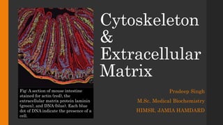
  • 1. Cytoskeleton & Extracellular Matrix Pradeep Singh M.Sc. Medical Biochemistry HIMSR, JAMIA HAMDARD Fig: A section of mouse intestine stained for actin (red), the extracellular matrix protein laminin (green), and DNA (blue). Each blue dot of DNA indicate the presence of a cell.
  • 3. Introduction  Eukaryotic cells contain protein filaments that are collectively called as cytoskeleton.  These protein filaments are involved in: ─ establishing cell shape ─ provide mechanical strength ─ help in locomotion of cell ─ chromosome separation ─ intracellular transport of organelles
  • 4.  The three major cytoskeleton filaments are: 1. Actin filaments 2. Microtubules 3. Intermediate filaments  In addition, a large number of accessory proteins, including the motor proteins, are also required for the properties associated with each of these filaments.
  • 6. 1. Actin • Actin was first isolated in 1942, from muscle cells in which it constitutes approximately 20% of the total cell protein. • The three-dimensional structures of both individual actin molecules and actin filaments were determined in 1990. • Actin filaments (F-Actin) also known as microfilaments, made up of small subunits called G-Actin. • G-Actin are globular proteins consisting of 375 amino acids. G-Actin is described as having a pointed end and a barbed end. Pointed End: Minus end Barbed End: Plus end
  • 7. The structure of an actin monomer and actin filament
  • 8. Assembly of Actin Filament (F-Actin) • G-Actin monomers polymerize to form actin filaments (F-Actin) having a diameter of about 8nm. • F-Actin is a helical structure. • Steps of formation of actin filament: a) Nucleation b) Elongation
  • 9. • Nucleation: For the formation of a new actin filament, subunits must assemble into a small aggregate or nucleus which then elongate rapidly by addition of new G-Actin subunits. This process is called filament nucleation. • Actin filament nucleate most frequently at the plasma membrane and nucleation is regulated by external signals. • Nucleation is the rate limiting step in the formation of a cytoskeletal polymer. • Elongation: G-Actin are added to the both ends but addition of new subunits is faster at plus end.
  • 10. The time course of actin polymerization in a test tube
  • 11. Concept of Critical Concentration • The number of monomers that are added to the polymer per second will be proportional to the concentration of the free subunit (KonC), but the subunits will leave the polymer end at a constant rate (Koff) that does not depend on C. • Polymerization: Polymerization is a reversible process which depends on the critical concentration (KonC) of the monomers. • At a concentration > KonC, filament grows at each end but growth is faster at the barbed end. • At a concentration < KonC, filament shrink at both ends.
  • 12. Nucleotide Hydrolysis • Each actin molecule carries a tightly bound ATP molecule that is hydrolysed to a tightly bound ADP molecule soon after its assembly into the polymer. • Hydrolysis of the bound nucleotide reduces the binding affinity of the subunit for neighbouring subunits and makes it more likely to dissociate from each end of the filament. It is usually the T-form that adds to the filament and D-form that leaves.
  • 13.
  • 14. Treadmilling • At the steady state, subunits undergo a net assembly of monomers at the plus end and a net disassembly at the minus end at an identical rate. • The polymers maintains a constant length, even though there is net flux of subunits through the polymer, known as treadmilling.
  • 15. Chemical Inhibitors of Actin • The functions of the actin filaments are inhibited by both polymer- stabilizing and polymer-destabilizing chemicals. • These chemicals are used as an important tools in the study of the filaments. Table: Chemical Inhibiters of Actin Chemical Effect on filaments Mechanism Original source Latrunculin Depolymerizes Binds actin subunits Sponges Cytochalasins B Depolymerizes Caps filament plus ends Fungi Phalloidin Stabilizes Binds along filaments Amanita mushroom
  • 16. Filament Elongation Is Modified By Proteins That Bind To The Free Subunits • In most non-muscle vertebrate cells, approximately 50% of the actin is in filament form and 50% in soluble form while the monomer concentration is 50-200 μM, well above the critical concentration (0.1 μM) . Question: why does so little of the actin polymerize into filaments? The actin is not polymerized as it is bound to special proteins, such as thymosin. Actin monomers bound to thymosin are locked where they cannot associate with either the (+) end or (-) end of the actin filament.
  • 18. Actin Binding Proteins • A number of different types of actin binding proteins remodel or modify existing filaments. Cellular Role Representative Proteins Monomer binding Thymosin, Profilin Filament initiation and polymerization Arp2/3, formin Filament stabilization Tropomyosin End capping CapZ, tropomodulin Filament severing Cofilin, gelsolin Filament cross-linking α-actinin, fimbrin, filamin Actin filament linkage to other proteins Spectrin
  • 19. A. Monomer Binding Proteins • Thymosin: Actin monomer bound to thymosin are in a locked state, where they cannot associate with either the plus end or minus ends of actin filaments and can neither hydrolyse nor exchange their bound nucleotide. • Profilin: Profilin binds to the actin monomer (G-Actin) opposite to the ATP- binding cleft, blocking the side of the monomer that would normally associate with the filament minus end, while leaving exposed the site on the monomer that binds to the plus end. • Profilin competes with thymosin for binding to individual actin monomer. Thus, by regulating the local activity of profilin, cells can control the length of actin filaments.
  • 20.
  • 21. B. Filament Initiation And Polymerization 1. Arp2/3: Arp stands for ‘Actin Related Proteins’. ― The Arp2/3 complex comprises 7 protein sub units. ― Both Arp2 and Arp3 are about 45% identical to actin. ― Arp2/3 complex nucleates actin filament growth from minus end, allowing rapid elongation at the plus end. ― The ARP complex can also attach to the side of another actin filament while remaining bound to the minus end of the filament that it has nucleated resulting in the formation of web like structure.
  • 22. Differences on the sides and minus end prevent the ARPs from forming filament on their own.
  • 23.
  • 24.
  • 25. 2. Formins: Formins are dimeric proteins that nucleate the growth of straight, unbranched filaments that can be cross linked by other proteins to form protein bundles. ― Formin dimer remains associated with the rapidly growing plus end while still allowing the addition of new subunits at that end. ― Formin dependent actin filament growth is strongly enhanced by the addition of profiling linked actin monomers.
  • 26.
  • 27. C. Filament Stabilization Proteins • Tropomyosin: Tropomyosin, an elongated protein that binds simultaneously to six or seven adjacent actin subunit along each of the two grooves of the helical actin filament. ― In addition to stabilizing and stiffening the filament, the binding of tropomyosin can prevent the actin from interacting with other proteins. ― Tropomyosin plays an important in the control of muscle contraction.
  • 28. D. End Capping Proteins • Tropomodulin: Tropomodulin is an end capping protein that binds tightly to the minus ends of the actin filaments that have have been coated and stabilized by tropomyosin. • CapZ: CapZ is named so because of its location in the muscle Z band. CapZ binds to the actin filament at the plus end, stabilizing the actin filament by greatly reducing the rates of filament growth and depolymerisation.
  • 29. E. Filament Severing Proteins • Sever means “breaking’. • These proteins breaks an actin filaments into many smaller fragments. • The fate of these fragments depends on the presence of other accessory proteins. • Under some conditions, newly formed ends acts as nucleus and promotes filament elongation. • Under other conditions, severing promotes the depolymerization of old fragments.
  • 30. 1. Gelsolin superfamily: These proteins are activated by high levels of cytosolic Ca2+ . ― Gelsolin interact with the side of the actin filament and contains two subdomains that binds to two different sites: one that is exposed on the surface of the filament and another that is hidden between the adjacent subunit. ― Gelsolin creates a small gap between neighbouring subunits and inserts itself into the gap to break into the filament. ― Gelsolin remains attached to the actin filament and caps the new plus end. Two superfamilies of severing proteins are: 1. Gelsolin 2. Cofilin
  • 32. 2. Cofilin superfamily: Also called actin depolymerizing factor. ― Cofilin binds along the length of the actin filament, forcing the filamentto twist a little more tightly. ― Mechanical stress generated weakens the contacts between the actin subunits in the filament, generating filament ends that undergo rapid disassembly. ― Cofilin binds to ADP-containing actin filaments rather than to ATP-containing filaments. ― Since, ATP hydrolysis is usually slower than filament assembly, the newest actin filament in the cell still contain mostlt ATP and are resistant to depolymerisation by cofilin. ― Cofilin therefore tends to dismantle the older filaments in the cell.
  • 34. F. Filament Cross Linking Proteins 1. α-Actinin: α-Actinin includes myosin and links oppositely charged actin filaments into contactile bundles. 2. Fimbrin: Fimbrin excludes myosin and links actin filaments into non contractile parallel bundles.
  • 35. 3. Filamin: Filamin promote the formation of a loose and highly viscous gel by clamping two actin filaments roughly at right angles.
  • 36. G. Actin Filaments Linking to Other Proteins • Spectrin: Spectrin was first identified in RBC. ― Spectrin is a long, flexible protein made out of four elongated polypeptide chains (two α subunits and two β subunits), arranged so that two actin binding sites are 200 nm apart. ― In RBC, spectrin is concentrated just beneath the plasma membrane. Spectrin has separate binding sites to actin filaments and other peripheral proteins of plasma membrane. ―Spectrin allows the RBC to get back to normal shape after squeezing through the capillary blood vessels.
  • 37. ACTIN BASED MOTOR PROTEINS • The first motor protein to be identified in the skeletal muscles was Myosin. • Consist of two heavy chains and two copies of each of two light chain. • Helps in the muscle contraction.
  • 38. Functions of Actin Filaments ― By forming a band under the plasma membrane, actin filaments allow cells to adopt different shapes and perform different functions ― Generate locomotion in cells such as white blood cells and amoeba. Villi Contractile bundles Sheet-like & Finger-like protrusions Contractile ring
  • 39. ― Link transmembrane proteins to cytoplasmic proteins ― Form contractile ring during cytokinesis in animal cells ― Cytoplasmic streaming ― Interact with myosin to provide force of muscular contraction
  • 41. 2. Microtubules • Microtubules are hollow cylindrical structure which are polymers of the protein tubulin. • The tubulin subunit is a heterodimer made up of two globular proteins called α-tubulin and β- tubulin. • The wall is composed of 13 parallel protofilaments, each composed of αβ-tubulin heterodimers stacked from head to tail. • The α- and β-subunits of the tubulin dimer can bind one molecule of GTP. The GTP in the α- tubulin subunit is never hydrolyzed while β- subunit may be in either GTP or the GDP form.
  • 42. The structure of a microtubule and its subunit
  • 43. Polymerization of tubulin to form microfilament • Microtubule growth is generally nucleated from specific locations with in the cell known as microtubule- organizing centre (MTOC). • Many animal cells have single MTOC called as centrosome, located near nucleus. • MTOC are highly enriched in another type of tubulin called γ-tubulin which form γ-tubulin ring complex (γ- TuRC).This γ-tubulin ring complex helps in the nucleation of microtubules.
  • 44. Mechanism: • Two accessory proteins bind directly to the γ-tubulin along with several other proteins that help to create a spiral ring of γ-tubulin molecules, which serves as a template that create a microtubule with 13 protofilaments. • The microtubule then grows by addition of free subunits. • After incorporation of a dimeric subunit into a microtubule, the GTP on the β-tubulin is hydrolysed to GDP CENTROSOME
  • 45. Microtubule nucleation by the γ-tubulin ring complex
  • 46. Treadmilling and Dynamic Instability Treadmilling is addition of subunit at one end and their loss at the other end. Dynamic instability is the oscillation between growth and shrinkage. Catastrophe: The change from growth to shrinkage. Rescue: The change from shrinkage to growth
  • 47. Centrosome • Centrosome in animal cells located near the nucleus. • It contain centrioles and a variable number of small dense bodies called as centriolar satellites. • It consist of an amorphous matrix of proteins containing the γ-tubulin ring complexes that nucleate microtubule growth. • They serve as basal bodies and sites of anchor for epithelial cilia.
  • 48. Centrioles and Cell Division • Duplication of the centrosomes takes place during ‘S’ phase cell division. To form two separate MTOCs at opposite poles of the mitotic spindle. • Two centrosomes then separate and move to opposite sides of the nucleus, forming the two poles of the mitotic spindle. • As the cell enters mitosis, the dynamics of microtubules assembly and disassembly also change dramatically. • First, the rate of microtubule disassembly increases about tenfold, resulting in overall depolymerisation and shrinkage of microtubules. • At the same time, the number of microtubules emerging from the centrosome increases by five-to-tenfold.
  • 49.
  • 50. • As mitosis proceeds, the two chromatids of each chromosome are then pulled to opposite poles of the spindle. This chromosome movement is mediated by motor proteins associated with the spindle microtubules. • In the final stage of mitosis, nuclear envelopes reform, the chromosome decondense, and cytokinesis takes place. • If mitotic cells are exposed to drugs like colchicine (binds to monomeric tubulin and prevent polymerization), vinblastine and taxol (disrupt microtubule dynamics), microtubule disappear and mitosis is arrested because of inadequate formation of the mitotic spindle. These drugs are useful in the treatment of certain cancers.
  • 51. Microtubules Associated Proteins (MAP) • Microtubule polymerization dynamics is very different in cells than in solutions of pure tubulin (in vitro). • Proteins that binds to microtubules are collectively called as microtubule-associated proteins. • MAP can be divided into two groups: 1. Microtubule stabilizing proteins 2. Microtubule destabilizing proteins.
  • 52. TABLE: Proteins That Modulate Microtubule (MT) Dynamics Protein Location Function Microtubule Stabilizing Proteins MAP1 Dendrites and axons; non- neuronal cells Assembles and stabilizes MTs MAP2 Dendrites Assembles and cross-links MTs to one another and to intermediate filaments. MAP4 Most cell types Stabilizes MTs Tau Dendrites and axons Assemble stabilizes and cross-links MTs Microtubule Destabilizing Proteins Stathmin Most cell types Binds tubulin dimers katanin Most cell types Microtubule Severing
  • 53. Microtubule Stabilizing Proteins • Microtubule stabilizing proteins helps in the organization of microtubule bundles. • MAPs have at least one domain that binds to the microtubule and another that project outwards. • The length of the projecting domain determine how closely MAP-coated microtubules pack together.
  • 54. Microtubule Destabilizing Proteins • One molecule of the small stathmin binds to two tubulin heterodimerers and prevents their addition to the ends of the microtubules. • Stathmin thus decreases the effective concentration of tubulin subunits that are available for polymerization. • Phosphorylation of stathmin inhibits its binding to tubulin. 1. Stathmin: Also known as tubulin- sequestering protein.
  • 55. 2. Katanin: katanin is also known as microtubule-severing protein. • Katanin is named after the Japanese word for “sword”. • Katanins release microtubules from their attachment to MTOC and contribute to rapid microtubule depolymerisation observed at the poles of spindles during mitosis. • Katanin is made up of two subunits: 1. Smaller Subunit - Hydrolyze ATP and perform severing. 2. Larger Subunit – Direct katanin to the centrosome.
  • 56.
  • 57. Microtubule Ends Binding Proteins Proteins that bind to the ends of microtubules can control microtubule positioning
  • 58. Motor Proteins Move Along Microtubules • Two major classes of microtubule- based motors are: 1. Kinesins – Move towards (+) end 2. Dyneins – Move towards (-) end • They use the energy of ATP hydrolysis to move along microtubules. • They mediate the sliding of filaments relative to one another and the transport of membrane- enclosed organelles along filament tracks.
  • 59. A. Kinesin • Kinsesin-1 has two kinesin motor head domains: • Rear or Lagging head domain: tightly bound to microtubule and ATP. • Front or leading head domain: loosely bound to microtubule with ADP in its binding site. • Most of them have the motor domain at the N-terminus of the heavy chain and walk toward the plus end of the microtubule. • The forward displacement of the rear motor head domain is driven by the hydrolysis of ATP and binding of ATP in the leading head domain. • They use “hand-over-hand” motion to walk over the microtubule.
  • 60.
  • 61. B. Dyneins • Dyneins are a family of minus end directed motor proteins. • The motor domain is present at the C-terminus of the heavy chain and walk toward the minus end of the microtubule. • They are composed of one, two or three heavy chains and a large number of light chains. • Dynein requires the presence of a large number of accessory proteins to associate with the membrane-enclosed oragnelles. • They are highly specialized for the rapid and efficient sliding movement of the microtubules that derive the beating of cilia and flagella.
  • 63. 3. Intermediate Filaments • Third major type of cytoskeletal protein. • Present particularly in the cytoplasm of cells which are subjected to mechanical stress. • Generally absent in animals that have rigid exoskeleton. • Structurally similar but biochemically distinct from actin and myosin filaments. • Intermediate filaments are extremely difficult to break and can be stretched to over 3 times their length.
  • 64. Structure of Intermediate Filaments • Monomer is consist of α-helical domain containing 40 or more hepted repeat motifs. • First Stage: Two monomers coil together to form the dimer. • Second Stage: A pair of parallel dimers associates in antiparallel fashion to form a staggered tetramer. Thus, they lack structural polarity. • Third Stage: Lateral association of 8 tetramers. • Fourth Stage: Addition of 8 tetramers to growing filament.
  • 65.
  • 66. Characteristics • Intermediate filaments are generally more stable than actin filaments or microtubules. • Intermediate filaments can be modified by phosphorylation which can regulate their assembly and disassembly.
  • 67. Table: Major Types of Intermediate Filament Proteins in Vertebrate Cells Types of Intermediate Filament Component Polypeptides Location Nuclear Lamins A,B and C Nuclear Lamina (Inner lining of nuclear envelope) Vimentin-like Vimentin Many cells of mesenchymal origin Desmin Muscle Peripherin Some neuron Epithelial Type I keratins (acidic) Epithelial cells and their derivatives.Type II keratins (neutral/basic) Axonal Neurofilament proteins (NF-L, NF-M, and NF-H) Neurons
  • 68. 1. Nuclear Lamins: Nuclear lamins are fibrous proteins providing structural function and transcriptional regulation in the cell nucleus. • Found in many eukaryotes but missing from unicellular organisms. • Forms a meshwork that lines the ineer membrane of the nuclear envelope. • Provide anchorage sites for chromosomes and nuclear pores. 2. Both Vimentin and Keratin attach to nuclear envelop serving to position and anchor nucleus within the cell. 3. Desmin connects individual actin-myosin assemblies of muscle cell to each other as well as to the plasma membrane. 4. Neurofilaments are major intermediate filaments of motor neurons.
  • 69. Cell Junctions • Intermediate filaments also form cell junctions. • There are generally two types of cell junction: 1. Cell-cell junction 2. Cell-matrix junction • Cell-cell junction formed by intermediate filaments – Desmosomes • Cell- Matrix Junction formed by intermediate filaments – Hemidesmosomes.
  • 70.
  • 71. Diseases of Intermediate Filament • Epidermis Bullosa Simplex: Mutations in the keratin gene form skin blister resulting from cell lysis after minor trauma.
  • 72. • Lou Gehrig’s Disease: Also known as Amyotrophic lateral sclerosis which leads to progressive loss of motor neurons which lead to muscle atrophy, paralysis and eventual death.
  • 74. “Half of the secrets of the cell are outside the cell.” Dr. Mina Bissell Oct. 17, 2007 Erlanger Auditorium
  • 75. Why do all multicellular animals have ECM? • Act as structural support to maintain cell organization and integrity (epithelial tubes; mucosal lining of gut; skeletal muscle fiber integrity) • Compartmentalize tissues (Pancreas: islets vs. exocrine component; skin: epidermis vs. dermis) • Provide hardness to bone and teeth (collagen fibrils become mineralized) • Present information to adjacent cells: • Inherent signals (e.g., RGD motif in fibronectin) • Bound signals (BMP7, TGFγ, FGF, SHH) • Serve as a highway for cell migration during development (neural crest migration), in normal tissue maintenance (intestinal mucosa), and in injury or disease (wound healing; cancer)
  • 76. Extracellular Matrix • Two broad categories of tissue that are found in all animals are: 1. Epithelial Tissue 2. Connective tissue • Tissues are not only made up of cells. They also contain a remarkably complex and intricate network of macromolecules constituting the Extracellular Matrix. • The matrix can become calcified to form the rock-hard structures of bone or teeth or it can form transmembrane substance of cornea etc. • In most connective tissue, the matrix molecules are secreted by cells called fibroblast.
  • 77. • The extracellular matrix is constructed from three major classes of macromolecules: 1. Glycosaminoglycas (GAGs) – Highly charged polysaccharide covalently linked to proteins to form proteoglycans 2. Fibrous Proteins – Collagen, Elastin 3. Glycoproteins
  • 78. 1. Glycosaminoglycan (GAGs) • Glycosaminoglycans are unbranched polysaccharide chains composed of repeating disaccharide units. • One of the two sugars in the repeating disaccharide is always an amino sugar (N-acetylglucosamine or N-acteylgalactosamine) which in most cases in sulfated. • GAGs are highly negatively charged because of the presence of sulfate and carboxyl groups on most of their sugars.
  • 79. • Four main groups of GAGs are distinguished by their sugars, the type of linkage between the sugars, and the number and location of sulfate groups: 1. Hyaluronan: D-Glucuronate + N-Acetyl-D-Glucosamine 2. Chondroitin sulfate and Dermatan sulfate • Chondroitin Sulfate: D-Glucuronate + N-Acetyl-D-Galactosamine-4-sulfate • Dermatan Sulfate: L-Iduronate + N-Acetyl-D-Galactosamine-4-sulfate 3. Heparan sulfate: L-Iduronate-2-sulfate + N-Sulfo-D-Glucosamine-6- sulfate 4. Keratan sulfate: D-Galactose + N-Acetyl-D-Glucosamine-6-sulfate
  • 80. Proteoglycans • Proteoglycans are composed of GAG chains covalently linked to proteins as proteoglycans. • Except for hyaluronan, all GAGs take part in the synthesis of proteoglycans. • Synthesis of Proteoglycans: Membrane bound ribosomes make the polypeptide chain or core protein which is then threaded into the lumen of the ER. • The polysaacharide chains are mainly assembled on this core protein in the Golgi apparatus before delivery to the exterior of the cell by exocytosis.
  • 81. • Examples of proteoglycans: 1. Aggrecan: ─ Major Component of cartilage ─ Contain over 100 GAG chains. 2. Decorin: ─ Secreted by fibroblasts, contain only 1-10 GAG chains ─ Decorin binds to collagen fibrils and regulate fibrils assembly and fibril diameter. • Function: The proteoglycan molecules in connective tissue typically form a highly hydrated, gel-like “ground substance” in which collagens and glycoproteins are embedded.
  • 82. 2. Fibrous Proteins 1. Collagen: ─ Collagens are a family of fibrous proteins ─ Collagens are the most abundant proteins in animals, where they constitute 25% of the total protein mass. • Structure: ─ Triple helix ─ Has 3.3 residue per turn ─ Every 3rd amino acid residue is a glycine residue ─ Gly-X-Y (X is commonly proline or hydroxyproline while Y can be any amino acid)
  • 83. Biosynthesis of Collagen 1. Synthesis of a chain of pre-procollagen on free ribosomes. A signal protein that directs them to RER. 2. Cleavage of signal protein forms free ribosomes. 3. Hydroxylation of lysine and proline 4. Glycosylation: Addition of galactose and glucose to some hydroxylysine residues. (Enzyme – Galactosyl transferase and Glycosyl transferase) m-RNA Signal protein
  • 84. 5. Assembly of three α-chains to form procollagen 6. Secretion of procollagen molecules by exocytosis into the extra cellular space. S S Registration peptides S S S S S S S S S S α-helices Procollagen
  • 85. 7. Cleavage of the additional peptides leads to formation of tropocollagen. 8. Self-assembly or polymerization of tropocollagen molecules form collagen fibrils Procollagen peptidase Procollagen peptidase
  • 86. Types of Collagen 1. Fibril Forming Collagens: • Type I,II and III are fibril forming collagens. • They have rope like structures. 2. Network Forming Collagens: • Type IV and VIII forms a three dimensional mesh, rather than distinct fibrils. 3. Fibril Associated Collagens • Type IX and XII binds to the surface of the collagen fibrils, linking these fibrils to one another and to other components in the ECM.
  • 87. Disease Associated with Collagen 1. Ehlers-Danlos Syndrome • It is an inheritable defect. • Caused by: A. Deficiency of collagen-processing enzymes a) Lysyl hydroxylase b) N-Procollagen peptidase B. Mutations in the amino acid sequences of collagen types I, III or V.
  • 88. 2. Scurvy • Caused due to deficiency of Vitamin-C. • Ascorbate is required for Propyl hydroxylase and Lysyl hydroxylase activities. • Acquired disease of fibrillary collagen • Manifestations: 1. Bleeding gums 2. Subcutaneous haemorrhage 3. Poor wound healing
  • 89. 3. Menke’s Syndrome  Characterized by kinky hair and growth retardation  Due to dietary deficiency of copper which is require by lysyl oxidase which catalyzes a key step in the formation of the covalent cross-links that strengthen collagen fibers.
  • 90. 2. Elastin • Connective tissue protein with rubber - like properties • Found in lungs, walls of large blood vessels and elastic ligaments • Can be stretched to several times their normal length, but recoil to their original shape when relaxed.
  • 91. • It is synthesized as a soluble monomer of 70 KDA called tropoelastin. • Composed primarily of small non polar amino acids residues (e.g. G, A, V) • Also rich in proline and lysine like collagen but not glycosylated, contains little hydroxyproline and hydroxylysine. • Long inelastic collagen fibrils are interwoven with elastic fibers to limit the extent of stretching and prevent the tissue from tearing.
  • 92. Genetic Abnormalities of Elastin William’s Syndrome • Deletion of elastin gene (7q 11.23) results in defective development of the connective tissues in various organs including the CVS. • Supravalvular aortic stenosis is a feature of this disorder. This severly impairs the blood flow to various organs. Pulmonary Emphysema • It is accompanied by fragmentation or decrease in elastin protein in lungs leading to destruction and lung cavity formation.
  • 93. COLLAGEN ELASTIN Triple helix No triple helix; random coil conformations permitting stretching (Gly-X-Y)n repeating structure No ( Gly-X-Y)n repeating structure Presence of hydroxylysine Carbohydrate-containing No or very little hydroxylysine No carbohydrate Intramolecular aldol cross-links Intramolecular desmosine cross-links Presence of extension peptides during bio-synthesis No extension peptides present during biosynthesis
  • 94. 3. Glycoproteins • Glycoproteins are proteins that contain oligosaccharide chains covalently attached to the polypeptide side-chains. • Glycoproteins have multiple domains, each with specific binding sites for other matrix macromolecules and for receptors on the surface of cells. • Major type of glycoproteins are: A. Fibronectins B. Fibrilin C. Laminin D. integrins
  • 95. A. Fibronectin • Fibronectin is a dimer composed of two very large subunits joined by di-sulphide bonds at their C-terminal ends. • Each subunit contains a series of small repeated domains separated by short stretches of polypeptide chain. • Each domain is usually encoded by a separate exon. • Both the Arg-Gly-Asp (RGD) and the “synergy” sequences are important for binding to integrins on cell surfaces.
  • 96. Fibronectin Binds to Integrins • Fibronectins can exist in both forms: 1. Soluble form – Circulating in the blood and other body fluids 2. Insoluble fibronectin fibrils – fibronectin dimer cross link to one another by additional disulphide bonds. • Fibronectin molecules assemble into fibrils only on the surface of cells where cells possess appropriate fibronectin-binding proteins particularly integrins. • Tight binding of integrins with the fibronectin requires more than just the RGD sequence.
  • 97. • Integrins provide a linkage from the fibronectin outside of the cell to the actin cytoskeleton inside it. • Integrin molecules allow fibronectin and actin filaments to bind directly to one another. Thus, intracellular cytoskeleton align with the extracellular fibronectin to determine cell shape. • Fibronectin molecules assemble where there is a mechanical need for them not in inappropriate locations such as bloodstream. • In many kinds of cancer, cells are unable to make fibronectins, loose shape and detach from the extracellular matrix to become malignant.
  • 98. • During cell movement (as during embryogenesis), pathways of fibronectins guide cells to their destinations. • Soluble plasma fibronectin promotes blood clotting by direct binding of fibrin. • Fibronectins guide immune cells to wounded areas and thus promote wound healing.
  • 99. B. Laminin • Laminin is a very large protein comprised of three proteins that form a cross. • Laminin and Type IV collagen are major components of the basal lamina. • The basal lamina acts as a selective barrier to the movement of cells, as well as a filter for molecules. • In the kidney, thick basal lamina helps to prevent the passage of macromolecules from the blood into the urine. • Basal lamina is also important in tissue regeneration after injury. • Progeria (early onset of aging), is possibly due to a defective laminin.
  • 100. C. Integrins • Group of transmembrane glycoproteins. • Allow cells to attach to ECM constituents (laminins and fibronectins mainly). • Most integrins are receptors for extracellular matrix proteins that helps in the cell signalling. • Integrins on the surface of leukocytes plays an important role in inflammation.
  • 101. Summary • Cytoskeleton and Extracellular matrix works in coordinated manner to regulate the normal functioning of the cell.