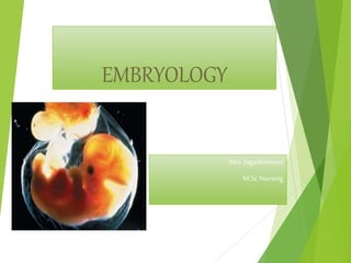
3. definition
A study of the development of an
individual before birth is called
EMBRYOLOGY.
Embryology is the study of the
formation and development of the
embryo(or fetus) from the moment
of its inception upto the time it is
born as an infant.
4. gestational period
Germinal period-Its starts from 1st week to the
3rdweek after fertilization ,during which the
zygote develops and forms the trilaminar
germ disc.
Embryonic period-Its starts from the 4th week to 8th
week during which there is differentiation and
formation of most of the tissues and organs of
the body.
Fetal period-Its starts from 9th week upto
termination of pregnancy during which there
is a rapid growth of the fetus and complete
development of the placenta.
5. gametogenesis
The process involved in the
maturation of the two highly
specialized cells ,spermatozoon
in male and ovum in female
before they unite to form
zygote is called gametogenesis.
9. spermatogenesis
The process involved in
the development of
spermatids from the
primordial male germ cells
and their differentiation
into spermatozoa is called
spermatogenesis
13. ovulation
It’s a process
whereby a secondary oocyte
is released from the ovary
following rupture of a
mature grafian follicle and
becomes available for
conception
14. Mechanism of ovulation
The process of
ovulation is a complex of
one preovulatory changes
occur both in the follicle
and the oocyte
17. causes
1] LH surge – secondary to sustained peak level of
estrogens in the late follicular phase. This will
cause completion of reduction division in the
oocyte and luteinisation of granulosa cells,
synthesise progesterone and prostaglandins.
2] FSH rise- leads to plasminogen and it helps in lysis
of follicle.
3] Stretching factor – Necrobiosis of wall due to
passive stretching
4] Contraction of micro muscles in theca externa
18. Effect of ovulation
Following ovulation the
follicle is changed to corpus
luteum.Ovum will be picked
up by fallopian tube and
may fertilise or degenerate.
21. fertilization
It begins with sperm egg collision
and ends with production of a mono
nucleated single cell called zygote.
Its objectives are …
1. To initiate the embryonic development of
the egg
2. To restore the chromosome number of the
species.
Almost always fertilization occur in
ampulla part of the uterine tube.
23. Approximation of the gametes
It involves the transport of the
sperms and ova in the female genital
tract to reach the uterine tube. The
ovum is picked up after ovulation by
tubal fimbrae and is by muscular or
kind of suction or ciliary action or
chemotaxis by tubal secretions.
Then transported to ampullary part.
24. Contact and fusion of the gametes
A direct contact between the spermatozoa and
the zona pellucida of the secondary oocyte is necessary
before actual fusion of the gametes.• The cells of corona
radiata provide obstacle to the penetration of the
sperms.• 200 to 300 million sperms emitted at single
ejaculation about 300 to 500 sperms reach the ovum,
only 1 unites with oocyte & rest engaged in
disintegration of corona radiata by secreting enzyme
hyalerinidase
Prior to penetration spermatozoa undergo
process of Capacitation and Acrosome reaction.
25. effect of fertilization
COMPLETION OF SECOND MEIOTIC DIVISION OF FEMALE
GAMETE: -It contains haploid number of chromosomes (22+ X).
The bigger one is female pronucleus & smaller polar body
pushed to peri-vitteline space. -Head and tail of sperm enter
the cytoplasm. Head and neck of spermatozoon number of
chromosome i.e. 22+X or 22+Y.
RESTORATION OF DIPLOID NOS OF CHROMOSOMES IN THE
ZYGOTE • Both male & female pronuclie meet near centre of
ovum, nuclear membrane disappear. This results in nuclear
fusion with restoration of diploid number of chromosome
44+2X or 44+xy
DETERMINATION OF THE CHROMOSOMAL SEX Out of the
total population in that half of them contain x bearing
chromosome and remaining half contain y bearing . If x
bearing spermatozoon fertilizes an ovum the zygote contain 2x
chromosome +44autosome & female child is formed & Zygote
containing 2y chromosome +44autosome male child is formed
26. Day 2 and 3: morulla/Cleavage
The original zygote divides about
30 hours after conception into two
daughter cells called blastomeres.
Continued subdivisions of the
original cell result in increasing
numbers of blastomeres.
During cell division the dividing
cells decrease in size. This type of
cell division is called cleavage.
By the time the zygote is ready to
enter the uterus, it contains a solid
ball of 12 to 16 blastomeres called
the morula (from the Latin word
for mulberry).
27. Day 4: Formation of the blastocyst
Fluid within the intercellular spaces of
the morula gradually increases, and
spaces on one side of the inner cell
mass come together, forming a single
cavity, the blastocele.
The outer layer organizes into the
trophoblasts, which give rise to the
placenta, and the inner layer of cells
form the embryo.
The cavity of the blastocele fills with
fluid, and the conceptus is now called
the blastocyst.
28. implantation/nidation –day 6
1. Apposition : most common occurs on
the upper posterior uterine wall
2. Adhesion
3. Penetration and Invasion
Implantation occurs 6th or
7th day after fertilization.
29. MAJOR EVENTS OF FIRST WEEK
NORMAL EVENTS
Fertilization and formation of
the zygote (30hours).
Cleavage of the zygote into 12
to 16 blastomeres- the morula
(day 2 and 3).
Formation of the blastocyst (
day 5-8).
POSSIBLE ABNORMAL EVENTS
Abnormal implantation
Maternal infection or a
genetic defect
Hydatidiform mole
Abortion
Ectopic implantation
30. trophoblast
The blastocyst attaches to the
uterine lining in the V-shaped.
When the trophoblast (the outer
cell layer) attaches to the
endometrium, it proliferates and
separates into an inner
cytotrophoblastic layer (fetal
side) and an outer
syncytiotrophoblastic (placental
side).
The outer layer develops finger
like projections that proliferate
and superficially attach the
blastocyst to the endometrium
within 6 days after conception.
31. Second week of life
A slit like amniotic cavity appears about day
8, and the yolk sac appears as a second
cavity on day 12. Bilaminar embryonic disc
is formed in between these two layers.
The endodermal disc becomes thicker at it’s
cephalad end, forming the prochordal
plate.
During early development of the nervous
system, the function of the prochordal
plate is to indicate the site of the mouth
and to form the membranes of the mouth
and throat.
The formation of the decidua, fetal
membranes, and placenta extends beyond
the second week, but their development
begins at this point.
32. THIRD WEEK OF LIFE
During the third week of life, the conceptus
develops rapidly. This period also coincides
with the first missed menstrual cycle of the
mother.
The primitive streak is formed during the third
week, and three germ layers develop.
This periods from approximately day 15 to
day 21, is called the “period of threes”; not
only do the three germ layers develop, but the
primitive streak, the notochord, and the
neural tube are formed.
34. GASTRULATION
Gastrulation is the process by
which the bilaminar embryo
becomes a trilaminar embryo.
On about day 15, the
cytotrophoblast cells
proliferate into the blastocyst
to form the extraembryonic
mesoderm, which later become
the extra-embryonic coelom.
The mesoderm lies between the
ectoderm and the endoderm,
completing the trilaminar disc
of the primitive streak. All
tissues and organs of the
embryo are developed from
these three layers.
36. Notochord
Days 16-18
Primitive node
epiblast cells
invaginate and
migrate anteriorly
with some endoderm
cells
Rod defining the body
axis is formed
Future site of the
vertebral column
37. Neurulation
Notochord signals overlying ectoderm
The neural tube is developed from the closure of the
neural plate and the neural fold- a process called
neurulation –at about 21 to 26 days.
38. Closure of neural tube: begins at end of week 3;
complete by end of week 4.
Extends cranially (eventually brain) and caudally
(spinal cord)
Neural crest, lateral ectodermal cells, pulled along
and form sensory nerve cells and other structures
39. MAJOR EVENTS OF
third WEEK
NORMAL EVENTS
Formation of blood vessels within
the chorionic villi ( day 13).
Gastrulation or conversion of the
bilaminar embryonic disc into the
three- layered trilaminar disc
(day 14).
Continued development of the
chorion with formation of tertiary
chorionic villi( day 15-20).
Development of the neural tube
(day 18).
Formation of somites (day 21).
Beginning of blood circulation
(day 24).
POSSIBLE ABNORMAL
EVENTS
Monozygous twins
result
Conjoined twins are
formed.
The heart is most
susceptible to
teratogens between
the 19th and the 41st
day.