Rapid Impact Assessment of Climatic and Physio-graphic Changes on Flagship G...
Grimmett et al., growth rate hypothesis
1. Does the growth rate hypothesis apply to aquatic
hyphomycetes?
I.J. GRIMMETT, K.N. SHIPP, A. MACNEIL, F. B€ARLOCHER*
Department of Biology, Mt. Allison University, Sackville, NB E4L 1G7, Canada
a r t i c l e i n f o
Article history:
Received 21 June 2013
Revision received 14 August 2013
Accepted 16 August 2013
Available online
Corresponding editor:
Petr Baldrian
Keywords:
Aquatic hyphomycetes
Carbon
DNA
Ecological stoichiometry
Ergosterol
Growth rate hypothesis
Nitrogen
Phosphorus
RNA
a b s t r a c t
The growth rate hypothesis states that in many organisms or tissues, the specific growth
rate m correlates with RNA concentrations. Since RNA often accounts for much of the
phosphorus content of cells, m may also correlate positively with P concentrations and
negatively with C:P and N:P ratios. We tested this hypothesis with broth cultures of five
aquatic hyphomycete species. Samples were harvested on eight occasions after 3e56 d of
incubation. Accumulation of biomass was fitted to a rectangular hyperbola, whose
parameters were used to estimate m. There were no consistent trends related to culture age
or m for C, N, P or ergosterol concentrations. RNA and DNA concentrations and RNA:DNA
ratios were significantly and negatively correlated with culture age. Only RNA concen-
trations were positively and linearly correlated with m. While RNA or DNA concentrations
are unsuitable as indicators for total biomass, levels of fungal RNA combined with markers
for fungal biomass may allow estimates of the extent to which the mycelia are metabol-
ically active.
ª 2013 Elsevier Ltd and The British Mycological Society. All rights reserved.
Introduction
Aquatic hyphomycetes are a polyphyletic group of higher
fungi that dominate microbial decomposition of autumn-shed
leaves in streams (Duarte et al., 2013; Gessner et al., 2007;
Krauss et al., 2011). Fungal colonization makes the leaves
more palatable and more nutritious to leaf-shredding inver-
tebrates. This conditioning effect is partly due to increased
nitrogen (protein) concentrations caused by the accumulation
of fungal mycelia on decaying leaves (Kaushik and Hynes,
1971). A similar enrichment can often be observed with
phosphorus (Webster et al., 2009; Grimmett et al., 2012). The
C:N and C:P ratios of colonized leaves are therefore closer to
the ratios of invertebrate consumers and provide a stoichio-
metrically more appropriate resource (Cross et al., 2005;
Hladyz et al., 2009). In conjunction with estimates of total
fungal biomass (Gessner et al., 2003), they are potential indi-
cators of the nutritional value of leaves at various stages of
decay.
Both fungal decomposer activities and growth are influ-
enced by substratum quality (leaf) and inorganic nutrients
(water column). They are further modulated by factors such as
temperature, pollutants and pH (Gessner et al., 2007; Krauss
et al., 2011). These factors are engaged in a complex
* Corresponding author. Tel.: þ1 506 364 3501; fax: þ1 506 364 2505.
E-mail address: fbaerlocher@mta.ca (F. B€arlocher).
available at www.sciencedirect.com
ScienceDirect
journal homepage: www.elsevier.com/locate/funeco
1754-5048/$ e see front matter ª 2013 Elsevier Ltd and The British Mycological Society. All rights reserved.
http://dx.doi.org/10.1016/j.funeco.2013.08.002
f u n g a l e c o l o g y 6 ( 2 0 1 3 ) 4 9 3 e5 0 0
2. feedback system with the composition of the fungal com-
munity (Gessner et al., 2010). Traditionally, fungal commun-
ities have been assessed by aerating colonized leaves in
nutrient-poor water (Gessner et al., 2003). This stimulates
the release of spores by aquatic hyphomycetes, which can be
counted and identified under the microscope. Non-
sporulating fungi are not detected. A more complete inven-
tory of the fungal community can be achieved by extracting
and amplifying fungal DNA and connecting it to specific taxa
(Nikolcheva et al., 2003; B€arlocher, 2010). However, the doc-
umentation of amplifiable DNA assigned to a given species
does not allow statements about the metabolic status (active
and growing, dormant, or dead) of that species. Additional
information may be provided by measuring RNA. It has long
been known that in non-marine bacteria the RNA:DNA ratio is
linearly related to growth rate whereas the DNA:dry mass
ratio remains almost constant (Dortch et al., 1983). The
extended growth rate hypothesis postulates that specific
growth rate m (rate of biomass addition per unit biomass), P
and RNA levels are all correlated (Elser et al., 2000). Active
growth generally requires allocation of P-rich RNA for protein
synthesis, and the ratios of RNA:biomass, RNA:protein, or
RNA:DNA have widely been used to assess the growth status
across diverse taxa (Elser et al., 2000, 2003; Cross et al., 2005).
However, very few studies have been done on fungi. In Pen-
icillium urticae (Bu’lock et al., 1965), RNA and P values were
highest in 24-hr old mycelia; in the next 12 hr, the values
declined rapidly to 27 % and 20 % (RNA and P, respectively). In
Rhizoctonia solani and Sclerotium bataticola, both RNA and DNA
concentrations, but not their ratio, declined during the first 5
d of growth in a liquid medium (Gottlieb and van Etten, 1966).
In four strains of saltmarsh fungi, N concentrations of 3
month old mycelia declined to between 36.7 and 63.6 % of
values at 2 weeks (Newell and Statzell-Tallman, 1982).
No studies on the growth rate hypothesis have been pub-
lished on aquatic hyphomycetes. The objective of the current
study was to fill this gap. We first determined the growth
dynamics of pure cultures of five aquatic hyphomycete spe-
cies during 56 d. On eight dates, we measured total mycelial
mass and concentrations of ergosterol (commonly used indi-
cator of mycelial biomass), carbon (C), nitrogen (N), and
phosphorus (P), as well as RNA and DNA. We expected a
positive correlation between P and RNA, with both declining at
later stages of incubation, when the growth rate m typically
approaches 0.
Materials and methods
Fungal cultures
Five species, all isolated from single conidia, were used
(Table 1). Cultures were maintained on malt extract agar in
Petri plates (1 % malt extract, Sigma 70146; 1.5 % agar, Sigma
A7002). For experiments, two 7-mm agar plugs were cut from
the growing edge of a colony and crushed with a hand-held
glass homogenizer in 5 ml of distilled, sterilized water. Two
ml of the resulting suspension were used as inoculum for each
250 ml Erlenmeyer flask filled with 50 ml of 2 % malt extract
broth (Sigma 70146). Separate suspensions from separate Petri
plates were used for each flask. The flasks were incubated in
an Environ-Shaker 3597 (Lab-Line Instruments, Melrose Park,
Illinois) at 16 Æ 1
C and 150 rpm. After 3, 5, 7, 10, 14, 21, 35 and
56 d the contents of a flask were filtered through a preweighed
glass microfibre filter (47 mm, Whatman GF/C), which con-
stituted one replicate value. One set of flasks (three replicates
per date, eight dates) was used for growth curves. Freeze-dried
mycelia of this set were then used for P analyses. Three
additional sets of 24 flasks were used for RNA/DNA, N and
ergosterol analyses.
Growth curves
The filter with fungal mycelium was freeze-dried and
weighed, and the initial weight of the filter was subtracted. At
each sampling date, the contents of three flasks were sacri-
ficed. For a crude measure of viability after 56 d, 25 pellets per
species (1 mm diameter) were plated on malt extract agar and
checked for growth after 7 d.
Ergosterol
Ergosterol was extracted from pre-weighed, freeze-dried
mycelia via microwave (Young, 1995; Nikolcheva et al., 2003).
Cleaned extracts with ergosterol were injected into a high-
performance liquid chromatography C18 column (Varian,
Palo Alto, Calif.) and eluted with methanol at 1.5 ml minÀ1
at
20
C (elution time, 5.3 min). Ergosterol content was estimated
by comparing peak areas with those of external standards
(Sigma 45480).
CNP analyses
Mycelium was removed from the filter and freeze-dried. C and
N content of mycelia were measured by a Vario EL II CHNOS
Elemental Analyzer (Elementar Analysensysteme GmbH,
Germany). Total P was measured after hydrolysis of mycelia
ground in liquid N following the procedures in Wetzel and
Likens (1991). The same analyses were performed on the
malt extract powder used for culture media.
Nucleic acid analyses
Mycelium was removed from the filter and placed on blotting
paper. All analyses proceeded with samples of this wet
Table 1 e Species of aquatic hyphomycetes used in this
study
Origin Abbreviation
Articulospora tetracladia
Ingold
Boss Brook, NS,
Canada
Arte
Heliscus lugdunensis
Sacc. Therry
Boss Brook, NS,
Canada
Helu
Lunulospora curvula
Ingold
Lous~a Stream,
Portugal
Lucu
Tricladium chaetocladium
Ingold
Bot~ao Stream,
Portugal
Trch
Varicosporium
elodeae Kegel
Boss Brook, NS,
Canada
Vael
494 I.J. Grimmett et al.
3. mycelium. To determine the ratio of wet to dry weight, 25
samples were collected from each species after 35 d of incu-
bation. The weight of wet samples was compared to the
remaining weight after freeze-drying.
For RNA and DNA analyses, 25e50 mg of wet weight were
used. RNA and DNA were extracted from the same sample
with Norgen’s RNA/DNA/Protein purification kits (Norgen
Biotek, Thorold, Canada). RNA and DNA concentrations in the
extracts were measured with a QubitÒ
Fluorometer 2.0 (Life
Technologies, Burlington, Canada), following manufacturer’s
instructions.
Statistical analyses
Prism 6.0c for Mac (www.graphpad.com) was used for curve-
fitting (Motulsky and Christopoulos, 2004). We fitted fungal
biomass harvested on eight dates to a rectangular hyperbola
(equivalent to Monod’s and MichaeliseMenten equation)
BT ¼ Bmax
T
T50 þ T
where BT ¼ biomass at time T, Bmax ¼ maximum biomass;
T ¼ time in days, and T50 ¼ time to reach 50 % of maximum
biomass. We also attempted to fit the data to the logistic and
to Boltzmann’s sigmoidal equation. Only the fit for the rec-
tangular hyperbola converged, and we used its parameters to
describe growth rates m, defined as per capita increase of
biomass per day.
Linear regressions were fitted to the various chemical
indicators (ergosterol, C, N, P, RNA, DNA) versus time and m. For
each data point, three replicate samples, originating from
three separate flasks, were used.
Two-way ANOVAs were used to analyze the values of the
chemical indicators, with Species (aquatic hyphomycete) and
Time (since inoculation) as factors (JMP 10.02 for Mac, www.
jmp.com).
Results
Growth curves
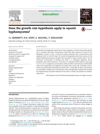
Accumulation of dry mass of the five species over time is
shown in Fig 1A. With non-linear curve-fitting, the parameters
of the logistic and Boltzmann sigmoidal equations did not
converge, those of a rectangular hyperbola (Monod or
MichaeliseMention equation) did (Table 2). The estimated
parameters were used to estimate per capita or specific
growth rate m (dayÀ1
), i.e., addition of fungal biomass per
existing biomass per day.
After 56 d of incubation, hyphae grew from all 25 pellets of
Heliscus lugdunensis, Lunulospora curvula and Tricladium chaeto-
cladium. No visible growth was found with Articulospora tetra-
cladia and Varicosporium elodeae.
Ergosterol
The average ergosterol concentration (all species, all dates, Æ1
SD) was 5.9 Æ 1.5 mg gÀ1
DM. Changes as function of incuba-
tion time are shown in Fig 1B.
ANOVA revealed significant effects of Time and Species
and their interaction on ergosterol concentrations ( p 0.013).
Linear regressions of ergosterol versus incubation time or m
were not significant (Table 3).
CNP analyses
The elemental concentrations (averages in %, Æ1 SD) of the
malt extract medium were 38.9 Æ 0.23 (C), 2.96 Æ 0.04 (N), and
0.64 Æ 0.03 (P), corresponding to a molar C:N:P ratio of 154:10:1.
Concentrations of C, N and P in fungal mycelia as a func-
tion of incubation time are shown in Fig 2AeC. For all three
variables, ANOVA revealed significant effects of Time and
0
200
400
600
800
0 10 20 30 40 50 60
Arte Helu Lucu Trch Vael
Drymass(mg)
0
2
4
6
8
10
Ergosterol(mgg-1
DM)
Time (days)
A
B
Fig 1 e Mean (±1 SD; n [ 3) biomass accumulation (A) and
ergosterol concentrations (B) of five fungal species over
time. Abbreviations as in Table 1.
Table 2 e Parameters of rectangular hyperbola fitted to
biomass accumulation by the five fungal species.
Bmax [ estimated maximum biomass in mg; T50 [ time
in days when biomass is 50 % of maximum.
Abbreviations as in Table 1
Arte Helu Lucu Trch Vael
Bmax 692 287 636 326 593
T50 8.0 3.1 25.1 6.4 8.5
R2
0.51 0.39 0.73 0.67 0.60
Does the growth rate hypothesis apply to aquatic hyphomycetes? 495
4. Species and their interaction ( p 0.037). Linear regressions of
C, N or P versus incubation time or m were not significant
(Table 3).
Average concentrations (in %; mean Æ1 SD) were 45.2 Æ 5.0
(C), 3.74 Æ 1.09 (N) and 0.608 Æ 0.005 (P). The data were con-
verted to molar equivalents to determine C:N, C:P and N:P
ratios. Ratios averaged over the experimental period and the
grand average for all species are listed in Table 4.
C, N, P or their ratios did not correlate significantly with
incubation period or the specific growth rate m dayÀ1
( p ! 0.39).
Nucleic acid analyses
DNA and RNA concentrations (Æ1 SD, in mg mgÀ1
DM) averaged
over all samples were 1.52 Æ 1.36 and 12.1 Æ 9.6, respectively.
Despite considerable variation, a gradual decline in con-
centrations of RNA and DNA with time seems to predominate
(Fig 3). Linear regressions of chemical indicators versus incu-
bation time were significant for RNA, DNA and the RNA:DNA
ratio (Table 3). Neither RNA nor DNA was significantly corre-
lated with C, N, P or ergosterol ( p ! 0.44).
The specific growth rate m (proportional increase per day)
was calculated as described above. A positive linear correla-
tion was only significant for RNA versus m (Table 3).
Discussion
The initial growth of microbial cultures or colonies is often
assumed to be zero (lag phase). It then exponentially accel-
erates to a maximum (Zwietering et al., 1990), which is fol-
lowed by a gradual decline and an asymptotic approach to
zero. In the final stage, the cell population may decline, i.e.,
the growth rate is negative. Without the final decline, the
general shape of these curves is sigmoidal, and Zwietering
et al. (1990) evaluated several equations of this type. A
hyperbolic model, connecting growth to a limiting nutrient,
was introduced by Monod (1949). We were unable to fit data
from five aquatic hyphomycete species to two common
sigmoidal models, while Monod’s equation provided a mod-
erately good fit. Nevertheless, there were some clear discrep-
ancies: biomass accumulation in L. curvula (Lucu, Fig 1) was
negligible until 10 d, and in A. tetracladia (Arte, Fig 2), there was
Table 3 e Statistical analyses of chemical indicators (Y).
Linear regressions versus time or specific growth rate m.
Data for all five species were combined
X Y Intercept Slope R2
p
Time Ergosterol 5.88 À0.005 0.003 0.54
C 45.8 À0.031 0.01 0.25
N 3.72 0.0029 0.002 0.62
P 6.0 À0.0008 0.00066 0.78
RNA 18.7 À0.39 0.41 0.0001
DNA 2.1 À0.033 0.25 0.0001
RNA:DNA 15.8 À0.17 0.076 0.019
m Ergosterol 5.98 À0.006 0.005 0.66
C 0.47 À0.0008 0.042 0.21
N 0.09 þ0.002 0.004 0.90
P 0.32 À0.04 0.003 0.28
RNA 6.5 þ56.4 0.41 0.0001
DNA 1.3 þ2.5 0.04 0.24
RNA:DNA 10.5 þ24.8 0.05 0.19
0
100
200
300
400
500
600
0 10 20 30 40 50 60
Arte Helu Lucu Trch Vael
C(mgg-1
DM)
0
10
20
30
40
50
60
70
0 10 20 30 40 50 60
N(mgg-1
DM)DM)
0
1
2
3
4
5
6
7
8
0 10 20 30 40 50 60
P(mgg-1
Time (days)
A
B
C
Fig 2 e Mean (±1 SD; n [ 3) carbon (A), nitrogen (B) and
phosphorus (C) concentrations in five fungal species as a
function of incubation time. Abbreviations as in Table 1.
Table 4 e Molar ratios averaged over experimental
period. Abbreviations as in Table 1
Species C:N C:P N:P C:N:P
Arte 16.2:1 178:1 11:1 178:11:1
Helu 12.4:1 186:1 15:1 186:15:1
Lucu 13.6:1 218:1 16:1 218:16:1
Trch 12.8:1 192:1 15:1 192:15:1
Vael 17:1 187:1 11:1 187:11:1
Average 14.4:1 192:1 13.6:1 192:13.6:1
496 I.J. Grimmett et al.
5. a substantial decline after 21 d. Such discrepancies between
actual growth parameters and those calculated from our
model may have affected the strength of support for the
growth rate hypothesis.
Today, ergosterol is the most widely used indicator for
biomass of higher fungi (Gessner et al., 2003; Suberkropp,
2008). For aquatic hyphomycetes, a ratio of 5.5 mg ergosterol
per mg fungal biomass is commonly assumed (Gessner and
Chauvet, 1993); our overall average (5.9 mg gÀ1
) is very close
to this standard. However, depending on culture conditions
and species, this ratio can vary by a factor of up to 14
(Charcosset and Chauvet, 2001; Raviraja et al., 2004).
Ergosterol is thought to be a measure of living biomass and
vulnerable to rapid degradation upon hyphal death and
membrane disruption. Since older cultures contain an
increasing proportion of dying or dead hyphae (on Day 56, no
growth occurred from pellets of A. tetracladia and V. elodeae),
we expected ergosterol concentrations to decline. While there
appeared to be a trend toward lower values after 14 d, the
decline was relatively modest (between 13 % in H. lugdunensis
and 25 % in T. chaetocladium). This supports observations by
Mille-Lindblom et al. (2004) who found ergosterol to be
remarkably resistant to degradation, both as pure substance
and when present in dead mycelium. At later stages of leaf
decay in streams, fungal biomass (estimated by ergosterol
levels) often seems to persist at high levels, while reproductive
output (conidia) declines drastically (B€arlocher, 2009). This has
been interpreted as a hedge-betting strategy. Conceivably, it
could be an artifact due to the persistence of ergosterol not
associated with living hyphae.
Elemental C, N and P were influenced by species, age of
culture and their interaction. Another factor is the supply of N
and P in the food resource e depending on their relative pro-
portions, N or P may limit growth and affect the composition
of the growing mycelium (Elser et al., 2003; Danger and
Chauvet, 2013). The C:N:P ratio of 154:10:1 in our study is
considered high; deciduous leaves, the usual food resource of
fungi, generally have C:N ratio of 40e80 and C:P ratios of
400e2 000 (Elser et al., 2003; Cross et al., 2005).
There are few comprehensive and systematic studies on
nutrient concentrations in the fungal mycelia. Based on the
limited information available, our data (3.74 % N, 0.61% P) are
close to values reported for other fungal groups. In saltmarsh
fungi, concentrations ranged between 2.2 and 6.0 % (Newell
and Statzell-Tallman, 1982); in hypogeous fungi it was an
average of 2.36% and in epigeous fungi 3.44% (Wallis et al.,
2012). P content in various basidiomycetes varied between
0.15 and 0.68% (Cromack et al., 1975) or between 0.1 and 0.2 %
(Wallander et al., 2003). Only two studies investigated N and P
contents of aquatic hyphomycetes. Leach and Gulis (2010)
reported a relatively low C:N:P ratio (90:9:1; compared to our
average of 192:13.6:1). Danger and Chauvet (2013) observed
highly variable ratios as a function of external nutrient supply.
For example, the C:P ratios for A. tetracladia and T. chaetocladium
varied between 76e1 166 and 89e1 175, respectively. Our
averages for the same two species were 178 and 192, respec-
tively. Overall, there is little evidence that aquatic hyphomy-
cetes are homeostatic with respect to N and P. For several soil
fungi, the C:N ratio varied between 5 and 17, while N:P was
close to 15 (Cleveland and Liptzin, 2007, data based on chloro-
form fumigation-extraction method), both close to our values.
Intraspecific variability of nutrient levels (N, P) was gen-
erally highest during the first 14 d, but there was no clear
temporal trend. At least the extended version of the growth
0
10
20
30
40
50
0 10 20 30 40 50 60
Arte Helu Lucu Trch Vael
RNA(µgmg-1
DM)
0
1
2
3
4
5
6
7
8
0 10 20 30 40 50 60
DNA(gmg
-1
DM)
Time (days)
0
10
20
30
40
50
0 0.05 0.1 0.15 0.2 0.25 0.3 0.35
RNA(µgmg-1
DM)
0
1
2
3
4
5
6
7
8
0 0.05 0.1 0.15 0.2 0.25 0.3 0.35
DNA(µgmg-1
DM)
µ (day-1
)
A
C
B
D
Fig 3 e Mean (±1 SD; n [ 3) RNA and DNA concentrations in
five fungal species as a function of incubation time (A, B) or
specific growth rate m (C, D). Abbreviations as in Table 1.
Does the growth rate hypothesis apply to aquatic hyphomycetes? 497
6. rate hypothesis (Sterner and Elser, 2002; Flynn et al., 2010),
which implies that organisms or tissues with higher nutrient
concentrations (N, P) grow more rapidly, was not supported by
our data.
Neither N nor P correlated with culture age or growth
rate. In the aquatic hyphomycete Tetrachaetum elegans, the
relationship between mycelial age and C:P, C:N and N:P
ratios was strongly influenced by the external nutrient
supply (Danger and Chauvet, 2013). For example, mycelial
C:P ratio increased with age when external P or both N and P
were depleted. No such effect was seen in NP-rich nor N-
depleted medium. On the other hand, the N:P ratio only
changed with age under P-depleted conditions (Danger and
Chauvet, 2013). Our conditions (N:P ¼ 10:1; total
N ¼ 0.65 g lÀ1
) approximated the NP-rich treatment in
Danger and Chauvet (2013). Overall, our results are con-
sistent with the assumption that with sufficient external P
supply, the link between RNA and P in tissues or cells can be
relaxed (Elser et al., 2003).
Flynn et al. (2010) discuss additional factors that may
weaken the correlation between N, P, RNA and instantaneous
growth rate in microalgae. These include difficulties of accu-
rately measuring elemental composition or growth rates, the
assumption that rate of protein synthesis by ribosomes is
fixed, or that RNA contains the majority of cellular P. The same
potential sources of error may apply to fungi. In three species
(ascomycete, yeast and zygomycete), the proportion of P
associated with nucleic acids varied between 19 and 44 %
(Beever and Burns, 1980).
A major concern with filamentous fungi is the fact that
hyphae grow from the tips. Harvested mycelia in our study
consisted for the most part of pellets, whose diameter was
variable (generally between 1 and 6 mm). Larger pellets gen-
erally have a hollow center; hyphae near the center are often
dead and growth is concentrated on the surface of the pellet
(Raviraja et al., 2004). Depending on the pellet’s size, the
contribution of the youngest (and presumably fastest-
growing) hyphal tips is variable. This pattern of fungal
growth has been shown to affect ergosterol content (Raviraja
et al., 2004). It may also influence the elemental composition
or nucleic acid concentrations.
Nucleic acids and their ratios are potentially more direct
indicators of growth status. The grand average of DNA data for
our five aquatic hyphomycetes (1.52 mg mgÀ1
) was reasonably
close to the average of 25 soil fungi (3.3 mg mgÀ1
, Anderson,
2008), as was the range (0.04e3.96 versus 0.92e6.32, respec-
tively). Similarly, the average RNA content in our study
(12.1 mg mgÀ1
, range: 0.2e41.6) did not deviate drastically from
earlier reports: Bu’lock et al. (1965) found values between 14
and 57 mg mgÀ1
(one species); averages were 54.1 (Gottlieb and
van Etten, 1966, two species) and 33.7 (Beever and Burns, 1980,
three species), respectively.
Both RNA and DNA content and their ratio declined with
mycelial age (Table 3), which confirms earlier reports (Bu’lock
et al., 1965: only RNA measured; Gottlieb and van Etten, 1966:
RNA and DNA; Cino and Tewari, 1976: RNA but not DNA). This
phenomenon may be influenced by at least two related pro-
cesses: fast growing cells require more RNA than slow growing
cells, while the DNA level may remain reasonably constant.
Filamentous fungi can be regarded “as a plasmodium moving
about in a system of tubes” (Beever and Burns, 1980). Nuclei,
including their DNA, may gradually be translocated to
younger cells, leaving behind a progressively vacuolated
mycelium. This implies a steeper RNA than DNA gradient
when comparing apical to distal hyphae, which will be further
amplified by the greater chemical stability of DNA compared
to RNA (Weaver, 2002). Not surprisingly, the regression slope
of nucleic acids versus age was steeper for RNA than for DNA
(Table 3).
The growth rate m as defined here (based on rectangular
hyperbola) increases monotonously with time, and, as
expected, RNA concentrations correlated positively with m.
DNA concentrations or the RNA:DNA ratio did not, implying
more complex patterns for these factors. The coefficient of
determination (R2
) reached a value of 0.41, indicating that the
model accounted for 41 % of the variability. It increased to
0.65, when the first datapoints (biomasses after 3 d) were
deleted from the analysis. This might indicate the presence of
an initial lag phase (which was clearly visible in L. curvula).
More detailed analysis of this earlier phase might further
strengthen the correlation between RNA levels and specific
growth rate.
Baldrian et al. (2013) cautioned against using copy num-
bers of fungal rDNA (estimated by qPCR) as biomarker for
mycelial mass in litter and soil. The high variability of total
DNA concentrations in our study, partly as a function of
culture age, support their conclusion. Wymore et al. (2013)
used bacterial and fungal rRNA (16S and 18S, respectively)
quantities to evaluate the relative abundances of these two
decomposer groups. In view of our results, they more accu-
rately reflect relative abundances of metabolically active
bacteria and fungi. This is often appropriate: we may be
interested primarily in active rather than dormant micro-
organisms. Additionally, “metabolically active” can indicate
fungal participation in leaf decomposition via exoenzymes or
fungal reproduction by sporulation (B€arlocher, 2010). In other
cases, total biomass may be more relevant, for example, if we
wish to estimate microbial contribution to invertebrate
nutrition.
Baldrian et al. (2012) described an approach that simul-
taneously addresses several aspects of fungal colonization
and litter decomposition. Community composition, based
on distribution of DNA sequences, was described through
pyrosequencing of fungal ITS regions. The same regions
were targeted in the precursor rRNA pool; their analysis gave
a description of active fungal taxa (Anderson and Parkin,
2007; Rajala et al., 2011). In addition, the soil metagenome
was searched for DNA sequences coding for cellobiohy-
drolase, an essential enzyme for cellulose decomposition
(Baldrian et al., 2012). These sequences were assigned to
fungal taxa. Some of the important conclusions were that
total and active fungal communities were quite different,
and that low-abundance species can make an important
contribution to decomposition processes. Clearly, there is
currently no single, all-encompassing method that
addresses all aspects of a fungal community and its eco-
logical functions. A combination of several complementary
methods is, therefore, often required for a comprehensive
interpretation of microbial ecology (B€arlocher, 2010;
Strickland and Rousk, 2010).
498 I.J. Grimmett et al.
7. Acknowledgments
Financial support by the Natural Science and Engineering
Research Council of Canada is gratefully acknowledged. We
thank Miranda Corkum for N analyses of our samples. Cristina
Canhoto and Ana Gonc¸alves provided us with two fungal
cultures. Two referees provided valuable suggestions.
r e f e r e n c e s
Anderson, T.-H., 2008. Assessment of DNA content of soil fungi.
Agriculture and Forestry Research 58, 19e28.
Anderson, I.C., Parkin, P.I., 2007. Detection of active soil fungi by
RT-PCR amplification of precursor rRNA molecules. Journal of
Microbiological Methods 68, 248e253.
Baldrian, P., Kolarık, M., Stursova, M., Kopecky, J., Valaskova, V.,
Vĕtrovsky, T., Zifcakova, L., Snajdr, J., Rıdl, J., Vlcek, C.,
Vorıskova, J., 2012. Active and total microbial communities in
forest soil are largely different and highly stratified during
decomposition. The ISME Journal 6, 248e258.
Baldrian, P., Vĕtrovsky, T., Cajthaml, T., Dobiasova, P.,
Petrankova, M., Snajdr, J., Eichlerova, I., 2013. Estimation of
fungal biomass in forest litter and soil. Fungal Ecology 6, 1e11.
B€arlocher, F., 2009. Reproduction and dispersal in aquatic
hyphomycetes. Mycoscience 50, 3e8.
B€arlocher, F., 2010. Molecular approaches promise a deeper and
broader understanding of the evolutionary ecology of aquatic
hyphomycetes. Journal of the North American Benthological
Society 29, 1027e1041.
Beever, R.E., Burns, J.W., 1980. Phosphorus uptake and utilization
by fungi. Advances in Botanical Research 8, 127e219.
Bu’lock, J.D., Hamilton, D., Hulme, M.A., Powell, A.J.,
Smalley, H.M., Shepherd, D., Smith, G.N., 1965. Metabolic
development and secondary biosynthesis in Penicillium urticae.
Canadian Journal of Microbiology 11, 765e778.
Charcosset, J.-Y., Chauvet, E., 2001. Effect of culture conditions on
ergosterol as an indicator of biomass in the aquatic
hyphomycetes. Applied and Environmental Microbiology 67,
2051e2255.
Cino, P.M., Tewari, R.P., 1976. Chemical composition of
Oidiodendron kalrai. Mycopathologia 58, 131e135.
Cleveland, C.C., Liptzin, D., 2007. C:N:P stoichiometry in soil: is
there a “Redfield ratio” for the microbial biomass?
Biogeochemistry 85, 235e252.
Cromack Jr., K., Todd, R.L., Monk, C.D., 1975. Patterns of
basidiomycete nutrient accumulation in conifer and
deciduous forest litter. Soil Biology Biochemistry 7, 265e268.
Cross, W.F., Benstead, J.P., Frost, P.C., Thomas, S.A., 2005.
Ecological stoichiometry in freshwater benthic systems:
recent progress and perspectives. Freshwater Biology 50,
1895e1912.
Danger, M., Chauvet, E., 2013. Elemental composition and degree
of homeostasis of fungi: are aquatic hyphomycetes more like
bacteria or plants? Fungal Ecology 6, 453e457.
Dortch, Q., Roberts, T.L., Clayton Jr., J.R., Ahmed, S.I., 1983. RNA/
DNA ratios and DNA concentrations as indicators of growth
rate and biomass in planktonic marine organisms. Marine
Ecology e Progress Series 13, 61e71.
Duarte, S., Seena, S., B€arlocher, F., Pascoal, C., Cassio, F., 2013. A
decade’s perspective on the impact of DNA sequencing on
aquatic hyphomycete research. Fungal Biology Reviews 27,
19e24.
Elser, J.J., Sterner, R.W., Gorokhova, E., Fagan, W.F., Markow, T.A.,
Cotner, J.B., Harrison, J.F., Hobbie, S.E., Odell, G.M., Weider, L.J.,
2000. Biological stoichiometry from genes to ecosystems.
Ecology Letters 3, 540e550.
Elser, J.J., Acharya, K., Kyle, M., Cotner, J., Makino, W., Markow, T.,
Watts, T., Hobbie, S., Fagan, W., Schade, J., Hood, J.,
Sterner, R.W., 2003. Growth rate e stoichiometry couplings in
diverse biota. Ecology Letters 6, 936e943.
Flynn, K.J., Raven, J.A., Rees, T.A.V., Finkel, Z., Quigg, A.,
Beardall, J., 2010. Is the growth rate hypothesis applicable to
microalgae? Journal of Phycology 46, 1e12.
Gessner, M.O., Chauvet, E., 1993. Ergosterol-to-biomass
conversion factors for aquatic hyphomycetes. Applied and
Environmental Microbiology 59, 502e507.
Gessner, M.O., B€arlocher, F., Chauvet, E., 2003. Qualitative and
quantitative analyses of aquatic hyphomycetes in streams. In:
Tsui, C.K.M., Hyde, K.D. (Eds.), Freshwater Mycology. Fungal
Diversity Press, Hong Kong, PRC, pp. 127e157.
Gessner, M.O., Gulis, V., Kuehn, K.A., Chauvet, E., Suberkropp, K.,
2007. Fungal decomposers of plant litter in aquatic
ecosystems. In: Kubicek, C.P., Druzhinina, I.S. (Eds.), The
Mycota: Environmental and Microbial Relationships, second
ed, vol. IV. Springer, Berlin, pp. 301e321.
Gessner, M.O., Swan, C.M., Dang, C.K., McKie, B.G.,
Bardgett, R.D., Wall, D.H., H€attenschwiler, S., 2010. Diversity
meets decomposition. Trends in Ecology and Evolution 25,
372e380.
Gottlieb, D., van Etten, J.L., 1966. Changes in fungi with age. I.
Chemical composition of Rhizoctonia solani and Sclerotium
bataticola. Journal of Bacteriology 91, 161e168.
Grimmett, I.J., Smith, K.A., B€arlocher, F., 2012. Tar-spot infection
delays fungal colonization and decomposition of maple
leaves. Freshwater Science 31, 1088e1095.
Hladyz, S., Gessner, M.O., Giller, P.S., Pozo, J., Woodward, G., 2009.
Resource quality and stoichiometric constraints on stream
ecosystem functioning. Freshwater Biology 43, 957e970.
Kaushik, N.K., Hynes, H.B.N., 1971. The fate of the dead leaves
that fall into streams. Archiv f€ur Hydrobiologie 68, 465e515.
Krauss, G.-J., Sole, M., Krauss, G., Schlosser, D., Wesenberg, D.,
B€arlocher, F., 2011. Fungi in freshwaters: ecology, physiology
and biochemical potential. FEMS Microbiology Reviews 35,
620e651.
Leach, D., Gulis, V., 2010. Microbial Stoichiometry and
Homeostasis of Nutrient Ratios in Fungi. Bridges 5 http://
www.coastal.edu/media/administration/honorsprogram/pdf/
Desiree%20Leach%202.pdf (accessed 01.06.13).
Mille-Lindblom, C., von Wachenfeldt, E., Tranvik, L.J., 2004.
Ergosterol as a measure of living fungal biomass: persistence
in environmental samples after fungal death. Journal of
Microbiological Methods 59, 253e262.
Monod, J., 1949. The growth of bacterial cultures. Annual Review
of Microbiology 3, 371e394.
Motulsky, H.J., Christopoulos, A., 2004. Fitting Models to
Biological Data Using Linear and Nonlinear Regression. A
Practical Guide to Curve Fitting. Oxford University Press,
New York, New York.
Newell, S.Y., Statzell-Tallman, A., 1982. Factors for conversion of
fungal biovolume values to biomass, carbon and nitrogen:
variation with mycelial ages, growth conditions, and strains of
fungi from a salt marsh. Oikos 39, 261e268.
Nikolcheva, L.G., Cockshutt, A.M., B€arlocher, F., 2003. Diversity of
freshwater fungi on decaying leaves e comparing traditional
and molecular approaches. Applied and Environmental
Microbiology 69, 2548e2554.
Rajala, T., Peltoniemi, M., Hantula, J., M€akip€a€a, R., Pennanen, T.,
2011. RNA reveals a succession of active fungi during the
decay of Norway spruce logs. Fungal Ecology 4, 437e448.
Raviraja, N.S., Nikolcheva, L.G., B€arlocher, F., 2004. Pellet size
affects mycelial ergosterol content in aquatic hyphomycetes.
Mycologia 96, 388e392.
Does the growth rate hypothesis apply to aquatic hyphomycetes? 499
8. Sterner, R.W., Elser, J.J., 2002. Ecological Stoichiometry: the
Biology of Elements from Molecules to the Biosphere.
Princeton University Press, Princeton.
Strickland, M.S., Rousk, J., 2010. Considering fungal:bacterial
dominance in soils e methods, controls, and ecosystem
implications. Soil Biology Biochemistry 42, 1385e1395.
Suberkropp, K., 2008. Fungal biomass and growth. In:
Sridhar, K.R., B€arlocher, F., Hyde, K.D. (Eds.), Novel Techniques
and Ideas in Mycology, Fungal Diversity Research Series, vol.
20, pp. 203e213.
Wallander, H., Mahmood, S., Hagerberg, D., Johansson, L.,
Pallon, J., 2003. Elemental composition of ectomycorrhizal
mycelia identified by PCReRFLP analysis and grown in contact
with apatite or wood ash in forest soil. FEMS Microbiology
Ecology 44, 57e65.
Wallis, O.R., Claridge, A.W., Trappe, J.M., 2012. Nitrogen content,
amino acid composition and digestibility from a nutritional
perspective in animal mycophagy. Fungal Biology 116,
590e602.
Weaver, R.F., 2002. Molecular Biology, second ed. McGraw-Hill,
New York, New York.
Webster, J.R., Newbold, J.D., Thomas, S.A., Valett, H.M.,
Mulholland, P.J., 2009. Nutrient uptake and mineralization
during leaf decay in streams e a model simulation.
International Review of Hydrobiology 94, 372e390.
Wetzel, R.G., Likens, G.E., 1991. Limnological Analyses, second ed.
Springer-Verlag, New York, New York.
Wymore, A.S., Compson, Z.G., Liu, C.M., Price, L.B.,
Whitham, T.G., Keim, P., Marks, J.C., 2013. Contrasting rRNA
gene abundance patterns for aquatic fungi and bacteria in
response to leaf-litter chemistry. Freshwater Science 32,
663e672.
Young, C.J., 1995. Microwave-assisted extraction of the fungal
metabolite ergosterol and total fatty acids. Journal of
Agriculture and Food Chemistry 43, 2904e2910.
Zwietering, M.H., Jongenburger, I., Rombouts, F.M., van’t Riet, K.,
1990. Modeling of the bacterial growth curve. Applied and
Environmental Microbiology 56, 1875e1881.
500 I.J. Grimmett et al.