Sonagachi Call Girls Services 9907093804 @24x7 High Class Babes Here Call Now
10 the influence of environmental bacteria in freshwater stingray
1. The influence of environmental bacteria in freshwater stingray
wound-healing
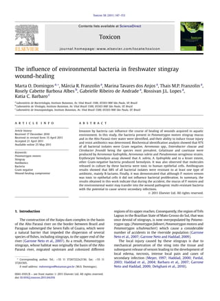
Marta O. Domingos a,*, Márcia R. Franzolin a
, Marina Tavares dos Anjos a
, Thais M.P. Franzolin a
,
Rosely Cabette Barbosa Albes b
, Gabrielle Ribeiro de Andrade a
, Rossivan J.L. Lopes a
,
Katia C. Barbaro c
a
Laboratório de Bacteriologia, Instituto Butantan, Av. Vital Brazil 1500, 05503-900 São Paulo, SP, Brazil
b
Laboratório de Virologia, Instituto Butantan, Av. Vital Brazil 1500, 05503-900 São Paulo, SP, Brazil
c
Laboratório de Imunopatologia, Instituto Butantan, Av. Vital Brazil 1500, 05503-900 São Paulo, SP, Brazil
a r t i c l e i n f o
Article history:
Received 17 December 2010
Received in revised form 15 April 2011
Accepted 21 April 2011
Available online 25 May 2011
Keywords:
Potamotrygon motoro
Stingray
Antibiotics
Bacteria
Gram negative
Wound-healing compromise
a b s t r a c t
Invasion by bacteria can influence the course of healing of wounds acquired in aquatic
environment. In this study, the bacteria present in Potamotrygon motoro stingray mucus
and in the Alto Paraná river water were identified, and their ability to induce tissue injury
and resist antibiotics was determined. Biochemical identification analysis showed that 97%
of all bacterial isolates were Gram negative, Aeromonas spp., Enterobacter cloacae and
Citrobacter freundii being the species most prevalent. Gelatinase and caseinase were
produced by Aeromonas hydrophila, Aeromonas sobria and Pseudomonas aeruginosa strains.
Erythrocyte hemolysis assay showed that A. sobria, A. hydrophila and to a lesser extent,
other Gram-negative bacteria produced hemolysin. It was also observed that molecules
released in culture by these bacteria were toxic to human epithelial cells. Antibiogram
results showed that 68% of all bacterial isolates were resistant to at least one type of
antibiotic, mainly B-lactams. Finally, it was demonstrated that although P. motoro venom
was toxic to epithelial cells it did not influence bacterial proliferation. In summary, the
results obtained in this work indicate that during the accident, the mucus of P. motoro and
the environmental water may transfer into the wound pathogenic multi-resistant bacteria
with the potential to cause severe secondary infections.
Ó 2011 Elsevier Ltd. All rights reserved.
1. Introduction
The construction of the Itaipu dam complex in the basin
of the Alto Paraná river on the border between Brazil and
Paraguai submerged the Seven Falls of Guaira, which were
a natural barrier that impeded the dispersion of several
species of fishes, including stingrays, to the upper end of the
river (Garrone Neto et al., 2007). As a result, Potamotrygon
stingrays, whose habitat was originally the basin of the Alto
Paraná river, migrated upstream and colonized different
regions of its upper reaches. Consequently, the region of Três
Lagoas in the Brazilian State of Mato Grosso do Sul, that was
once devoid of stingrays, is now overpopulated by Potamo-
trygon spp. (Potamotrygon falkneri, Potamotrygon motoro and
Potamotrygon schuhmacheri) which cause a considerable
number of accidents in the riverside population (Garrone
Neto et al., 2007; Garrone Neto and Haddad, 2009).
The local injury caused by these stingrays is due to
mechanical penetration of the sting into the tissue and
subsequent release of venom leading to the development of
local edema, necrosis, intense local pain and cases of
secondary infection (Meyer, 1997; Haddad, 2000; Pardal,
2003; Haddad et al., 2004; Barbaro et al., 2007; Garrone
Neto and Haddad, 2009; Dehghani et al., 2010).
* Corresponding author. Tel.: þ55 11 37267222x2136; fax: þ55 11
37261505.
E-mail address: mdomingos@butantan.gov.br (M.O. Domingos).
Contents lists available at ScienceDirect
Toxicon
journal homepage: www.elsevier.com/locate/toxicon
0041-0101/$ – see front matter Ó 2011 Elsevier Ltd. All rights reserved.
doi:10.1016/j.toxicon.2011.04.016
Toxicon 58 (2011) 147–153
2. It is postulated that the local inflammatory reaction and
necrosis in freshwater stingray accidents is due to the
release into the wound of several proteins with enzymatic
activity produced by the protein secretory cells that covers
the sting (Barbaro et al., 2007; Pedroso et al., 2007;
Magalhães et al., 2008; Antoniazzi et al., 2011).
The protein secretory cells are overlaid by a fin layer of
mucus which also covers the entire surface of the stingray
and separates the cutaneous tissue from direct contact with
the environmental water (Pedroso et al., 2007).
It has been reported that some Gram-negative bacteria
such as Photobacterium damsela, Vibrio alginolyticus, Cit-
robacter freundii, Aeromonas hydrophila and Pseudomonas
aeruginosa, that are commonly encountered in environ-
mental water and on the surface of aquatic animals, have
been isolated from wounds acquired during stingray acci-
dents (Fenner et al.,1989; Ho et al.,1998; Polack et al.,1998;
Baldinger, 1999; Barber and Swygert, 2000).
The involvement of these bacteria, especially Aeromonas
spp. and P. aeruginosa on the development of severe and
persistent secondary infection after tissue injury is well
documented (McManus et al., 1985; Semel and Trenholme,
1990; Gang et al., 1999). In addition, many other types of
bacteria present in the soil and aquatic environment can be
involved in secondary infections (van Elsas et al., 2011), and
the extent of infection cause by them can be determined by
how many of them are present, their ability to survive on
damaged tissue and to produce toxins able to induce cytokine
release and destroy host cells (Bhakdi et al., 1986; Lallier and
Higgins, 1988; Paraje et al., 2005; Markov et al., 2007;
Domingos et al., 2009).
Because of the considerable number of accidents caused
by Potamotrygon spp. stingrays in the region of Três Lagoas,
and the increasing importance of environmental Gram-
negative bacteria as emergent pathogens responsible for
secondary infections acquired in aquatic settings, the species
of bacteria encountered in the mucus of P. motoro stingrays
and in the Alto Paraná river water were determined and their
capacity to release toxins, cause injury to epithelial cells,
resist antibiotics and survive in the presence of stingray
venom was evaluated.
2. Material and methods
2.1. Venom, mucus and environmental water samples
Mucus and tissue extract samples were obtained from
twenty four P. motoro stingrays collected in the upper end
of the Alto Paraná river, in the region of Três Lagoas, Mato
Grosso do Sul state (BR). Briefly, the stingrays were
restrained and samples of the mucus that covers their
external surface were collected with sterile swabs from
three different regions of their dorsal area. The tissue
extracts were obtained from integumentary tissue covering
the sting as previously described (Barbaro et al., 2007). The
protein content of tissue extract pools (from now on
referred to as venom) utilized in this work was determined
by bicinchoninic acid albumin method (Smith et al., 1985),
using bovine serum albumin (BSA) as a standard. The
procedures involving animals were conducted in
conformity with national laws and policies (protocol
number CGEN 02001.005111/2008, SISBIO 15702-1).
The environmental water samples were collected from
the surface and the bottom of the Alto Paraná river at the
same points where P. motoro stingrays were restrained for
mucus sampling.
2.2. Cell line
The HEp-2 cell line used in this study was obtained from
Institute Adolfo Lutz, São Paulo, Brazil, previously acquired
from the American Type Culture Collection (CCL2).
2.3. Bacterial strains isolation and identification
The mucus samples were collected with sterile swabs,
placed in Cary-Blair transportation media and after 18 h of
incubation at 37
C, the bacterial strains were isolated in
blood-agar plates. Only the predominant colonies were
selected for identification by standard biochemical identi-
fication tests (Koneman et al., 2000), including one
commercially available biochemical identification system
(API 20E and API 20NE, Biomerieux, France).
2.4. Antibiogram
Antimicrobialsusceptibilityofall Gram-negative bacterial
strains isolated either from the environmental water or from
the mucus of P. motoro stingrays was determined by the
standard disk diffusion method (Bauer et al., 1966) utilizing
commercially available sensitivity discs and Mueller-Hinton
Agar. The results were evaluated according to the NCCLS,
2004 guidelines. The following antibiotics were tested:
amikacin(AMI), amoxicillin/clavulanicacid(AMC), ampicillin
(AMP), cephalotin (CFL), ceftazidime (CAZ), ciprofloxacin
(CIP), chloramphenicol (CLO), trimethoprim/sulfamethox-
azole (SUT), streptomycin (EST) and tetracycline (TET). For
quality control the test was run against the following ATCC
strains: Escherichia coli 25922 and P. aeruginosa 27853.
2.5. Blood-agar culture
Blood-agar culture plates were prepared according to
Beutin et al. (1989). Briefly, 1.5 g of TSA (Tryptic Soy Agar)
re-suspended in a 10 mM solution of CaCl2 was autoclave.
When the temperature of the agar fell to 45
C, goat red
cells previously washed three times in PBS pH 7.2 were
then added to the agar until a final concentration of 5% was
reached. The agar was then added to petri dish plates
(20 mL per plate), left to solidify and kept at 4
C until use.
2.6. Identification of hemolysin-producing bacterial samples
Forty microliters of bacterial culture previously grown
in TSB (Tryptic Soy Broth) for 18 h at 37
C were added in
triplicates to 3 mL of TSB and incubated overnight at 37
C.
After incubation,100 mL of each bacterial culture was added
to blood-agar plates in aliquots of 10 mL each. The plates
were then incubated for 18 h at 37
C and the presence of
hemolysin was determined by the formation of a halo of
lysed erythrocytes around the bacterial growth.
M.O. Domingos et al. / Toxicon 58 (2011) 147–153148
3. 2.7. Identification of caseinase-producing bacterial samples
Bacterial isolates cultured in TSB were centrifuged at
12,000 g for 15 min at 4
C and filtered through a Millipore
0.45 mm pore-diameter syringe filter. Clarified supernatant
was tested for proteolytic activity on casein agar plates.
Casein agar plates consisted of 25 mM Tris (pH 7.2),150 mM
NaCl, 0.6% casein (Sigma technical grade) and 1% TSA.
Aliquots (10 mL) of culture supernatants were placed in
3 mm diameter wells cut in the casein agar and incubated
at 37
C for 18 h. The plates were overlaid with 3% acetic
acid, and proteolytic activities were noted as a clear zone
around the sample well. Trypsin (1 mg/mL) was used as
a positive control standard.
2.8. Identification of gelatinase-producing bacterial samples
Gelatinase production was determined by API 20E and
API 20NE biochemical identification kit from Biomerieux,
France.
2.9. Inhibition of bacterial proliferation
Forty microliters of bacterial culture previously grown
in TSB at 37
C for 18 h (106
cell/mL) were added in tripli-
cate to 3 mL of TSB in the presence of either 5,1 or 0.5 mg of
P. motoro venom and incubated for 18 h at 37
C. As control,
the bacterial strains were grown in the presence of TSB
alone. After incubation, the absorbance was determined at
600 nm in a spectrophotometer (Spectronic 20, Genesys 1).
2.10. Cytotoxicity assay
The cytotoxic effect of P. motoro venom, mucus and
bacterial culture supernatants on human epithelial cells
(HEp-2) was determined by the MTT method which
measures the viability of cells in terms of their mitochon-
drial metabolic rate. Accordingly, 100 mL of DMEM (Dul-
becco’s Modified Eagle’s Medium) containing 106
cells was
added to each well of 96 well cell culture plates and incu-
bated for 24 h at 37
C in a 5% CO2 incubator. After incu-
bation, the medium was discarded and either 100 mL of
different concentrations of tissue extract (5 mg, 1 mg,
0.5 mg and 0.1 mg), 100 mL of mucus (v/v) or 100 mL of
bacterial culture previously grown for 18 h in DMEM were
added to the plates and incubated overnight at 37
C in a 5%
CO2 incubator. After incubation the supernatant was dis-
carded and 20 mL of a 5% solution of MTT in PBS was then
added into each well and the plates were incubated for 2 h
at 37
C. One hundred microliters of Triton (1%) was used as
positive control. Subsequently, 100 mL/well of methanol
(100%) was added to the plate and then incubated for
further 10 min. After incubation, the absorbance of each
sample was determined at 570 nm in a Spectronic 20
Genesys 1 spectrophotometer.
2.11. Statistic analysis
Results were expressed as mean Æ SD. Single criterion
ANOVA followed by Bonferroni’s test was used to analyze
the data, using SigmaStat 3.0 software. Values with p 0.05
were considered statistically significant.
3. Results
3.1. Bacterial strains isolated from the mucus of P. motoro
stingrays and the Alto Paraná river water
In order to determine the species of bacteria present in
the mucus of P. motoro rays or environmental water, 89
bacterial strains obtained either from the mucus of P.
motoro rays (n ¼ 24) or from the Alto Paraná river water
were isolated and identified. The results showed that only
3.4% of all isolates were Gram positive and they were found
only in the mucus. A total of fifteen different species of
Gram-negative bacteria were identified, however, Acineto-
bacter spp., P. aeruginosa, Klebsiella pneumoniae, Klebsiella
oxytoca, Serratia spp., Shigella spp. and Enterobacter spp.
were encountered only in the mucus whereas Plesiomonas
shigelloides and Citrobacter koseri were found only in the
water. Six bacterial species, A. hydrophila, Aeromonas sobria,
Pseudomonas putida, C. freundii, E. coli and Enterobacter
cloacae were encountered in both, water and mucus
samples (Table 1).
3.2. Proteases released by bacterial strains
The API 20E and 2API 20NE kits, casein agar and
erythrocyte hemolysis assays were utilized to determine
the ability of all Gram-negative bacterial isolates to produce
gelatinase, caseinase and hemolysin respectively. The
results showed that all A. sobria, A. hydrophila and P. aeru-
ginosa strains produced gelatinase. All A. sobria and to
a lesser extent, other Gram-negative strains produced
hemolysin. Caseinase was produced only by A. sobria, A.
hydrophila, P. aeruginosa and C. freundii strains (Table 2).
3.3. Antimicrobial drug profile of the bacterial isolates
The antimicrobial profile of each Gram-negative bacte-
rial isolate was determined by the standard disk diffusion
Table 1
Bacterial species isolated from the mucus of P. motoro stingrays and the
Alto Paraná river water.
Bacteria Number of isolates
Water Mucus Total
Aeromonas hydrophila 6 8 14
Aeromonas sobria 4 4 8
Pseudomonas aeruginosa 0 3 3
Pseudomonas putida 2 3 5
Acinetobacter spp. 0 6 6
Citrobacter freundii 3 9 12
Escherichia coli 1 8 9
Enterobacter cloacae 7 7 14
Klebsiella pneumoniae 0 5 5
(Others) 3 7 10
Gram positive 0 3 3
Total 26 63 89
Others: Water: Plesiomonas shigelloides (2); Citrobacter koseri (1). Mucus:
Serratiaspp. (3); Shigellaspp.(1);Enterobacterspp. (2); Klebsiella oxytoca(1).
M.O. Domingos et al. / Toxicon 58 (2011) 147–153 149
4. method. The results obtained showed that only 32% of all
bacterial samples were sensitive to all antibiotics tested,
whereas, 23% was sensitive to only one antibiotic and 45%
was sensitive to 2 or more antibiotics. The bacterial isolates
showed more resistance to three groups of antibiotics:
ampicillin, amoxicillin/clavulanic acid and cephalotin.
However, some pathogens such as P. aeruginosa, P. putida,
and E. cloacae were also resistant to other classes of anti-
biotics. E. coli was the only specie sensitive to all antibiotics
tested (Table 3).
3.4. Influence of P. motoro venom on bacterial growth
The influence of P. motoro venom on the proliferation of
all Gram-negative bacterial strains isolated in this work
was determined by incubating the bacterial isolates in TSB
for 18 h in the presence of 5, 1 or 0.5 mg/mL of venom and
subsequent determination of the absorbance at 600 nm.
The results obtained in this experiment showed that the
proliferation of all bacterial strains tested were not influ-
enced by the venom even in a concentration as high as
5 mg/mL (Fig. 1). Fig. 1 presents the results of one experi-
ment only, however, similar results were obtained from all
isolates tested.
3.5. Influence of P. motoro venom and mucus on cell viability
Human epithelial cells were incubated in the presence
of mucus or different concentrations of venom to deter-
mine their cytotoxic effect by measuring the mitochondrial
metabolic rate in terms of MTT bioreduction. The results
obtained in this experiment showed that P. motoro venom
(Fig. 2a) and P. motoro mucus (Fig. 2b) are both toxic to
epithelial cells.
3.6. Toxic effect of bacterial culture supernatants on human
epithelial cells
The toxic effect of all A. hydrophila, A. sobria and P. aer-
uginosa culture supernatants on human epithelial cells was
measured by the MTT method. The results showed that all
culture supernatants tested were toxic to epithelial cells
(Fig. 3).
4. Discussion
It is common knowledge that open wounds raise the
chance for infection, becoming one of the most prevalent
causes of non-healing of wounds. It is also known that
injuries induced by aquatic animals such as stingrays and
catfish can be infected by environmental microorganisms
such as A. hydrophila, Pseudomonas spp. Vibrio spp.
(Broderick et al., 1985; Ho et al., 1998; Polack et al., 1998;
Table 2
Proteases released by bacterial samples isolated from the mucus of
P. motoro stingray and the Alto Paraná river water.
Bacteria Hemolysin* Caseinase Gelatinase
Acinetobacter spp. 1/6 0/6 0/6
Aeromonas hydrophila 9/14 6/14 14/14
Aeromonas sobria 8/8 5/8 8/8
Citrobacter freundii 5/12 2/12 0/12
Enterobacter cloacae 0/14 0/14 0/14
Escherichia coli 1/9 0/9 0/9
Klebsiella pneunomiae 0/5 0/5 0/5
Pseudomonas aeruginosa 2/3 3/3 3/3
Pseudomonas putida 1/5 0/5 0/5
Others 2/10 0/10 0/10
Others: Water: *Plesiomonas shigelloides (1/2); Citrobacter koseri (1);
Mucus: *Serratia spp. (1/3); Shigella spp. (1); Enterobacter aerogenes (1);
Enterobacter spp. (1); Klebsiella oxytoca (1).
Table 3
Antimicrobial drug susceptibility of bacterial strains isolated from the mucus of P. motoro stingrays and the Alto Paraná river water.
Sensitive
to all
antibiotics
Number of strains resistant to antibiotics
AMI CAZ CIP AMC AMPa,e
CFLc
AMC-
AMP
AMC-
CFLb
AMP-
CFLd
AMC-
AMP-
CFL
AMP-
CFL-
SUT
AMC-
AMP-
CFL-
CLO-
SUT
AMP-
CLO
CFL-
EST
AMC-
AMP-
CFL-
TET
AMC-
AMP-
CFL-
SUT
P. aeruginosa 1/3 0/0 0/0 0/0 0 0 0 0 0 0 0 0 0 0 0 2/3 0
P. putida 0/5 0/0 0/0 0/0 0 1/5 0 0 0 0 0 2/5 2/5 0 0 0 0
Acinetobacter spp. 3/6 0/0 0/0 0/0 0 0 0 0 0 1/6 0 0 0 1/6 1/6 0 0
A. hydrophila 0/14 0/0 0/0 0/0 0 0 1/14 0 1/14 2/14 10/14 0 0 0 0 0 0
A. sobria 1/8 0/0 0/0 0/0 0 4/8 0 2/8 0 1/8 0 0 0 0 0 0 0
C. freundii 4/12 0/0 0/0 0/0 0 1/12 3/12 0 0 2/12 2/12 0 0 0 0 0 0
E. coli 9/9 0/0 0/0 0/0 0 0 0 0 0 0 0 0 0 0 0 0 0
E. cloacae 3/14 0/0 0/0 0/0 2/14 0 1/14 0 2/14 1/14 3/12 0 0 0 0 1/14 1/14
Klebsiella
pneumoniae
1/5 0/0 0/0 0/0 0 4/5 0 0 0 0 0 0 0 0 0 0 0
Others 5/10 0/0 0/0 0/0 0 2/10 1/10 0 1/10 1/10 0 0 0 0 0 0 0
Total 27/86 0/86 0/86 0/86 2/86 12/86 6/86 2/86 4/86 8/86 15/86 2/86 2/86 1/86 1/86 3/86 1/86
AMI: amikacin, AMC: amoxicillin/clavulanic acid, AMP: ampicillin, CFL: cephalotin, CAZ: ceftazidime, CIP: ciprofloxacin, CLO: chloramphenicol, SUT:
trimethoprim/sulfamethoxazole, EST: streptomycin and TET: tetracycline.
a
Plesiomonas shigelloides (1/2-AMP).
b
Citrobacter koseri (1/1-AMC-CFL).
c
Serratia spp. (1/3-CFL); Shigella spp. (1).
d
Enterobacter spp. (1/2 AMP-CFL).
e
Klebsiella oxytoca (1/1-AMP).
M.O. Domingos et al. / Toxicon 58 (2011) 147–153150
5. Baldinger, 1999). The capacity of environmental bacteria to
cause tissue damage, however, is determined by their
ability to colonize the tissue, produce toxins that damage
host cells and invade the organism. Their degree of path-
ogenicity is also influenced by the number of virulent
factors released by them which varies between strains of
the same bacterial species. Consequently, it is possible to
encounter non-pathogenic and pathogenic strains in the
same species. A good example is A. hydrophila, whose
ability to produce hemolysis is not enough for pathoge-
nicity which requires highly hemolytic and highly proteo-
lytic activities (Cipriano, 2001). In contrast, the results
obtained in this work indicate that most strains of A.
hydrophila encountered either in the mucus or in the Alto
Paraná river water have the potential to be pathogenic and
cause severe secondary infection since they are both highly
hemolytic and highly proteolytic against different
substrates. In addition, zymographic analysis demonstrated
that some of these strains were also able to release several
molecules with the same proteolytic activity, such as
gelatinase (data not shown).
Environmental bacteria considered to display low viru-
lence, however, such as Acinetobacter spp. encountered in
the mucus of P. motoro, can also become a threat to the
patient if delivered into the wound, due their ability to
survive in damaged tissue and resist antibiotic treatments
(Sebeny et al., 2008; Dallo and Weitao, 2010). For this
reason, these bacteria are even more dangerous to immune-
compromised people who cannot fully fight the infection
that can develop with serious consequences. In addition,
severe secondary infection by environmental bacteria can
also progress in immune-competent hosts, as demonstrated
by Markov et al. (2007) in a clinical report that describes
a case of necrotizing fasciitis (Thompson et al., 1993) in an
immune-competent patient due to A. hydrophila acquired in
brackish water. Necrotizing fasciitis due to V. alginolyticus
and P. damsela have also been reported in immune-
competent patients after marine stingray accidents, both
organisms being rarely associated with human infections,
and nearly always encountered in immune-compromised
hosts (Barber and Swygert, 2000; Ho et al., 1998). Other
bacterial species such as C. freundii, which in this work was
encountered both in P. motoro mucus and in environmental
water, has also been isolated from a wound acquired during
a stingray accident (Fenner et al., 1989). In addition to
bacterial infections, invasive fusariosis due to Fuscarium
solani is also associated with injury acquired in a stingray
accident (Hiemenz et al.,1990). The clinical cases previously
described highlight the importance of both bacterial and
fungal wound-infections in stingray accidents.
It is also important to take into consideration the fact
that most environmental bacteria are multi-drug resistant
(Grobusch et al., 2001; Rennie et al., 2003; Valencia et al.,
2004; Horii et al., 2005; Flattau et al., 2008; Shak et al.,
2011). A. hydrophila resistant to amikacin, tobramycin and
multiple ceplalosporins has been isolated from a poly-
microbial infection acquired during a fall into freshwater
(Shak et al., 2011). Also, P. damsela with intermediate
Bacterial isolates
1 2 3 4 5 6 7 8
Absorbance600nm
medium
5 mg
1 mg
0.5 mg
1.2
0.8
0.6
1.0
0.4
0.2
0
Fig. 1. Influence of P. motoro venom on bacterial growth. Bacterial isolates
from the mucus of P. motoro stingrays and from the environmental water,
both collected in the Alto Paraná river, region of Três Lagoas, Mato Grosso do
Sul (BR) were grown for 18 h at 37
C in TSB (medium) in the presence of
different concentration of P. motoro venom. After incubation, their absor-
bance was determined at 600 nm in a Spectronic 20 Genesys 1 spectro-
photometer. 1 – Shigella spp., 2 – Serratia spp., 3 – E. cloacae, 4 – K.
pneumoniae, 5 – P. putida, 6 – C. freundii, 7 – A. sobria, 8 – A. hydrophila.
a
b
Fig. 2. Effect of P. motoro venom and mucus on human epithelial cell
viability. HEp-2 cells (106
/mL) were seeded in a 96 well cell culture plate
(100 mL/well) and incubated at 37
C in a CO2 chamber overnight with either
different concentrations of P. motoro venom (a) or mucus diluted (v/v) in
DMEM (b). After incubation, the toxicity was determined by the MTT
method. Triton (1%) was used as positive control. *Statistically significant
(p 0.05) difference between experimental and control (cells incubated
only with DMEM) groups.
M.O. Domingos et al. / Toxicon 58 (2011) 147–153 151
6. resistance to amikacin has been isolated from a wound
acquired in a stingray accident (Barber and Swygert, 2000).
In our work, none of the strains isolated was resistant to
this antibiotic, but 68% of all Gram-negative isolates were
highly resistant to other types of beta-lactam antibiotics,
indicating that they were able to produce beta-lactamases,
which in case of mixed infections can be released into the
wound and protect susceptible bacteria against this cate-
gory of antibiotic (Brook et al., 1983, 1984; Brook, 2009).
Bacteria resistant to other categories of antibiotic such as
tetracycline have been isolated from fish (Schmidt et al.,
2000; Nawaz et al., 2006; Jun et al., 2010) and clinical
wound samples (Nwankwo and Shuaibu, 2010). In the
present work, a small number of bacterial strains resistant to
tetracycline was also encountered. In addition, opportunistic
pathogens such as P. putida and Acinetobacter spp., resistant
to streptomycin and trimethoprim/sulfamethoxazole, were
also found. It is worth noting that bacterial strains isolated
from seven P. falkneri stingrays captured in the region of Três
Lagoas were also characterized and the results were similar
to those obtained from P. motoro (data not shown). These
results indicate that the wound caused by either species of
stingray is exposed to the same bacterial milieu.
In relation to P. motoro mucus, it was verified in this
work that, apart from carrying pathogenic bacteria, the
mucus alone was toxic to human epithelial cells. Similar
results were obtained by Magalhães et al. (2006) who
demonstrated in vivo that local necrosis induced by Pota-
motrygon spp. venom is increased by the presence of
mucus. Nevertheless, despite being toxic to human
epithelial cells, it was demonstrated herein that P. motoro
venom did not affect the survival of any bacterial strain,
including some, such as K. pneumonia, that were also able
to produce mucus (data not shown).
In summary, this work has shown that both the mucus
of P. motoro, and the Alto Paraná river water, carry patho-
genic multi-resistant bacterial strains with the potential to
cause severe secondary infection in wounds acquired
during stingray accidents.
Acknowledgments
This work was supported by FAPESP (07/55272-4). The
authors thank Mr Silvio Marciano da Silva Jr for the
statistical analysis, Dr Denise Horton and Dr João Luiz
Cardoso for their support and Dr. Roger Randal Charles New
for revising the manuscript. The authors also thank the
fishermen Marcos and Antenor for helping in the capture of
stingrays and Marcela S. Lira, José Pedro Prezotto Neto and
Dr. Domingos Garrone Neto for their support. Katia C.
Barbaro (304800/2007-4) was supported by a grant from
CNPq.
Conflict of interest
The authors declare that there are no conflicts of
interest.
References
Antoniazzi, M.M., Benvenuti, L.A., Lira, M.S., Jared, S.G., Neto, D.G., Jared, C.,
Barbaro, K.C., 2011. Histopathological changes induced byextracts from
the tissue covering the stingers of Potamotrygon falkneri freshwater
stingrays. Toxicon 57, 297–303.
Baldinger, P.J., 1999. Treatment of stingray injury with tropical beca-
plermin gel. J. Am. Podiatr. Med. Assoc. (US) 89, 531–533.
Barbaro, K.C., Lira, M.S., Malta, M.B., Soares, S.L., Garrone, D.N., Cardoso, J.
L.C., Santoro, M.L., Haddad Jr., V., 2007. Comparative study on extracts
from the tissue covering the stingers of freshwater (Potamotrygon
falkneri) and marine (Dasyatis guttata) stingrays. Toxicon 50, 676–687.
Barber, G.R., Swygert, J.S., 2000. Necrotizing fasciitis due to Photo-
bacterium damsela in a man lashed by a stingray. New Engl. J. Med.
342, 824.
Bauer, A.W., Kirby, W.M., Sherris, J.C., Turck, M., 1966. Antibiotic suscep-
tibility testing by a standardized single disc method. Am. J. Clin.
Pathol. 45, 493–496.
Beutin, L., Montenegro, M.A., Orskov, I., Orskov, F., Prada, J.,
Zimmermann, S., Stephan, R., 1989. Close association of verotoxin
(Shiga-like toxin) production with enterohemolysin production in
strains of Escherichia coli. J. Clin. Microbiol. 27, 2559–2564.
Bhakdi, S., Mackman, N., Nicaud, J.M., Holland, I.B., 1986. Escherichia coli
hemolysin may damage target cell membranes by generating trans-
membrane pores. Infect. Immun. 52, 63–69.
Broderick, A., Perlnan, S., Deitz, F., 1985. Pseudomonas bursitis inoculation
from catfish. Pediatr. Infect. Dis. 4, 693–694.
Brook, I., Pazzaglia, G., Coolbaugh, J.C., Walker, R.I., 1983. In vivo protection
of group A beta-haemolytic streptococci from penicillin by beta-
lactamase-producing bacteroides species. J. Antimicrob. Chemother.
12, 599–606.
Brook, I., Pazzaglia, G., Coolbaugh, J.C., Walker, R.I., 1984. In vivo protection
of penicillin-susceptible Bacteroides melaninogenicus from penicillin
by facultative bacteria which produce beta-lactamase. Can. J. Micro-
biol. 30, 98–104.
Brook, I., 2009. The role of beta-lactamase-producing-bacteria in mixed
infections. BMC Infect. Dis. 9, 202.
Cipriano, C.R., 2001. Aeromonas hydrophila and Motile Aeromonad
Septicemias of Fish. United States Department of the Interior Fish and
Wildlife Service Division of Fishery Research, Washington D.C. http://
koiclubsandiego.org/library/FHB68.pgf.
Dallo, S.F., Weitao, T., 2010. Insights into acinetobacter war-wound
infection, biofilms, and control. Adv. Skin Wound Care 23, 169–174.
Dehghani, H., Sajjadi, M.M., Parto, P., Rajaian, H., Mokhlesi, A., 2010.
Histological characterization of the special venom secretory cells in
the stinger of rays in the northern waters of Persian Gulf and Oman
Sea. Toxicon 55, 1188–1194.
Domingos, M.O., Andrade, G.R., Barbaro, K.C., Borges, M.M., Lewis, D.J.,
New, R.R.C., 2009. Influence of the A and B subunits of cholera toxin
(CT) and Escherichia coli toxin (LT) on TNF-alpha release from
macrophages. Toxicon 53, 570–577.
Flattau, A., Schiffman, J., Lowy, F.D., Brem, H., 2008. Antibiotic-resistant
gram-negativebacteria in deeptissuecultures. Int. Wound J.5, 599–600.
Fenner, P.J., Williamson, J.A., Skinner, R.A., 1989. Fatal and non-fatal
stingray envenomation. Med. J. Aust. 151, 621–625.
Absorbance570nm
A. hydrophila A. sobria P. aeruginosa Triton DMEM
1.2
1.0
0.8
0.6
0.4
0.2
0
*
*
*
*
Fig. 3. Cytotoxic effect of bacterial supernatants on human epithelial cells.
Bacterial culture supernatants grown overnight in DMEM were exposed to
HEp-2 cells for 18 h and subsequently tested for toxicity by the MTT method.
Triton (1%) was used as positive control. * Statistically significant (p 0.05)
difference between experimental and control (cells incubated only with
DMEM) groups.
M.O. Domingos et al. / Toxicon 58 (2011) 147–153152
7. Gang, R.K., Bang, R.L., Sanyal, S.C., Mokaddas, E.M., Lari, A.R., 1999. Pseu-
domonas aeruginosa septicaemia in burns. Burns 25, 611–616.
Garrone Neto, D., Haddad Jr., V., Vilela, M.J.A., Uieda, V.S., 2007. Registro
de ocorrência de duas espécies de Potamotrígonídeos na região do
Alto do Rio Paraná e algumas considerações sobre biologia. Biota
Neotrop. 7, 205–208.
Garrone Neto, D., Haddad Jr., V., 2009. Acidentes por raias. In: Cardoso, J.L.
C., França, F.O.S., Wen, F.H., Málaque, C.M.S., Haddad Jr., V. (Eds.),
Animais peçonhentos no Brasil. Biologia, Clínica e Terapêutica dos
Acidentes. Sarvier, São Paulo, pp. 295–312.
Grobusch, M.P., Göbels, K., Teichmann, D., 2001. Cellulitis and Septicemia caused
by Aeromonas hydrophila acquired at home. Infection 29,109–110.
Haddad Jr., V., 2000. Atlas de animais perigosos do Brasil: guia médico de
identificação e tratamento. Roca, São Paulo.
Haddad Jr., V., Garrone, N.D., de Paula, N.J.B., Marques, F.P.L., Barbaro, K.C.,
2004. Freshwater stingrays: study of epidemiologic, clinic and ther-
apeutic aspects based on 84 envenomings in humans and some
enzymatic activities of the venom. Toxicon 43, 287–294.
Hiemenz, J.W., Kennedy, B., Kwon-Chung, K.L., 1990. Invasive fusariosis
associated with an injury by a stingray barb. J. Med. Vet. Mycol. 28,
209–213.
Ho, P.L., Tang, W.M., Lo, K.S., Yuen, K.Y., 1998. Necrotizing fasciitis due to
Vibrio alginolyticus following an injury by a stingray. Scand. J. Infect.
Dis. 30, 192–193.
Horii, T., Morita, M., Muramatsu, H., Monjii, A., Muyagishima, D., Kanno, T.,
Maekawa, M., 2005. Antibiotic resistance in Aeromonas hydrophila
and Vibrio alginolyticus isolated from a wound infection: a case report.
J. Trauma 58, 196–200.
Jun, J.W., Kim, J.H., Gomez, D.K., Choresca Jr., C.H., Han, J.E., Shin, S.P.,
Park, S.C., 2010. Occurence of tetraclycline-resistant Aeromonas
hydrophila infection in korean cyprinid loach (Misgurnus anguilli-
caudatus). Afr. J. Microbiol. Res. 4, 849–855.
Koneman, E.W., Allen, S.D., Janda, W.M., Schreckenberger, P.C., Winn Jr., W.C.,
2000. Diagnostic Microbiology. Color Atlas and Textbook, fifth ed.
Lippincott, Philadelphia, Pennsylvania.
Lallier, R., Higgins, R., 1988. Biochemical and toxigenic characteristics of
Aeromonas spp. isolated from diseases mammals, moribund and
healthy fish. Vet. Microbiol. 18, 63–71.
Magalhães, K.W., Lima, C., Piran-Soares, A.A., Marques, E.E., Hiruma-
Lima, C.A., Lopes-Ferreira, M., 2006. Biological and biochemical
properties of the Brazilian Potamotrygon stingrays: Potamotrygon cf.
scobina and Potamotrygon gr. orbignyi. Toxicon 47, 575–583.
Magalhães, M.R., Silva Jr., N.J., Ulhoa, C.J., 2008. A hyaluronidase from
Potamotrygon motoro (freshwater stingrays) venom. Isolation and
characterization. Toxicon 51, 1060–1067.
Markov, G., Kirov, G., Lyutskanov, V., Kondarev, M., 2007. Necrotizing fasciitis
and myonecrosis due to Aeromonas hydrophila. Wounds 19, 223–226.
McManus, A.T., Mason Jr., A.D., McManus, W.F., Pruitt Jr., B.A., 1985.
Twenty-five year review of Pseudomonas aeruginosa bacteremia in
a burn center. Eur. J. Clin. Microbiol. 4, 219–223.
Meyer, P.K., 1997. Stingray injuries. Wilderness Environ. Med. 8, 24–28.
Nawaz, M., Sung, K., Khan, S.A., Khan, A.A., Steele, R., 2006. Biochemical
and molecular characterization of tetracycline-resistant Aeromonas
veronii isolates from catfish. Appl. Environ. Microbiol. 72, 6461–6466.
NCCLS, 2004. NCCLS document M100-S14. Performance Standards for
Antimicrobial Susceptibility Testing; Fourteenth Informational
Supplement, vol. 24. National Committee of Clinical Laboratory
Standards, Wayne, PA. no. 1.
Nwankwo, E.O.K., Shuaibu, S.A., 2010. Antibiotic susceptibility patterns of
clinical isolates of Pseudomonas aeruginosa in a tertiary health insti-
tution in Kano, Nigeria. J. Med. Biomed. Sci., 37–40.
Paraje, M.G., Barnes, A.I., Albesa, I., 2005. An Enterobacter cloacae toxin
able to generate oxidative stress and to provoke dose-dependent lysis
of leukocytes. Int. J. Med. Microbiol. 295, 109–116.
Pardal, P.P.O., 2003. Ictismo por Arraias. In: Cardoso, J.L.C., França, F.O.S.,
Wen, F.H., Málaque, C.M.S., HaddadJr., V. (Eds.), Animais peçonhentos
no Brasil. Biologia, Clínica e Terapêutica dos Acidentes. Sarvier, São
Paulo, pp. 279–285.
Pedroso, C.M., Jared, C., Charvet-Almeida, P., Almeida, M.P., Garrone
Neto, D., Lira, M.S., Haddad Jr., V., Barbaro, K.C., Antoniazzi, M.M.,
2007. Morphological characterization of the venom secretory
epidermal cells in the stingers of marine and freshwater stingrays.
Toxicon 50, 688–697.
Polack, F.P., Coluccio, M.M.D., Ruttimann, R.M.D., Gaivironsky, R.A.M.D.,
Polack, N.R.M.D., 1998. Infected stingray injury. Ped. Infect. Dis. J. 17,
349–360.
Rennie, R.P., Jones, R.N., Mutnick, A.H., 2003. Occurrence and antimicro-
bial susceptibility patterns of pathogens isolated from skin and soft
tissue infection report from the SENTRY antimicrobial surveillance
program (United States and Canada 2000). SENTRY program study
group (North America). Diagn. Microbiol. Infect. Dis. 45, 287–293.
Sebeny, P.L., Riddle, M.S., Petersen, K., 2008. Acinetobacter baumannii skin
and soft-tissue infection associated with war trauma. Clin. Infect. Dis.
47, 444–449.
Semel, J.D., Trenholme, G., 1990. Aeromonas hydrophila water-associated
traumatic wound infections: a review. J. Trauma-Injury Infect. Crit.
Care 30, 324–327.
Schmidt, A.S., Bruun, M.S., Dalsgaard, I., Pedersen, K., Larsen, J.L., 2000.
Occurrence of antimicrobial resistance in fish-pathogenic and envi-
ronmental bacteria associated with four danish rainbow trout farms.
Appl. Environ. Microbiol. 66, 4908–4915.
Shak, J.R., Witaker, J.A., Ribner, B.S., Burd, E.M., 2011. Aminiglycoside-resis-
tant Aeromonas hydrophila as part of a polymicrobial infection following
a traumatic fall into freshwater. J. Clin. Microbiol. 49, 1169–1170.
Smith, P.K., Krohn, R.I., Hermanson, G.T., Mallia, A.K., Gartner, F.H.,
Provenzano, M.D., Fujimoto, E.K., Goeke, N.M., Olson, B.J., Klenk, D.C.,
1985. Measurement of protein using bicinchoninic acid. Anal. Bio-
chem. 150, 76–85.
Thompson, C.D., Brekken, A.L., Kuttech, W.H., 1993. Necrotizing fasciitis:
a review of management guidelines in a large obstetrics and gyne-
cology teaching hospital. Inf. Dis. Obstet. Gynecol. 1, 16–22.
Valencia, I.C., Kirsner, R.S., Kerdel, F.A., 2004. Microbiologic evaluation of
skin wounds: alarming trend toward antibiotic resistance in an
inpatient dermatology service during a 10-year period. J. Am. Acad.
Dermatol. 50, 845–849.
van Elsas, J.D., Semenov, A.V., Costa, R., Trevors, J.T., 2011. Survival of
Escherichia coli in the environment: fundamental and public health
aspects. ISME J. 5, 173–183.
M.O. Domingos et al. / Toxicon 58 (2011) 147–153 153