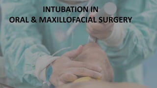
3. Introduction
In oral and maxillofacial surgery, maintainence of airway is an essential
step, while performing under general anesthesia.
Endotracheal intubation provides an artificial medium between the
atmosphere and the patient’s trachea for the purpose of alveolar gas
exchange.
Asok kumar RS OMFS
4. History
Hippocrates (460-380 BC) - Described tracheal intubation.
Vesalius in 1543- Described the rhythmic inflation of the lungs by passing a tube the trachea
of an animal
Manuel Garci- Used blind or tactile techniques to visualize the larynx using indirect
laryngoscopy.
1546 : First well-documented tracheostomy by Antonius Musa Brasavola
William Macewen in 1879 - Performed the first elective oral intubation .
1900- Kuhn introduced metal endotracheal tube and gave the first detailed description of
orotracheal intubation
1913 - Jackson first anaesthetic laryngoscope and modified by Sir Robert Machintosh in
1943 Asok kumar RS OMFS
5. 1921: Chevaliar Jackson – standardized the technique of the tracheostomy .
1960 - Retrograde endotracheal intubation described by Butler and Cirillo.
1963 - Water described passing a plastic tube through cricothyroid membrane and using
it as a guide to intubate patients
1967 - Murphy described Fiberoptic bronchoscope intubation
1969 : Modern percutaneous tracheostomy (PCT) developed by Toye and Weinstein
1970- Preformed orotarcheal tube described by Ring ,Adair And Elwyn (RAE tube)
1986- Submental intubation was first reported by Francisco Hernandez Altemir
1993 – Stoll simultaneously advocated a submandibular route.
Asok kumar RS OMFS
14. Oral and Nasotracheal intubation
Involves passing an endotracheal tube through nose or
mouth or surgically made tract (Eg: Submental) into the
trachea.
Provides a patent airway
Method of choice in emergency care.
Provides an airway for patients who cannot maintain an
adequate airway on their own and needs mechanical
ventilation.
Asok kumar RS OMFS
15. Technique
Place the patient in a “sniffing” position
Place a folded towel or bath blanket under the head
Administer topical anaesthesia
Preoxygenate with 100% oxygen for 3 minutes
Average male size, 8.0-9.0 mm
Average female size, 7.0-8.0 mm Asok kumar RS OMFS
16. ETT is lubricated
Elevate the epiglottis anteriorly to expose the vocal cords
using laryngoscope
Visualize the vocal cords
ETT is inserted 5 to 6 cm beyond the vocal cords, and the
cuff is inflated
After proper tube placement, tube is secured to prevent
dislodgement
Asok kumar RS OMFS
17. Indications
1. Maxillofacial trauma
2. Securing the airway in questionable cervical spine or
severe degenerative cervical spine disease
3. Surgical removal of pathological lesions
4. Correction of structural and congenital abnormalities.
Asok kumar RS OMFS
18. Absolute contraindications
1. Suspected inflammation of the epiglottis
2. Communited midface fracture
3. Coagulopathy
4. Suspected fractures in base of the skull
Relative contraindications
1. Large nasal polyps
2. Suspected nasal foreign bodies
3. Recent nasal surgery
4. Upper respiratory tract infection
5. Epistaxis
6. Prosthetic heart valve (increased risk of bacteremia during the intubation)
Asok kumar RS OMFS
20. Submental intubation
• Submental intubation was first reported by Francisco
Hernandez Altemir in 1986
• Simple, safe and convenient technique in maxillofacial
trauma, where oral and nasal endotracheal intubation cannot
be performed.
• Alternative to tracheostomy for the concomitant restoration
of occlusion and reduction of facial fractures
Asok kumar RS OMFS
21. Technique
1.5 cm incision was made in the median region
of the submental area adjacent to the lower
border of the mandible.
Orotracheal intubation done
The muscular layers (platysma and mylohyoid
muscles) were traversed by blunt dissection
using a Kelly forceps
Asok kumar RS OMFS
22. 2 cm incision parallel to the mandible made
over the distal end of the forceps over lingual
mucosa on the floor of the mouth
The forceps were then opened and a tunnel is
created.
Width of the submental access should be
adequate for the passage of the tube without
any interference
Asok kumar RS OMFS
23. The tube is secured with stay sutures and
connected to boyle’s apparatus
Make sure that the tube has not been
displaced during its passage through the
submental tunnel
Asok kumar RS OMFS
24. Submandibular intubation
Invented by a spanish maxillofacial surgeon,
Francisco Hernandez Altemir
Modification of submental intubation given by Stoll
et al.(1993) simultaneously advocated a
submandibular route.
Alternative to tracheostomy and thus it keeps the
endotracheal tube away from the field of surgery.
The technique of both these approaches are similar.
Asok kumar RS OMFS
25. Technique
Blunt dissection performed through the platysma, the deep
cervical fascia and mylohyoid muscle;
Tunnel created in close proximity to the lingual cortex of the
mandible
The pilot balloon is pulled out first, then the proximal end of
the orotracheal tube is grasped, exteriorized and secured to
skin
1.5 cm incision is made through the skin in the right or left
anterior submandibular region parallel to the inferior border
of the mandible
Asok kumar RS OMFS
26. Indications
• Panfacial fracture
• Fractured base of the skull with a possible fracture of the cribriform
plate.
• Midface fractures (Le fort II or III) associated with skull base
fractures.
• Mandible fracture
• Naso-ethmoidal bone fracture
Asok kumar RS OMFS
27. Advantage
Cost-effective
Fewer complications
Shorter duration of hospital stay
Reduces the need for a high dependency unit caring for the tracheostomy
tube.
Scar left by the S-MEN/ S-MAN incision is much less obvious
Asok kumar RS OMFS
28. Disadvantage
Hypertrophic scar formation
Accidental extubation
Hemorrhage
Infection
Mucocele formation
Damage to salivary gland ducts in floor of mouth
Asok kumar RS OMFS
29. Retromolar intubation
Non-invasive technique of securing airway in
patients with panfacial trauma, fracture of base of
skull, fracture of naso-orbital-ethmoid complex, etc
Alternative to submental intubation and
tracheostomy
Asok kumar RS OMFS
30. Techniques
Orotracheal intubation is done initially with a
flexometallic tracheal tube using standard
general anaesthesia technique
The orotracheal tube is grasped and placed
into the retromolar space (space behind the
last upper and lower erupted molar teeth)
Asok kumar RS OMFS
31. or
The tube is then fixed by a ligature wire to the
molar/premolar tooth figure of eight” fashion.
The tube is fixed by suturing it to buccal
mucosa
Asok kumar RS OMFS
32. Advantages
Cost effective
Non invasive
Alternative to tracheostomy
Disadvantages
Intraoperative IMF cannot be done in patients with reduced retromolar space.
Interfere in the main surgical field.
Interfere with fixation in case of bilateral maxillary/ mandibular fractures.
Asok kumar RS OMFS
33. Fiber optic intubation
One of the most important advances in airway management to
occur in the past thirty years.
Plays a crucial role in the management of the anticipated
difficult airway.
Procedure includes the placement of an endotracheal tube
while the patient is awake or sedated or anaesthetized
Asok kumar RS OMFS
35. Oral or nasal route is preferred
Jaw thrust maneuver done to improve visualization
Broncoscope is lubricated and loaded with
endotracheal tube
Scope is introduced strictly in the midline following the base of the
tongue, pass the uvula posterior to epiglottis and between the vocal
cords.
Once after the carina is visualized endotracheal
tube is introduced. Asok kumar RS OMFS
36. Fiberoptic intubation
Indications:
1. Predicted difficult intubation (upper airway abnormality)
2. Cases where neck extension is not desirable / not possible.
3. Limited mouth opening
Contraindications:
1. Bleeding
Asok kumar RS OMFS
37. Retrograde intubation
Lignocaine 2% nebulisation for 10 min
Gargle with 10 ml of viscous 4% lignocaine
Neck preparation with 10% povidone iodine, the
cricothyroid puncture site was infiltrated with lignocaine 2%,
0.5 ml
Bilateral superior laryngeal nerve block Asok kumar RS OMFS
38. Initial percutaneous puncture made through the
cricothyroid membrane at an 30 to 40 degrees angle to the
skin in a cephalad direction
The J-tip of the wire passed up the trachea until retrieved
from the nose or mouth
The catheter sheath at the skin removed and the wire is
clamped.
The guiding catheter advanced anterograde over the wire,
into the trachea till the cricothyroid access site
Asok kumar RS OMFS
39. Guiding catheter removed, as the endotracheal tube further
advanced into final position
The flexometallic endotracheal tube then passed over
guiding catheter into position below the level of the vocal
cords
Asok kumar RS OMFS
40. Indication:
Blood and secretions in the airway
Limited mouth opening
Ankylosing spondylitis
TMJ ankylosis
Airway tumors
Failed direct laryngoscopic intubation
Advantage :
Less invasive
Shorter procedural duration
Lower risk of subglottic odema and stenosis
Asok kumar RS OMFS
42. Tracheostomy
Surgical procedure involves the creation of a stoma
at the skin surface which leads into the trachea
One of the oldest surgical procedures that involve
placement of an artificial airway in the subglottic
region to bypass the airway in the patient with upper
airway obstruction.
Asok kumar RS OMFS
43. Indications
Maxillofacial trauma ( pan facial fractures, NOE fracture, midface fracture, gun shoot injuries
etc)
Orofacial neoplasm (tumors of the tongue base, tonsils, and oral and pharyngeal regions)
Infection (ludwig angina., Cervicofacial cellulitis)
Edema of the oropharynx and glottis.
Patients with respiratory disorder requiring constant suction of airway secretions.
Failed nasotracheal and orotracheal intubation
Asok kumar RS OMFS
44. Supine position with a pillow under the shoulders to extend
the neck
After draping and the thyroid cartilage, cricoid cartilage,
and sternal notch are marked
Infiltration of local anesthetic and a vasoconstrictor
Horizontal skin incision is made halfway between the marks
on the cricoid cartilage and sternal notch .
Surgical Technique
Subcutaneous tissue dissection done vertically in the
midline. Asok kumar RS OMFS
45. Retractors are placed superiorly, inferiorly, and laterally to
expose the sternohyoid and sternothyroid muscle .
Dissect and retract the muscles
A cross-shaped incision is made in the anterior tracheal wall
with a no. 11 scalpel blade
The 4 tracheal flaps of the cross-shaped incision are
removed with a conchotome to create a large stoma
Adequate-sized tracheostomy tube is placed in the airway
under the control of an aspiration probe Asok kumar RS OMFS
47. References
Intubation Techniques: Preferences of Maxillofacial Trauma Surgeons. Mehul R. Jaisani , Leeza Pradhan,
Balkrishna Bhattarai .J. Maxillofac. Oral surg. (Apr–june 2015) 14(2):501–505
Submandibular intubation.Jafar H Faraj, M Al Khalil Qatar medical journal vol. 2012 / no. 2 / 2012
Miller’s Anesthesia, 7th Edition by Ronald D. Miller.
Retrograde intubation: an old–new technique. D Vieira, N Lages et al., OA anaesthetics 2013 nov 01;1(2):18.
Hall CEH, Shutt LE (2003) Nasotracheal intubation for head and neck surgery. J anesth 58:249–256
A brief history of tracheostomy and tracheal intubation, from the Bronze Age to the Space Age.Petr Szmuk.
Intensive care med (2008) 34:222–228.
Tracheostomy in Maxillofacial Surgery: A Simple and Safe Technique for Residents in Training. Attilio Carlo
Salgarelli, MD DDS, Marco Collini, MD DDS et al. J Craniofac Surg 2011;22: 243-246
Changing Indications for Tracheostomy in Maxillofacial Trauma Shlomo Taicher DMD, Navot Givol DMD et al. J
Oral Maxillofac Surg. 54:292-295, 1996
Asok kumar RS OMFS