
Recommended
More Related Content
What's hot
What's hot (20)
Similar to Iridodialysis repair
Similar to Iridodialysis repair (20)
Recently uploaded
Recently uploaded (20)
Iridodialysis repair
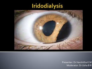
- 1. Presenter: Dr Harshitha K M Moderator: Dr Usha B R
- 2. Iridodialysis is a separation of the iris from its attachment to the ciliary body Also known as coreodialysis. The iris root is one of the weakest parts of iris that could be easily impaired. Most common cause-Trauma- either blunt or penetrating trauma Iatrogenic (during cataract surgery)
- 3. Asymptomatic if there is a small superior iridodialysis- covered by the upper lid Temporal and inferio temporal, inferior iridodialysis are symptomatic. Symptoms- Glare, photophobia, monocular diplopia. Signs- ‘D shaped pupil’
- 4. Iris incarceration- Paton Mc Cannel- Mc cannel’s suture. Double armed suture/ re attachment of iris to sclera. Sewing machine technique Modified SMT Single suture customised loop technique Cobbler’s technique Knotless technique Riveting Technique
- 5. The McCannel technique is usually done for repairing iris lacerations. A limbal paracentesis is made over the iris discontinuity. Then a long Drews needle with 10-0 polypropylene is passed through the peripheral cornea, the edges of the iris, and the peripheral cornea opposite, and the suture is cut.
- 6. A Sinskey hook, introduced through the paracentesis and around the suture peripherally, is drawn back out through the paracentesis. The suture is securely tied. After the suture is secure, it is cut, and the iris is allowed to retract. This technique is used for iridodialysis repair also
- 8. Also known as Hang back technique. It is the principal of repairing peripheral iris defect using horizontal mattress suture. A double-armed suture is employed. The first bite is taken at 1/3rd and 2/3rd junction of the iris defect. Second bite- 1½ clock hours apart. The mattress suture brings the iris back to its origin, if possible ,or closes a peripheral defect using available adjacent iris tissue.
- 9. The knot is tied with just enough tension so the iris is replaced in its anatomical position and does extend further beyond.. The knot is tied externally but then rotated below the surface so that only a smooth loop of external suture remains. By using this technique of suture rotation and burying the knot, identical to the concept used in transscleral suturing of secondary posterior chamber (PC) IOLs ,only a smooth loop of suture material remains.
- 11. A scleral flap does not need to be dissected, and conjunctiva alone provides adequate coverage of the suture material. Alternatively, if it is desired that both suture material and knot lie below the scleral surface, then creating a scleral groove with a beaver blade before placement of the sutures can be helpful.
- 12. This allows the suture material to lie in a trench beneath the scleral surface when the knot is tied. The knot can similarly be rotated into the sclera. A large iridodialysis will require several adjacent horizontal mattress sutures. The size of each suture “bite” of iris should be about 1½ clock hours. This technique provides an excellent functional and cosmetic result while minimizing occlusion of the trabecular meshwork.
- 13. Dr K V Ravi Kumar in 2013 Retrograde threading of 10/0 prolene suture into 26G needle. Creation of partial thickness scleral tunnel parallel to and all along the iris dialysis (1.5 to 2 mm away from the limbus). Pupil constriction by 0.5% pilocarpine injection through a side port made with 20G MVR blade 180° degree opposite to iris dialysis.
- 14. Create suture loops all along the dialysis by passing prethreaded 26G needle with the suture through the root of iris dialysis and scleral tunnel from inside out through a paracentesis on the opposite side. Cut the loops of prolene and tying the adjacent free ends to each other so that the knots get buried into scleral tunnel. Finally, closure of conjunctiva using 10/o nylon or vicryl or using bipolar cautery .
- 16. The SMT was further modified using 30G 25-mm long dental needle instead of 26G needle. The dental needle with scalpel tip design has better penetration with minimal trauma to tissue and suture can be threaded from both sides of the hub.
- 17. After creating suture loops as described earlier, first free end of the suture is passed through the loops and tied with the second free end of suture so that only one knot is sufficient for entire iris dialysis repair.
- 18. Modified SMT a 26-gauge needle.
- 19. Under topical anaesthesia, a limbal conjunctival peritomy is performed at the site of the iridodialysis. A paracentesis is created on the opposite side of the cornea. One end of a double-armed 10-0 prolene suture on a straight needle is introduced into the anterior chamber via the paracentesis. The needle is then driven through the iris base. Penetration through the iris tissue and externalization of the needle are facilitated by introducing a bent 27-gauge needle through the sclera 1.5 mm posterior to the limbus.
- 21. Then the second arm of the prolene suture is introduced into the anterior chamber via the same paracentesis and passed through a second point of the iris base again using the 27-gauge needle. The second arm of the prolene suture is introduced into the anterior chamber through the sclera and the iris base and then retrieved through the paracentesis. The above 2 steps are repeated as necessary to achieve complete apposition of the iris base. At the end, the prolene suture is secured in the sclera using a zigzag-shaped intrascleral suture (Z-suture). Five passes are needed, and each pass starts directly adjacent to the exiting site, then the suture is cut without any knot.
- 22. Creation of partial thickness triangular scleral flap centered over the dialysis area using a crescent knife Introduction of the first straight needle of a double-armed polypropylene suture through the cornea and then through one end of the dialysis, and then using a 27- gauge needle introduced below the scleral flap 1.0 mm posterior to the limbus as a guide track Introduction of the second polypropylene suture straight needle through the cornea facing the other end of the huge dialysis Withdrawing the two threads from the AC through the main phaco wound using a Sinskey hook, forming two loops arising from the main wound
- 24. Two loops are cut and the cut ends are tied together; The two ends of the polypropylene loop are pulled from beneath the scleral flap to reposit the iris back to its root; One of the two suture ends is pulled to extrude the knot from the ac; Pull on the other suture in the opposite direction to correct any wrinkles in the iris periphery that might have been caused by the first pull; Tie the free ends of the sutures below the scleral flap; and Suture the scleral flap with single nylon 10-0 suture at the apex.
- 25. Iridodialysis repair is performed using double- flanged polypropylene suture. This method was first described by Drs. Mami Kuasaka and Masayuki Akimoto from Japan. A scleral grove is created along the length of the iridodialysis. A flange is created on one side of a 6-0 prolene suture.
- 26. The bulbs are flattenned and a 30-gauge needle is used to pull the suture through the anterior chamber and iridodialysis. Additional flanges are made on the remaining end of the suture to prevent slippage.
- 27. Thank you!