Poster - determining the effects of tau on synaptic density in a mouse model ...
Veritas Silver
1. Introduction Conclusion
Figure 1: Piriform Cortex Cellular ComparisonAbstract
Kainic Acid is commonly used to induce recurrent seizures
in animal model experimentation. The consequence is
selective limbic system damage and neuron degradation.
Kainic acid is a non-NMDA receptor agonist, and is 30-fold
more toxically potent than glutamate. (Zhang and Zhu)
Glutamate excitotoxicity is triggered by an excessive influx of
extracellular calcium, occurring after overstimulation of a
glutamate receptor. Excitotoxicity from recurrent glutamate
overstimulation is behind the neuronal pathology of diseases
such as Alzheimer’s disease (AD) and Parkinson’s disease
(PD). Kainic Acid (KA) acts upon ionotropic (fast) glutamate
receptors, which initiates postsynaptic potentials directly to
ion channels. Kainic acid will bind, with greater affinity than
glutamate, to Gluk4 and Gluk5. These genes are synonymous
with kainate, a glutamate receptor subtype family.
Kainic Acid’s successful binding to these receptors is
known to cause brain damage and extensive central nervous
system (CNS) pathology. Necrosis or cell death can be
observed after status epilepticus is initiated post KA injection
intra-peritoneally (IP). Neuro pathological assessment of
brain regions effected will be wide spread through limbic
affiliated structures such as Piriform/Entorhinal Cortex (EC),
Hippocampal fields (CA1, CA2, CA3), and Dentate Gyrus
which receives neuronal input from the EC and projects to
CA3.
A Special Thank You to the Family of
FRANK P. PALOPOLI
(Inventor of the fertility drug, Clomid)
Thanks for the support!
Gregory Wright• Julia Paulus • JaQuay Wheatley. • David Zuzga Ph. D. • Gerald Ballough Ph.D.
Consequential Cerebral Insult Via Kainic Acid Exposure:
Assessing Pathological Variation in Glutamate Excitotoxic Models
Department of Biology, La Salle University, Philadelphia, Pennsylvania
Results
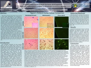
Figure 1: Each picture is recorded from 4ųm coronal sections of non-treatment control, or kainic acid experimental rattus
norvegicus cerebrum. Slides A, B, and C show control piriform cortex at low magnification in three different stains, Hematoxylin
and Eosin (H&E) (A), Bielschowsky Silver (B), and Fluoro-Jade B (C). A and B show the left piriform cortex where as C displays the
right. Although the Hematoxylin and Eosin (H&E) staining in figure A displays darkly stained neurons(blue arrows), these are only
“Dark Neuron” artifacts consequential from some H&E methods. Picture D contains the typical healthy neurons of control
piriform cortex at high magnification. The high mag. control pictures (D, E, and F) display neurons from controls’ in each of the
three different stains. In these higher magnifications of the controls, the Bielschowsky silver nitrate staining method (E) better
distinguishes nuclei than H&E (D). The cerebral sections, from kainic acid exposed rats, in the second row, display the
spongiform cellular morphology (G, H), and positive Fluoro-Jade B (I) indicative of neuronal degeneration in the experimental rat
cerebrum. These features are highlighted in the high magnification in the third row (J,K, and L). The picture in (C) demonstrates
the expected lack of positive fluorescence in the piriform, below it, (I) is a piriform from a rat exposed to IP injection of kainic
acid. (L) shows the highly magnified positive neurons which express degeneration via fluorescence, as well as degenerative
neuron swelling/shrinkage. Eosinophilic “red dead” neurons can be observed in slide J (arrows), where the neurons’ cytoplasm
stains densely with eosin and the nucleus condenses to demonstrate pyknosis and thus cellular injury. Nuclear condensation or
pyknosis also occurs in slide K (arrows), where two neurons have cause extracellular vacuolation due to excessive
swelling/shrinking. Staining; H&E (A, D, G, J), Bielschowsky (B, E, H, K), Fluoro-jade B (C, F, I, L) Final Mag.; 100x( C, I), 400x (A, B,
G, H), 600x (F, L), 1000x (D, E, J, K)
A
ED
LKJ
IHG
CB
F
Hematoxylin & Eosin Bielschowsky’s Fluoro-Jade B
CONTROLKAINICACIDKAINICACID
Neuro pathological assessments can prove extremely
difficult, but there are multiple ways to assess neuron
pathology using tissue staining and light microscopy. The
damage present in the CNS after kainic acid injection, should
overlay most of the limbic system brain regions. In this
study, the piriform cortex is chosen to be the limbic region of
assessment because of its connections with other major
limbic regions. H&E neuron assessment can show
consequential damages to cellular nuclei and surrounding
matrix. Pyknotic (chromatin condensation), karyorrhectic
(nuclear fragmentation), and brightly stained eosinophilic
cytoplasm all are irrefutable signs of cellular damage and
pending cellular death. Although the mechanism is yet to be
defined, fluoro-jade B positive cellular fluorescence is
evidence of degeneration and is accepted as a marker for cell
death. Bielschowsky’s silver is specific for neuronal fibers,
axons and neurofibrillary tangles; which will show detailed
cytoarchitecture compared to the most common method of
staining, H&E. By viewing a control piriform cortex along
with a cerebrum exposed to the excitotoxic effects of
kainate, there should be a clear culmination of cellular injury
due to the widespread necrotic cell death affiliated with
glutamate excitotoxic seizures.
Nuclear Pyknosis can be seen in picture K, under high
magnification of Bielschowsky staining. Also high
magnification H&E displays eosinophilic “red dead”
cytoplasm, indicative of neuronal degeneration.
Excessive swelling and shrinking of neurons, post
processing, can be seen in pictures G, H, I, K, and L are
often closely associated with neuronal damage. Positive
Fluoro-Jade B fluorescence is present throughout
piriform (I), where brightly stained eosinophilic cells are
present as well (G, J). The lower magnification pictures
show damage is present throughout piriform toward
perirhinal cortex (G, H, I). The non-treated control rat
displays normal piriform histology, without fluoro-jade
fluorescence or excessive vacuolation.
Definitive Neuronal Degeneration has been
uncovered from the Kainic Acid Injections. The Fluoro-
jade B method of staining is highlighting neurons in
areas of significant pathology shown by the congruent
H&E and Bielschowsky stains with sufficient cytoplasmic
eosinophilia and pyknosis. Not only has damage been
demonstrated through nuclear changes but we have
observed morphological change (spongiform
appearance) in an entire brain region, the piriform. A
comparative look at the limbic accessory region, the
piriform, proves our lab is capable of accessing
irreversible neuro pathology via IP injection of a known
glutamate excitotoxic molecule, while also identifying
histological neuronal artifacts similar to dead neurons.
These methods, along with the other data collection
from the Veritas team members, should be able to
produce similar results with other neuro toxic
molecules (I.e. ethanol in binge exposure), so that our
lab can further analyze the depth of neuronal insult in
all brain regions, with sound knowledge of “bona fide”
neuronal cell death analysis.
Materials/Methods
Hilltop lab animals, Inc. (Scottsdale, PA) provided the
rats for experimentation. The animals were kept alive
for 8 days prior to deliverance of kainic acid (12mg/kg)
to the experimental group, and were euthanized the
next day after grand mal seizures had occurred. Tissues
were cleared of blood with 10% buffered formalin via
transcardial perfusion. All injections were done IP.
Tissues were embedded with paraffin wax after
processing, and a microtome was used to section
tissues at 4ųm, and all stained sections were taken near
bregma -3.30.