
Recommended
More Related Content
What's hot
What's hot (20)
Similar to Uro gynaecology- anatomy- pelvic floor
Similar to Uro gynaecology- anatomy- pelvic floor (20)
More from GovtRoyapettahHospit
More from GovtRoyapettahHospit (20)
Recently uploaded
Recently uploaded (20)
Uro gynaecology- anatomy- pelvic floor
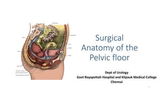
- 1. Surgical Anatomy of the Pelvic floor Dept of Urology Govt Royapettah Hospital and Kilpauk Medical College Chennai 1
- 2. Moderators: Professors: • Prof. Dr. G. Sivasankar, M.S., M.Ch., • Prof. Dr. A. Senthilvel, M.S., M.Ch., Asst Professors: • Dr. J. Sivabalan, M.S., M.Ch., • Dr. R. Bhargavi, M.S., M.Ch., • Dr. S. Raju, M.S., M.Ch., • Dr. K. Muthurathinam, M.S., M.Ch., • Dr. D. Tamilselvan, M.S., M.Ch., • Dr. K. Senthilkumar, M.S., M.Ch. Dept Of Urology, KMC and GRH, Chennai 2
- 3. Pelvic Floor The bony pelvis form a basin whose bottom is covered by what is called as the pelvic floor. Dept Of Urology, KMC and GRH, Chennai 3
- 4. Sciatic Foramens • 2 ligaments and 2 foramens • Sacrospinous ligament: From sacrum to ischial spine • Sacrotuberous ligament: From sacrum to ischial tuberosity • Greater schiatic foramen: Above sacrospinous ligament • Lesser sciatic foramen: Below sacrospinous ligament Dept Of Urology, KMC and GRH, Chennai 4
- 5. Sacrotuberous and Sacrospinous ligaments Dept Of Urology, KMC and GRH, Chennai 5
- 6. Pelvic Outlet The pelvic outlet is diamond-shaped, with the apices defined by bony landmarks— the symphysis pubis anteriorly, the ischial tuberosities laterally, and the tip of the coccyx posteriorly. Dept Of Urology, KMC and GRH, Chennai 6
- 7. Pelvic Outlet • It can be further dissected into: • Urogenital triangle/Anterior triangle defined by the symphysis and two tuberosities and • Anal triangle/posterior triangle defined by the coccyx and two tuberosities Dept Of Urology, KMC and GRH, Chennai 7
- 8. Pelvic Floor • The pelvic floor is a compound structure consisting of • Pelvic floor muscles, • Fascia and ligaments. It is enclosed by the bony scaffolding of the pelvis, formed by 2 innominate bones which articulate with the sacrum posteriorly and each other anteriorly Dept Of Urology, KMC and GRH, Chennai 8
- 9. Pelvic Floor Muscles • Pelvic diaphragm • Piriformis • Obturator Internus Dept Of Urology, KMC and GRH, Chennai 9
- 10. Obturator Internus and Piriformis Dept Of Urology, KMC and GRH, Chennai 10
- 11. Pelvic Diaphragm Dept Of Urology, KMC and GRH, Chennai 11
- 12. Levator Ani Subdivided, from medial to lateral into • Puborectalis, • Pubococcygeus, and • Iliococcygeus. Dept Of Urology, KMC and GRH, Chennai 12
- 13. Puborectalis and Pubococcygeus • Originate from the inner aspect of the pubic rami on either side of the midline at the level of the pubic symphysis. • The muscle fibers pass laterally to the vagina and rectum, creating a U-shaped sling surrounding the genital hiatus medially. Dept Of Urology, KMC and GRH, Chennai 13
- 14. Iliococcygeous • Origin from arcus tendineus levator ani or muscle white line. • The muscles of the iliococcygeous pass laterally to the pubococcygeus muscles, fanning out to create the pelvic floor posteriorly and laterally. • Insertion into the lower aspect of lateral sacrum. Dept Of Urology, KMC and GRH, Chennai 14
- 15. Coccygeus/Ischiococcygeus • The coccygeus is also termed the ischiococcygeus muscle. • It is attached medially to the lateral margins of the coccyx and fifth sacral segment and laterally to the ischial spine. Dept Of Urology, KMC and GRH, Chennai 15
- 16. Levator plate • The levator plate is formed by the midline fusion of the levator ani muscles between the rectoanal junction and the coccyx. • The uterus and cervix, as well as the upper two-thirds of the vagina, are suspended over the levator plate. • Most important source of indirect mechanical support for these organs. Dept Of Urology, KMC and GRH, Chennai 16
- 17. Perineal Body • The perineal body, located under the pelvic floor, is a fibromuscular mass of tissue between vagina and anus in females, bulb and anus is males. • It is formed by the attachments of several muscles. Dept Of Urology, KMC and GRH, Chennai 17
- 18. Perineal Body - Muscles • Superficial transverse perineus • Bulbospongiosus • Sphincter urethrae in the male • Sphincter urethrovaginalis and deep transverse perineus muscles in the female • Superficial part of the external anal sphincter • Levator prostatae and pubovaginalis of the levator ani Dept Of Urology, KMC and GRH, Chennai 18
- 19. Fibre types • Majority type I (slow twitch) muscle fibers that maintain a constant resting tone over time. • Also contains a smaller proportion of type II (fast twitch) fibers, permitting them to respond quickly during sudden increases in intra- abdominal pressures. • Contraction of the pelvic diaphragm closes the genital hiatus and provides a horizontal levator plate on which the pelvic viscera lie. Dept Of Urology, KMC and GRH, Chennai 19
- 20. Pelvic Floor Muscles Dept Of Urology, KMC and GRH, Chennai 20
- 21. Foramen and Hiatus • Obturator foramen • Urogenital hiatus • Rectal Hiatus Dept Of Urology, KMC and GRH, Chennai 21
- 22. Fasciae The pelvic fascia is divided into: • Parietal pelvic fascia, which forms the coverings of the pelvic muscles, and • Visceral pelvic fascia, which forms the coverings of the pelvic organs and their neurovascular supply Dept Of Urology, KMC and GRH, Chennai 22
- 23. Parietal Pelvic Fascia Forms the coverings of the pelvic muscles, • Mechanically dense matrix of connective tissue consisting predominantly of collagen fibers • They coalesce into thick bundles that are then interwoven into a strong, three-dimensional sheet. Dept Of Urology, KMC and GRH, Chennai 23
- 24. Parietal Pelvic Fascia It includes • Obturator fascia, • Fasciae over piriformis, and • Fasica over levator ani (the pelvic diaphragm) and • Presacral fascia. Dept Of Urology, KMC and GRH, Chennai 24
- 25. Arcus Tendineus Levator Ani • Thickened portion of Obturator fascia. • Anterior – Pubic rami, Posterior- Ischial spine. • Origin of iliococcygeus muscle Dept Of Urology, KMC and GRH, Chennai 25
- 26. Arcus Tendineus Fasciae Pelvis • Condensation of obturator fascia and the visceral fascia enveloping the anterior and posterior vagina. • Medial to and runs nearly parallel to the arcus tendineus levator ani. • Anterior- Pubic rami, • Posterior- Joins arcus tendineus levator ani • Provides the lateral anchoring sites for the anterior vaginal wall and posterior vaginal wall. Dept Of Urology, KMC and GRH, Chennai 26
- 27. Presacral Fascia • Part of the parietal layer of pelvic fascia. • Located posterior to the retrorectal space. • Thick and strong, it covers the concave surface of the sacrum. • Multiple veins, several arteries, and nerves reside beneath this fascia. Dept Of Urology, KMC and GRH, Chennai 27
- 28. Visceral fascia/Endopelvic fascia • It is a loose, three-dimensional meshwork of collagen, elastin, and smooth muscle with a richer vascular supply. • Connective tissue that encapsulates the individual organs within the pelvis. • Named according to the organ it covers, such as vesical, rectal, or prostatic. • Where the organ passes through the pelvic floor, the visceral fascial capsule fuses with the adjacent parietal fascia of the floor. Dept Of Urology, KMC and GRH, Chennai 28
- 29. Visceral / Endopelvic fascia Dept Of Urology, KMC and GRH, Chennai 29
- 30. Endopelvic Fascia-Specialisations Dept Of Urology, KMC and GRH, Chennai 30
- 31. Uterosacral Ligament • Fan shaped origin from sacrum S2-S4 narrowing to its smallest width just proximal to the cervix. • This broader attachment disperses the suspensory forces acting on the uterosacral ligament Dept Of Urology, KMC and GRH, Chennai 31
- 32. Rectovaginal septum or Denonvillier’s Fascia • Rectovaginal septum separates the rectum from the posterior vaginal wall. • Extends from one ischial spine to the other. • Attached inferiorly to the perineal body (perineal center) and floor of the pelvis. • Bilaminar; potential space (of Proust) lies between the two laminae. Dept Of Urology, KMC and GRH, Chennai 32
- 33. Cardinal Ligament/Transverse Cervical Ligament • Condensation of the endopelvic fascia. • It is a thickening around the uterine vessels. • Extends from the sidewall of the pelvis laterally to the cervix. Dept Of Urology, KMC and GRH, Chennai 33
- 34. Pubocervical Fascia • Thickened anterior portion of the fibromuscular coat that surrounds the vaginal epithelium. • Extends from underneath the urethra, laterally to the fascial white lines, and posteriorly to the pericervical ring of endopelvic fascia around the cervix. • Forms a horizontal hammock upon which the bladder rests. • Purpose of the intact pubocervical fascia is to prevent cystocele Dept Of Urology, KMC and GRH, Chennai 34
- 35. Pubocervical Fascia Dept Of Urology, KMC and GRH, Chennai 35
- 36. Hammock Hypothesis • Distal portion of the pubocervical fascia supports the urethra and UV junction. • Provides back drop against which urethra is compressed during straining. Dept Of Urology, KMC and GRH, Chennai 36
- 37. Paravaginal Defects Dept Of Urology, KMC and GRH, Chennai 37
- 38. DeLancey Classification • DeLancey introduced the concept of dividing the connective tissue support in the pelvis into three levels. • Levels I, II, and III represent apical, middle, and distal vaginal support, respectively Dept Of Urology, KMC and GRH, Chennai 38
- 39. DeLancey Support • Level I - Cardinal and uterosacral ligaments Dept Of Urology, KMC and GRH, Chennai 39
- 40. DeLancey Support • Level II is the support of the midvagina- • Produced by the lateral attachments of the anterior and posterior endopelvic fascia to the pelvic sidewalls. (ATFP, ATLA) Dept Of Urology, KMC and GRH, Chennai 40
- 41. DeLancey Support • Level III support results from the fusion of these same sheets with the pubic symphysis anteriorly and perineal body posteriorly. (Perineal membrane and Urogenital diaphragm) Dept Of Urology, KMC and GRH, Chennai 41
- 42. Materials Used for Midurethral slings Dept Of Urology, KMC and GRH, Chennai 42
- 43. Ideal Sling Material • Chemically and physically inert, • Sterile, • Noncarcinogenic, • Mechanically strong, • Not physically modified by the body tissue, • Readily available, • Inexpensive, and have Dept Of Urology, KMC and GRH, Chennai 43
- 44. Ideal Sling Material • Once healed the graft should restore normal anatomy and function to the pelvis, • Be equally durable to autologous tissue. • In addition the material should remain long enough for incorporation of the surrounding host tissue. • It should withstand mechanical stress and shrinkage, be pliable and easily manipulated during surgery, causing minimal surrounding reaction. Dept Of Urology, KMC and GRH, Chennai 44
- 45. Types • Natural Biomaterials • Synthetic Biomaterials Dept Of Urology, KMC and GRH, Chennai 45
- 46. Natural Biomaterials • Autologous grafts (tissue harvested from the patient), • Allografts (tissue obtained from a source other than the recipient but from the same species), and • Xenografts (tissue obtained from a species different from the recipient). Dept Of Urology, KMC and GRH, Chennai 46
- 47. Autologous grafts The commonly used materials here are • Rectus fascia and • Tensor fascia lata. Dept Of Urology, KMC and GRH, Chennai 47
- 48. Rectus Fascia • Easily harvested, even in patients with multiple abdominal operations. • Durable and rarely causes urethral erosion. • Disadvantages: longer operative time, higher surgical morbidity, postoperative pain and longer recovery. • Rectus fascia may be scarred and thickened owing to prior operations. Dept Of Urology, KMC and GRH, Chennai 48
- 49. Fascia Lata • Easily obtainable long graft. • Generally unscarred and of uniform thickness. • The length means it is easier to achieve adequate tension on the sling. • As there is no abdominal incision, recovery time is less, and there is no risk for abdominal hernias. • Disadvantage: Longer operative time. Dept Of Urology, KMC and GRH, Chennai 49
- 50. Allograft • Cadaveric human fascia lata, dura or dermis has been used. • It decreases operative time and avoids the possible morbidity associated with a second surgical site. • Risk of infectious disease transmission, in particular Creutzfeldt-Jakob disease and other prion transmission-related illnesses Dept Of Urology, KMC and GRH, Chennai 50
- 51. Xenograft • Porcine dermis and small intestine submucosa has been used. • Acellular graft made up of collagen and elastin fibers which provide a matrix on which new tissue and cells are supported. • Comparatively less immune response. Dept Of Urology, KMC and GRH, Chennai 51
- 52. Synthetic Biomaterials-Advantages • Lack of potential infectious disease transmission • High tensile strength, • Readily available, • Cost-effective, • Do not require harvesting, therefore reduce operative risks. Dept Of Urology, KMC and GRH, Chennai 52
- 53. Synthetic Mesh Types Classified on the basis of pore size and the filamentous nature of the material. • Type I meshes are macroporous (>75mm) and monofilament. • Type II meshes are microporous with pore sizes less than 10 mm. • Type III meshes are macroporous meshes with multifilamentous components. • Type IV meshes are ‘coated’ biomaterials that have submicronic (less than 1 mm) pore size. Dept Of Urology, KMC and GRH, Chennai 53
- 54. Polyethylene Terapthalmate Mesh • First used mesh, not used now. • Long chain polymer of the monomer ethylene, • Classified into several categories based on density and branching. • Merisilene was a famous Type III mesh. • With the introductionof polypropylene mesh, it has been abandoned. Dept Of Urology, KMC and GRH, Chennai 54
- 55. Polypropylene Mesh • It is a thermoplastic polymer which has a variety of applications including food packaging and car components. • When used in a mesh, this material is composed of loosely woven strands of synthetic material. • If pore size is greater than 80 μm supposedly allows the passage of macrophages which may result in improved host tissue ingrowth. • Eg: TVT, Ethicon Dept Of Urology, KMC and GRH, Chennai 55
- 56. Polytetrafluoro ethylene (PTFE) • It is a microporous material. Classified as Type II mesh • Soft and pliable multifilament with fewer adhesions and a less obvious inflammatory response. • Eg: Gore-Tex • Not widely used to problems of erosion and high rejection rates (37.5%) Dept Of Urology, KMC and GRH, Chennai 56
- 57. Thank You Dept Of Urology, KMC and GRH, Chennai 57