
Recommended
Recommended
More Related Content
What's hot
What's hot (20)
Similar to Image guided EBRT and MRI-based BT in cervical cancer
Similar to Image guided EBRT and MRI-based BT in cervical cancer (20)
Recently uploaded
Recently uploaded (20)
Image guided EBRT and MRI-based BT in cervical cancer
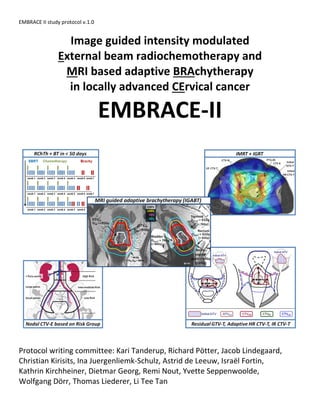
- 1. EMBRACE II study protocol v.1.0 Image guided intensity modulated External beam radiochemotherapy and MRI based adaptive BRAchytherapy in locally advanced CErvical cancer EMBRACE-II Protocol writing committee: Kari Tanderup, Richard Pötter, Jacob Lindegaard, Christian Kirisits, Ina Juergenliemk-Schulz, Astrid de Leeuw, Israël Fortin, Kathrin Kirchheiner, Dietmar Georg, Remi Nout, Yvette Seppenwoolde, Wolfgang Dörr, Thomas Liederer, Li Tee Tan
- 2. CONTENTS 1 2 Contents __________________________________________________________________________________ 1 3 1 Abbreviations___________________________________________________________________________ 6 4 2 Summary ______________________________________________________________________________ 8 5 2.1 Background __________________________________________________________________________________8 6 2.2 Interventions, aims and hypotheses ______________________________________________________________8 7 2.3 Type of design ________________________________________________________________________________9 8 2.4 Patients to be included _________________________________________________________________________9 9 2.5 Treatment of patients in the trial_________________________________________________________________9 10 2.6 Quality assurance ____________________________________________________________________________10 11 2.7 Outcome measures ___________________________________________________________________________10 12 2.8 Evaluation of outcome measures________________________________________________________________10 13 2.9 Sample size and data maturity __________________________________________________________________11 14 3 Introduction ___________________________________________________________________________ 12 15 3.1 Background _________________________________________________________________________________12 16 3.2 Tumor and target CONCEPTS FOR RESPONSE ADAPTED RADIOTHERAPY in cervix cancer: residual GTV-T, 17 adaptive CTV-THR and CTV-TIR_________________________________________________________________________13 18 3.3 Evidence from the retroEMBRACE and EMBRACE studies ____________________________________________17 19 3.3.1 Local control and D90 to CTVHR, GTV and CTVIR___________________________________________________________17 20 3.3.2 Overall treatment time _____________________________________________________________________________19 21 3.3.3 Urinary morbidity and bladder D2cm3 ___________________________________________________________________19 22 3.3.4 Rectal bleeding and rectum D2cm3 _____________________________________________________________________20 23 3.3.5 Bowel morbidity and sigmoid/bowel D2cm3 ______________________________________________________________20 24 3.3.6 Vaginal morbidity and ICRU recto-vaginal dose __________________________________________________________21 25 3.3.7 Gastrointestinal/urinary morbidity and intermediate dose levels related to EBRT _______________________________21 26 3.3.8 Patterns of spread and PROGNOSTIC PARAMETERS for nodal pelvic and para-aortic recurrences___________________22 27 3.3.9 Administration of chemotherapy______________________________________________________________________24 28 3.4 Internal target motion_________________________________________________________________________25 29 4 Interventions and Aims __________________________________________________________________ 26 30 4.1 Interventions ________________________________________________________________________________26 31 4.1.1 increased use of IC/IS technique in BT__________________________________________________________________26 32 4.1.2 Reduction of vaginal source loading ___________________________________________________________________27 33 4.1.3 Systematic Utilisation of IMRT________________________________________________________________________27 34 4.1.4 Utilisation of daily IGRT (set-up according to bony structures) ______________________________________________28 35 4.1.5 EBRT target concept related to the primary tumour (CTV-T) and internal motion; Concepts for OAR contouring_______28 36 4.1.6 EBRT dose prescription and reporting__________________________________________________________________28 37 4.1.7 Adaptation of EBRT nodal elective CTV according to risk of nodal and systemic recurrence _______________________28 38
- 3. 2 4.1.8 Systematic application of simultaneous chemotherapy ____________________________________________________29 39 4.1.9 Reduction of overall treatment time ___________________________________________________________________29 40 4.2 Aims of the EMBRACE II study __________________________________________________________________30 41 4.2.1 General aims______________________________________________________________________________________30 42 4.2.2 Specific aims______________________________________________________________________________________30 43 5 Study design, endpoints and hypotheses ____________________________________________________ 31 44 5.1 Study design_________________________________________________________________________________31 45 5.2 Estimate of patient accrual and study period ______________________________________________________31 46 5.3 Hypotheses and endpoints _____________________________________________________________________31 47 5.3.1 General hypothesis on overall survival _________________________________________________________________32 48 5.3.2 Specific hypotheses on technique, dose and volumes:_____________________________________________________32 49 5.3.3 Specific hypotheses on clinical endpoints _______________________________________________________________33 50 6 EMBRACE Outline_______________________________________________________________________ 37 51 7 Staging and patient work-up______________________________________________________________ 38 52 8 Patient selection________________________________________________________________________ 39 53 8.1 Inclusion criteria _____________________________________________________________________________39 54 8.2 Exclusion criteria _____________________________________________________________________________39 55 9 External Beam Radiotherapy______________________________________________________________ 40 56 9.1 Introduction_________________________________________________________________________________40 57 9.1.1 Aims of external Beam Radiotherapy (compare ch 3-5) ____________________________________________________40 58 9.1.2 Nodal targets based on risk group allocation for nodal spread ______________________________________________40 59 9.2 Preparations for treatment planning _____________________________________________________________42 60 9.3 Tumor and Target definition and contouring: Initial GTV, initial HR CTV-t, initial LR CTV-T, ITV-T; GTV-N, CTV-N, 61 CTV-E; PTV________________________________________________________________________________________44 62 9.3.1 GENERAL Overview ________________________________________________________________________________44 63 9.3.2 Initial GTV and CTV related to primary tumour (GTV-Tinit, CTV-Tinit (HR, LR)) ____________________________________46 64 9.3.3 GTV and CTV for pathologic lymph nodes (GTV-N, CTV-N) __________________________________________________50 65 9.3.4 CTV for nodal regions with assumed microscopic disease (CTV-E)____________________________________________50 66 9.3.5 ITV (ITV-T)________________________________________________________________________________________51 67 9.3.6 Strategies to derive the ITV-T LR ______________________________________________________________________51 68 9.3.7 Generating the ITV45 _______________________________________________________________________________52 69 9.3.8 PTV _____________________________________________________________________________________________52 70 9.4 Contouring of organs at risk, reference points _____________________________________________________54 71 9.5 Contouring of Tumour, Targets and OARs based on MRI and CT _______________________________________54 72 9.6 Dose and fractionation for PTV45 _______________________________________________________________55 73 9.7 Dose and fractionation for PTV-N (nodal boosting) _________________________________________________55 74 9.8 Technique and procedures for EBRT including daily image guidance ___________________________________56 75 9.8.1 Angulation of the pelvis in relation to the lumbar spine____________________________________________________57 76
- 4. 3 9.9 Planning aims for targets and organs at risk _______________________________________________________57 77 9.10 Reporting of EBRT parameters ________________________________________________________________58 78 10 BRACHYTHERAPY _____________________________________________________________________ 59 79 10.1 Introduction and specific aims for brachytherapy_________________________________________________59 80 10.1.1 Overall Schedule for EBRT and BT and chemotherapy _____________________________________________________59 81 10.1.2 Pre-application treatment planning____________________________________________________________________60 82 10.2 Applicator insertion for brachytherapy _________________________________________________________60 83 10.3 Imaging for brachytherapy ___________________________________________________________________61 84 10.4 Applicator reconstruction and dose points for OARS ______________________________________________62 85 10.5 Contouring for brachytherapy: OARS, GTVres, adaptive CTVHR, CTVIR __________________________________64 86 10.5.1 Contouring of organs at risk__________________________________________________________________________64 87 10.5.2 Contouring of target volumes ________________________________________________________________________65 88 10.6 Treatment planning for brachytherapy _________________________________________________________66 89 10.6.1 Evidence obtained from retroEMBRACE and EMBRACE I ___________________________________________________66 90 10.6.2 Planning aims and dose prescription for EMBRACE II ______________________________________________________67 91 10.6.3 Dose optimisation for brachytherapy __________________________________________________________________67 92 10.6.4 Intracavitary treatment plans should be based on iterative steps ____________________________________________68 93 10.6.5 Vaginal dose de-escalation___________________________________________________________________________68 94 10.6.6 PTV and dose conformality considerations in relation to brachytherapy_______________________________________69 95 10.7 Dose and volume recording and reporting_______________________________________________________70 96 11 Systemic treatment____________________________________________________________________ 71 97 11.1 Aims for chemotherapy______________________________________________________________________71 98 11.2 Concomitant chemotherapy __________________________________________________________________71 99 11.3 Adjuvant chemotherapy _____________________________________________________________________71 100 12 Outcome assessment __________________________________________________________________ 73 101 12.1 Oncological outcome and salvage treatment ____________________________________________________73 102 12.2 Morbidity _________________________________________________________________________________74 103 12.3 Quality of Life (QoL)_________________________________________________________________________75 104 13 Translational research _________________________________________________________________ 76 105 13.1 Prognostic markers _________________________________________________________________________76 106 13.2 Predictive markers for radiotherapy related morbidity ____________________________________________76 107 14 Patient material, benchmarking, validation, evaluation and statistics___________________________ 78 108 14.1 Benchmark of outcome and treatment related parameters_________________________________________78 109 14.2 Validation of dose and volume effects__________________________________________________________78 110 14.3 Exploration and Evaluation of dose and volume effects____________________________________________78 111
- 5. 4 14.4 Identification of prognostic and predictive parameters ____________________________________________78 112 14.5 Statistics __________________________________________________________________________________78 113 15 Accreditation, dummy run, data monitoring, quality assurance and continuous education __________ 80 114 15.1 Commitment letter, compliance questionnaire and process document _______________________________80 115 15.2 Dummy run _______________________________________________________________________________80 116 15.2.1 Training, registration, and submission__________________________________________________________________81 117 15.2.2 Evaluation by study coordinators _____________________________________________________________________81 118 15.3 Data monitoring____________________________________________________________________________81 119 15.4 Continuous education _______________________________________________________________________82 120 16 Patient enrollment procedure ___________________________________________________________ 83 121 17 Case record forms, procedures for data collection, EMBRACE II database ________________________ 84 122 18 Ethical considerations__________________________________________________________________ 85 123 18.1 PATIENT PROTECTION ____________________________________________________________________85 124 18.2 SUBJECT IDENTIFICATION _________________________________________________________________85 125 18.3 INFORMED CONSENT _____________________________________________________________________85 126 18.4 ADVANTAGES AND DISADVANTAGE FOR THE PATIENTS _____________________________________85 127 19 PUBLICATION OF DATA_________________________________________________________________ 86 128 20 Study office, Study coordinators, Study structure, communication ______________________________ 86 129 20.1 Study-office EMBRACE II Vienna (at present: 09/2015): ____________________________________________87 130 20.2 Study Coordination:_________________________________________________________________________87 131 21 EMBRACE RESEARCH GRoup ____________________________________________________________ 88 132 22 AppendiCES __________________________________________________________________________ 89 133 22.1 Appendix 1 Standard Clinical Diagram ________________________________________________________89 134 22.2 Appendix 2 Gyn GEC ESTRO Recommendations I-IV, ICRU 88 _______________________________________91 135 22.3 Appendix 3. Compliance questionnaire _________________________________________________________93 136 22.4 Appendix 4. Clinical cases for contouring________________________________________________________97 137 22.4.1 Cases from Vienna, Utrecht and Aarhus, contouring tables _________________________________________________97 138 22.5 Appendix 5: EBRT contouring atlas (complement to chapter 9) _____________________________________98 139 22.5.1 Introduction ______________________________________________________________________________________98 140 22.5.2 Clinical Target Volumes related to the primary tumor _____________________________________________________98 141 22.5.3 Fixed margin approach______________________________________________________________________________99 142 22.5.4 individualized approach ____________________________________________________________________________103 143 22.5.5 Clinical Target Volumes for nodal metastases and nodal regions____________________________________________105 144 22.5.6 Para-aortic nodes _________________________________________________________________________________109 145 22.5.7 Inguinal nodes ___________________________________________________________________________________109 146
- 6. 5 22.5.8 Planning Target Volumes (PTV) ______________________________________________________________________110 147 22.5.9 nodal boost______________________________________________________________________________________111 148 22.5.10 Primary target contouring summary with diagnostic MRI* ______________________________________________112 149 22.5.11 Primary target contouring summary with an MRI in treating position______________________________________113 150 22.5.12 Contouring of organs at risk_______________________________________________________________________117 151 22.5.13 For para-aortic irradiation in addition _______________________________________________________________119 152 22.6 Appendix 6: Measurement and reporting of SUV ________________________________________________122 153 22.7 Appendix 8: CRFs (ch 16) ____________________________________________________________________122 154 22.8 Appendix 9: Principals and structures of EMBRACE RESEARCH GROUP_______________________________122 155 22.9 Appendix 10: Patient Information ____________________________________________________________124 156 23 References__________________________________________________________________________ 128 157 158
- 7. 6 1 ABBREVIATIONS 159 160 161 18 F-FDG Fluorine 18 - Fluorodeoxyglucose 162 2/3/4D Two/Three/Four-dimensional 163 ACT Addenbrooke’s Contouring Tool 164 ATRAB Applied and Translational Radiobiology (Medical University Vienna) 165 AUC Area Under the Curve 166 BL Baseline 167 BT Brachytherapy 168 CBCT Cone beam computed tomography 169 CHT Chemotherapy 170 COP Coverage Probability 171 CR Complete Remission 172 CRF Case Report Form 173 CRT Conformal Radiotherapy 174 CSS Cancer Specific Survival 175 CT Computed Tomography 176 CTCAE Common Terminology Criteria for Adverse Events 177 CTV Clinical Target Volume 178 CuSO4 Copper sulphate 179 D90 The isodose that includes 90% of the target 180 D100 The isodose that includes 100% of the target 181 D2cm3 Minimum dose in the most exposed 2 cm3 of an OAR 182 DFS Disease Free Survival 183 DNA Deoxyribonucleic acid 184 DVH Dose Volume Histogram 185 EANM European Association of Nuclear Medicine 186 EBRT External Beam Radiotherapy 187 EMBRACE The European and International study on MRI-guided Brachytherapy in locally Advanced 188 Cervical Cancer 189 EORTC European Organisation for Research and Treatment of Cancer 190 EPID Electronic Portal Imaging Device 191 ESTRO European Society for Therapeutic Radiology and Oncology 192 EQD2 Equivalent dose in 2 Gy fractions 193 FIGO Fédération Internationale de Gynécologie et d’ Obstétrique 194 FTE Full Time Equivalent 195 Fx Fraction 196 G (Morbidity) Grade 197 GEC Groupe Européen de Curiethérapie 198 GFR Glomerula Filtration rate 199 GI Gastro-Intestinal 200 GTV Gross Tumor Volume 201 Gy Gray 202 HDR High Dose Rate 203 HPV Human Papilloma Virus 204 HR High Risk 205 IC Intracavitary 206
- 8. 7 ICH International Conference on Harmonisation of Technical Requirements for Registration of 207 Pharmaceuticals for Human Use 208 ICRU International Commission on Radiation Units and Measurements 209 IMRT Intensity Modulated Radiotherapy 210 IGRT Image Guided Radiotherapy 211 IR Intermediate Risk 212 IS Interstitial 213 ITV Internal Target Volume 214 IV Intravenous 215 kV Kilovoltage 216 LACC Locally Advanced Cervical Cancer 217 LN Lymph Nodes 218 LR Low Risk 219 MRI Magnetic Resonance Imaging 220 MVCT Megavoltage Computed Tomography 221 N0/N- Lymph Node Negative 222 N1/N+ Lymph Node Positive 223 OAR Organs at Risk 224 OS Overall Survival 225 OTT Overall Treatment Time 226 PAN Para-Aortic Lymph Nodes 227 PDR Pulsed Dose Rate 228 PET-CT Positron Emission Tomography- Computed Tomography 229 PFS Progression Free Survival 230 PI Principal Investigator 231 PIBS Posterior-Inferior Border of Symphysis 232 PTV Planning Target Volume 233 QoL Quality of Life 234 RT Radiotherapy 235 SD Standard Deviation 236 SIB Simultaneous Integrated Boost 237 SPSS Statistical Package for Social Sciences 238 SUVmax Maximum Standardized Uptake Value 239 TNM Tumor (Lymph)Nodes Metastasis 240 TPS Treatment Planning System 241 TRAK Total Reference Air Kerma 242 uCR Uncomplete Remission 243 US Ultrasound 244 VMAT Volumetric Modulated Arc Therapy 245 WHO World Health Organization 246 247
- 9. 8 2 SUMMARY 248 249 2.1 BACKGROUND 250 The standard treatment of locally advanced cervical cancer is radio-chemotherapy including external beam radiotherapy (EBRT), 251 brachytherapy (BT) and concomitant chemotherapy with weekly Cisplatin. Image Guided Adaptive Brachytherapy (IGABT), with 252 repetitive MRI regarded as gold standard, is increasingly recognized as the new paradigm replacing 2D BT and spreading throughout the 253 world. This spread is at present predominantly in Europe, North America and in many places in Asia. The Gyn GEC ESTRO 254 Recommendations I-IV have been used as the conceptual frame for these developments during the last decade and are now embedded 255 into the new ICRU/GEC ESTRO report 88 which is being published in 2015. 256 Beside increasing mono-institutional clinical experience – also reported in literature – there is increasing clinical evidence and analyses 257 from multi-institutional studies, in particular RetroEMBRACE (n=731) and EMBRACE (n>1350) about dose volume effects and outcome. 258 The mature RetroEMBRACE clinical outcome data and dose volume effect analysis for disease outcome show an improved excellent 259 local and pelvic control and survival and significant dose volume effects for IGABT. Overall treatment time was found to have significant 260 impact on local control, and in addition, volume effects of EBRT were found (IMRT vs. 3D CRT) with impact on morbidity and quality of 261 life. Furthermore, dose effects of chemotherapy (≥5 cycles) were found to have impact on survival in advanced disease. Comprehensive 262 analyses from both large patient cohorts reveal further relevant treatment parameters with major impact on disease outcome, 263 morbidity and quality of life. In the international community the results from the EMBRACE studies are regarded as benchmark for 264 future clinical research in this field. 265 Based on the large success of the RetroEMBRACE and EMBRACE studies, the EMBRACE study and research group decided to continue 266 the clinical research work and to initiate a consecutive EMBRACE II study with interventions derived from the evidence collected within 267 the EMBRACE studies. 268 269 2.2 INTERVENTIONS, AIMS AND HYPOTHESES 270 The EMBRACE II interventions address local, nodal and systemic treatment as well as exposure of organs at risk: 271 272 • Increased use of IC/IS technique in BT 273 • Reduction of vaginal source loading 274 • Systematic utilisation of IMRT 275 • Utilisation of daily IGRT (set-up according to bony structures) 276 • EBRT target concept related to the primary tumour; concepts for OAR contouring 277 • EBRT dose prescription and reporting 278 • Adaptation of EBRT nodal elective CTV according to risk of nodal and systemic recurrence 279 • Systematic application of simultaneous chemotherapy 280 • Reduction of overall treatment time 281 282 The general aims of the EMBRACE II study are: 283 284 To systematically apply IMRT with daily IGRT as well as advanced image guided adaptive BT in a prospective multi-centre 285 setting 286 To systematically implement a dose prescription protocol for IGABT 287 To implement systematic contouring, prescription and reporting for EBRT CTV and OARs. 288 To administer EBRT in different targets which are adapted to the risk of nodal and systemic failure: to improve para-aortic and 289 systemic control in high risk patients and not to decrease lymph node control in low risk and intermediate risk patients 290
- 10. 9 To systematically administer simultaneous chemotherapy to EBRT to reach prescribed dose in as many patients as possible, in 291 particular in high risk patients 292 To benchmark an outstanding high level of local, nodal and systemic control as well as survival with application of advanced 293 EBRT, BT and chemotherapy within limited overall treatment time 294 To benchmark a low incidence of intermediate and major morbidity as well as a high level of quality of life with application of 295 advanced EBRT, BT and chemotherapy 296 297 Beside these general aims, there is a significant number of specific aims which refer to the prospective validation of dose volume 298 parameters from the EMBRACE analyses (e.g. dose escalation for large tumors with increased application of IC/IS techniques), to 299 explore and evaluate dose volume parameters for EBRT and to identify prognostic parameters. 300 301 General and specific hypotheses were formulated for the various interventions (BT, EBRT, chemotherapy) and endpoints (disease, 302 morbidity, quality of life). 303 304 2.3 TYPE OF DESIGN 305 The study is a multicenter prospective interventional study with some areas for observational research (e.g. DVH for IMRT). Reporting 306 on the key patient, tumor, treatment and outcome parameters is mandatory including disease, morbidity and quality of life. Sub-studies 307 as on adaptive IMRT and translational research are optional for cooperation between individual departments. Patient registration and 308 reporting will be performed by the individual investigator via the internet to a central database. 309 310 2.4 PATIENTS TO BE INCLUDED 311 Patients with newly biopsy proven squamous carcinoma, adenocarcinoma or adeno-squamous carcinoma of the uterine cervix, FIGO 312 stage IB, IIA, IIB, IIIA, IIIB and IVA (and nodal status according to TNM) in whom definitive radio-chemotherapy with curative intent is 313 planned are qualified for the study. Treatment has to include IGABT with MRI and IMRT with IGRT and ≥5 cycles of cis-Platin. Patients 314 with para-aortic metastatic nodes (stage IVB) to the level of L2 are also eligible but patients with further dissemination are not (M0). 315 Patient work up and staging includes as a minimum patient characteristics with performance status and blood tests (e.g. haemoglobin, 316 lymphocytes), tumor status (biopsy), gynaecological examination, MRI of the pelvis, abdominal CT or MRI, whole body FDG PET-CT 317 (preferably) or at least chest CT. Further investigations are applied if necessary (e.g. cystoscopy, rectoscopy) or done according to 318 institutional practice (e.g. laparoscopic lymph node assessment). Baseline morbidity scoring and quality of life questionnaire are 319 mandatory. 320 321 2.5 TREATMENT OF PATIENTS IN THE TRIAL 322 All patients will receive both EBRT and concomitant chemotherapy and BT. Summation of EBRT and BT doses will be performed by 323 calculation of a biologically equivalent dose in 2 Gy per fraction (EQD2) using the linear-quadratic model with / = 10 Gy for tumour 324 effects and / = 3 Gy for late normal tissue damage. The repair half time is assumed to be 1.5 hrs. 325 EBRT has to be delivered as IMRT/VMAT with daily cone beam CT (IGRT) in 25 fractions with 1.8 Gy to a total dose of 45 Gy given in 5 326 weeks. Target definition is MRI based (initial GTV) for the CTV-T with an initial HR and LR CTV-T and an ITV-T. CT or MRI based nodal 327 Target (CTV-E) is according to risk of nodal spread “Small Pelvis”, “Large Pelvis” or “Large Pelvis + Para-aortic Region”. Overall CTV/ITV 328 to PTV margin is 5 mm. Involved nodes are boosted preferably based on PET CT with 10-15 Gy and treated as simultaneous integrated 329 boost within 5 weeks (2.2-2.4 Gy per fraction). A range for DVH parameters for the various OARs - contoured according to specific 330
- 11. 10 protocols - is taken into account for treatment planning. The LR CTV-T and the CTV-E will be treated with 45 Gy by use of EBRT (PTV45). 331 Maximal treatment time including both EBRT and BT is 50 days. 332 Brachytherapy is prescribed with dose escalation for advanced disease with large adaptive CTV-THR including IC/IS techniques and dose 333 de-escalation for limited size CTV-THR to spare organs at risk and in particular the upper vagina. The primary imaging method is MRI with 334 the applicator in place which enables definition of the relevant volumes of interest directly on the images for treatment planning: 335 GTVres, adaptive CTVHR, CTVIR and organ volumes. The applicator and the reference points are reconstructed in the same image series. 336 All treatment plans have to be optimized to achieve defined planning aims for dose and volume parameters for tumor (D98 for GTVres) 337 and target volumes (e.g. D90-95 Gy for adaptive CTV-THR) and for 2cm3 reference volumes for OARs (e.g. <80 Gy for bladder, <65 Gy for 338 rectum) and for vaginal reference points (recto-vaginal point < 65 Gy, PIBS). If the planning aims cannot be achieved, limits for the 339 finally prescribed dose levels are defined for GTVres, CTVHR, CTVIR, point A, bladder, rectum, sigmoid bowel and vagina. Planning aim 340 doses and limits for the finally prescribed dose levels are based on the experience of the previous retroEMBRACE and EMBRACE trials. 341 For chemotherapy weekly concomitant Cisplatin (40 mg/m2) for 5-6 courses is standard unless chemotherapy is precluded by patient 342 age, co-morbidity and toxicity. Aim is to apply minimum 5 cycles of cis-Platin, in particular in advanced disease. 343 344 2.6 QUALITY ASSURANCE 345 Only approved departments and investigators can enroll patients into the protocol. This approval is the under the responsibility of the 346 study coordinators. The approved departments are at present those that have contributed continuously to EMBRACE in a considerable 347 number of patients. These departments have to go additionally through a QA procedure for IMRT/IGRT. 348 New departments will have to go through a QA procedure both for IGABT and IMRT/IGRT. Approval requires a compliance 349 questionnaire, successful training, registration and submission of cases and positive evaluation by the study coordinators for each 350 centre. 351 There is no formal on site monitoring, but patient files and treatment plans must be kept at least until closure of the protocol and final 352 analysis of the results is obtained. Continuous data monitoring is performed through the study offices in Vienna and Aarhus and 353 through Utrecht for the centres in the Netherlands. 354 Continuous education will be offered through ACT and annual workshops and EMBRACE meetings. 355 356 2.7 OUTCOME MEASURES 357 Local and nodal (pelvic) control within the specific EBRT and BT targets (HR-CTV-T, IR-CTV, LR CTV-T; CTV-E, CTV-N) and morbidity 358 related to OAR in the pelvis and the para-artic region as well as overall survival, cancer specific survival and systemic control are the 359 primary outcome measures. All endpoints will be evaluated by actuarial statistics. Morbidity will be scored by use of the Common 360 Terminology Criteria for Adverse Events (CTCAE v3.0/4.0). QoL will also be systematically recorded in all patients. 361 362 2.8 EVALUATION OF OUTCOME MEASURES 363 Tumor and nodal remission status (complete, uncertain complete, partial, stable & progressive disease) will be evaluated 3 months 364 after treatment by pelvic (para-aortic, CT) MRI and gynaecological examination. Regular follow-up including gynaecological examination 365 will then be instituted with planned appointments 6, 9, 12, 18, 24, 30, 36, 48 and 60 months after treatment. Pelvic (para-aortic, CT) 366
- 12. 11 MRI will be repeated at 12 months after treatment or in case of suspected recurrence. Morbidity and quality of life will be scored 367 systematically at base line and at each time point during follow-up. 368 369 2.9 SAMPLE SIZE AND DATA MATURITY 370 The study aims at recruiting 1000 patients in 4 years and to follow them for at least 5 years to allow for a meaningful assessment of the 371 endpoints by univariate and multivariate analysis. 372 373 374 375 376
- 13. 12 3 INTRODUCTION 377 378 3.1 BACKGROUND 379 The standard treatment for locally advanced cervical cancer is currently radio-chemotherapy consisting of EBRT, intracavitary BT and 380 concomitant chemotherapy with Cisplatin. During the last decade, the utilisation of MRI guided brachytherapy has grown based on the 381 GEC ESTRO recommendations (Haie-Meder C. et al. 2005, Pötter R. et al. 2006, Hellebust TP. et al. 2010, Dimopoulos JC. et al. 2012) and 382 the cervix is among the first cancer sites where response-adaptive radiotherapy has been successfully implemented in clinical practice. 383 The novel target concepts involved in response-adaptive radiotherapy are described further in section 3.2. Acquisition of MRI at the 384 time of brachytherapy allows the brachytherapy boost to be individually tailored according to the residual tumour volume after 385 typically 40-50 Gy of external beam radiation therapy (EBRT). This approach has changed patterns of clinical practise with regard to 386 dose administration, and significant improvements in clinical outcome have been reported from mono-institutional settings with regard 387 to local control, overall survival and morbidity (Pötter R. et al. 2007, Pötter R. et al. 2011, Lindegaard JC. et al. 2013). 388 In 2008, the GEC-ESTRO Gyn network initiated the “International Study on MRI-Based Brachytherapy in Cervical Cancer” (EMBRACE, 389 www.embracestudy.dk). EMBRACE has recruited >1300 patients by 2015 from 27 international centers performing MRI-guided 390 brachytherapy. The purpose of the EMBRACE study is to evaluate and benchmark MRI-guided brachytherapy in a prospective 391 multicenter study. In 2010, the GEC-ESTRO Gyn network also initiated the retrospective study retroEMBRACE, in which 852 patients 392 treated with image-guided brachytherapy prior to initiation of EMBRACE accrual have been included to provide long-term outcome 393 data for image-guided brachytherapy while the EMBRACE study data is still maturing (www.retroembrace.com). 394 Data from retroEMBRACE shows that overall local control is excellent with 89% at 5 years with 98% in stage IB and 91% in IIB tumours. 395 However, in stage IIIB tumours there is still a significant challenge with regard to local control which is 75% at 5 years (Sturdza A. et al. 396 in submission 2015). Nodal and systemic control also remains challenging with levels of 87% and 77% at 5 years, respectively (all stages) 397 (RetroEMBRACE 01/2015 work in progress). Furthermore, treatment related urinary and gastrointestinal late morbidity is still a 398 significant problem with the 3 year actuarial incidence of intermediate to major morbidity (G≥2) being 30% and 29% for urinary and 399 gastrointestinal side effects, respectively, according to EMBRACE data. Major morbidity (G≥3) is seen in 7% and 8%, respectively 400 (EMBRACE 2014, work in progress). Patient reported symptoms are equally high with 30-40% of patients reporting significant urinary 401 and gastrointestinal bother according to quality of life data from the EMBRACE study (EMBRACE 2015, work in progress). Sexual side 402 effects are still poorly understood although almost 30% of patients develop significant narrowing and shortening of the vagina 403 (Kirchheiner K. et al. 2014). Further development of both BT and EBRT is needed to improve on local control, regional control as well as 404 on treatment related morbidity and quality of Life. 405 Adjuvant and neo-adjuvant chemotherapy has been proposed to improve systemic control, and is currently being evaluated in a 406 randomized phase III study (OUTBACK, https://www.rtog.org/ClinicalTrials/ProtocolTable/StudyDetails.aspx?study=1174; INTERLACE 407 www.cancerresearchuk.org). However, local and nodal disease also has impact on systemic disease, and therefore improvement on 408 loco-regional treatment is equally important. Recent developments in advanced image guidance for both EBRT and BT have potential to 409 improve local as well as nodal and also systemic control. Furthermore, the new technologies has potential to decrease organ doses as 410 well as well as the overall burden of treatment, with the promise to significantly reduce treatment related organ symptoms and overall 411 quality of life. 412 Advances in image guided adaptive brachytherapy include improved individualisation of brachytherapy applicators as well as 413 individualised dose optimisation. Dose optimisation using intracavitary (IC) applicators has shown to significantly decrease OAR dose 414 and morbidity (Charra-Brunaud C. et al. 2012). Dose optimisation based on IC may be used to improve target dose coverage in tumours 415 of limited size at BT, but for large residual tumours or in case of unfavourable topography, IC BT has limited possibilities to cover the 416 CTVHR to doses larger than e.g. 85Gy (Tanderup K. et al. 2010). Combined intracavitary-interstitial (IC/IS) applicators have been 417 developed for targeting tumours which are not well covered by intracavitary (IC) applicators (Dimopoulos JC. et al. 2006, Kirisits C. et al. 418 2006). The IC/IS applicators allow for improved dose conformality, and target dose escalation and/or dose de-escalation in organs at 419
- 14. 13 risk can be carried out (Fokdal L. et al. 2013). Furthermore, in the process of moving from standard loading to 3D image guided 420 optimisation there has so far been reluctance to change the loading drastically in ovoids and ring, in order to stay as close as possible to 421 previous clinical practise. However, EMBRACE data have demonstrated that dose to the ICRU recto-vaginal point correlate with the 422 probability of G≥2 vaginal morbidity (Kirchheiner K. et al. in submission 2015). This observation is a strong motivation to explore new 423 approaches to dose optimisation which spare vaginal mucosa and decreases the dose to the ICRU recto-vaginal point. 424 Pelvic EBRT is currently delivered with different techniques: 3D conformal EBRT, intensity modulated radiotherapy (IMRT), volumetric 425 arc techniques (VMAT), and tomotherapy. Application of IMRT in cervix cancer significantly reduces the volume of tissue irradiated to 426 intermediate doses such as 30-40Gy for bladder, rectum, sigmoid and bowel (Forrest J. et al. 2012). The progress from 3D conformal 427 EBRT to IMRT has demonstrated a reduction of treatment related morbidity in mono-institutional and retrospective settings (Mundt AJ. 428 et al. 2003, Xu KM. et al. 2015). Furthermore, EMBRACE quality of life data has shown a significantly lower incidence of bowel 429 symptoms in patients treated with IMRT as compared to 3D conformal EBRT with the four-field box technique (see Figure 3.6 and 3.7). 430 During the last decade, a variety of techniques, such as kV x-ray, cone beam CT (CBCT) or megavolt CT (MVCT), have been developed to 431 improve the possibilities to perform on board image guidance in EBRT. With imaging devices mounted on or in a fixed relationship to 432 the accelerator, it is now possible to perform daily imaging with the patient in the treatment position. The on-board images can be 433 fused with the treatment planning scan and a couch correction can be applied to correct for translational setup errors. In the case of 434 cervix cancer the daily imaging can be used for visualisation and fusion of bony anatomy. By using daily image guided set-up in cervix 435 cancer, the precision of the elective lymph node clinical target volume (CTV-E) can be significantly improved (Laursen LV. et al. 2012), 436 and thereby planning target volume (PTV45 Gy) margins can be reduced. A further step is to use daily image guidance (CBCT) to 437 visualise soft tissue such as bladder and uterus in order to further reduce the PTV-T margins which are applied to take into account the 438 motion of the primary gross tumour volume (GTV), the CTV-T and the uterus (see chapter 9). Such approaches have been developed 439 and involve adaptive EBRT where daily library plans are applied (Heijkoop ST. et al. 2014). Decrease of PTV margins as well as 440 implementation of IMRT has potential to reduce morbidity, in particular bowel morbidity. 441 The primary aim of EMBRACE II is to implement a risk adaptive dose prescription protocol in locally advanced cervical cancer. The 442 individualised dose prescription is based on evidence of dose and effect relationships for target and OARs from the EMBRACE and 443 retroEMBRACE studies and involves a set of new dose planning aims. The ability to reach these dose planning aims is based on 444 interventions in terms of advanced BT and EBRT technology. Advanced BT involves increased utilisation of IC/IS applicators as well as 445 vaginal dose de-escalation. Advanced EBRT involves IMRT as well as daily image guidance utilising margin reduction. This approach will 446 enable delivery of increased focal doses to gross disease (primary tumour and positive lymph nodes) as well as reduction of high and 447 intermediate dose to OARs. The improved dose administration is hypothesised to benchmark an outstanding high level of local, nodal 448 control, and systemic control as well as a low incidence of intermediate and major morbidity. Through this well-controlled prospective 449 interventional study we aim to achieve the composite aims listed in section 4.2. 450 3.2 TUMOR AND TARGET CONCEPTS FOR RESPONSE ADAPTED RADIOTHERAPY IN CERVIX CANCER: 451 RESIDUAL GTV-T, ADAPTIVE CTV-THR AND CTV-TIR 452 The target concept for response-adapted radiotherapy is focussed on the primary tumour change (GTV-T) and the change of the CTV-T 453 during upfront chemo-radiation. These changes are essential for selecting the appropriate target for brachytherapy (see chapter 5.4, 454 ICRU report 88). Therefore new terms and concepts have been introduced as compared to ICRU 50, 62 and 83 which correspond to 455 those of the Gyn GEC ESTRO Recommendations I and II (Haie-Meder C. et al. 2005, Pötter R. et al. 2006). These terms and concepts are 456 further elaborated in the ICRU/GEC ESTRO report 88. Therefore, in the following, a short summary is given, taken from the recent 457 ICRU/GEC ESTRO report 88 (chapter 5): 458 “Residual GTV-T (GTV-Tres) is defined as the residual macroscopic tumor at the time of (brachytherapy) boost after treatment assumed 459 sufficient to control microscopic disease. GTV-Tres still bears clinical and/or imaging characteristics similar to the initial GTV-Tinit and may 460 represent macroscopic and/or microscopic and/or even no residual disease. 461
- 15. 14 Residual pathologic tissue may surround the residual GTV-T and bears different clinical and/or imaging characteristics (e.g. edema, 462 fibrosis) compared to the initial GTV-T. It is always located within the region of the initial GTV-T. 463 Adaptive CTV-T (CTV-Tadapt) can be defined after any treatment phase and includes in any case the GTV-Tres and the residual 464 surrounding pathologic tissue, if present. The adaptive CTV-T is a sub-volume of the initial CTV-T, except in case of tumor progression. 465 Adaptive High Risk CTV-T (CTV-T HRadapt) is defined as a specific form of the adaptive CTV-T for cervix cancer radiotherapy following the 466 GEC ESTRO recommendations. CTV-T HRadapt includes the GTV-Tres and the whole cervix and adjacent residual pathologic tissue, if 467 present. It is the volume bearing the highest risk for recurrence. The CTV-T HRadapt for cervix cancer is selected by clinical examination 468 and imaging at the time of brachytherapy, after 40-45 Gy EBRT plus chemotherapy in advanced cervical cancer.* 469 Intermediate Risk CTV-T (CTV-T IR) represents the area of the GTVinit as superimposed on the topography at the time of brachytherapy 470 and a margin surrounding the anatomical cervix borders (CTV-T HRadapt) in areas without an initial GTV-T. The CTV-T IR therefore always 471 includes the CTV-T HRadapt and margins as appropriate. 472 Adaptive Low Risk CTV-T (CTV-T LRadapt) represents compartmental areas at risk for potential contiguous or incontiguous microscopic 473 spread from the primary tumor. CTV-T LRadapt comprises in advanced cervix cancer the whole parametria, the whole uterus, the upper 474 part of the vagina and the anterior/posterior spaces towards bladder and rectum. This CTV-T LR always includes the CTV HR/IR, 475 respectively. The CTV-T LR is defined at diagnosis (initial CTV-T LR) and maybe adapted during EBRT and also at brachytherapy (adaptive 476 CTV-T LR).*” (ICRU 88, 2015) 477 * in EMBRACE II an initial CTV-T HR (CTV-T HRinit) and an initial CTV-T LR (CTV-T LRinit) are defined for EBRT which correspond to the 478 adaptive CTV-Ts as defined for brachytherapy (see chapter 9). 479 Examples, variations and uncertainties for selection and contouring of the initial and residual GTV-T and the initial and adaptive CTV-T 480 are described in detail in ICRU 88, in chapter 9 and 10, and in the appendix. Most research work has focussed so far on the adaptive 481 CTV-T. Uncertainties vary with method of investigation (e.g. imaging modality such as MRI, CT, US) with MRI and clinical examination at 482 present regarded as gold standard. For this reason, MRI and clinical examination are mandatory tools for EMBRACE II at diagnosis and 483 during treatment, in particular at the time of brachytherapy. 484 In the following, typical examples for contouring are given for brachytherapy in schematic diagrams for contouring of GTV-Tres, adaptive 485 CTV-T HR, CTV-T IR and adaptive CTV-T LR taking into account various disease extensions and stages at diagnosis and various forms of 486 response (taken from ICRU report 88). The 9 comprehensive examples in the Appendix of ICRU 88 are also of major interest. 487 488
- 16. 15 489 Figure 3.1 (compare figure 9.4 for EBRT). “Schematic diagram for cervical cancer, limited disease, stage IB1, with initial GTV-T, initial 490 CTV-THR (cervix) and initial CTV-TIR (margins around cervix)* and initial CTV-TLR (margins for whole parametria, whole uterine corpus, 491 upper third of vagina, utero-bladder and cervix-rectum space) for initial brachytherapy combined with EBRT: coronal, transversal and 492 sagittal view (see also Appendix example 1, Paris)” (Fig. 5.8 from ICRU report 88 in press). *only considered for brachytherapy in 493 EMBRACE II. 494 495 Figure 3.2 (compare figure 9.5 for EBRT). “Schematic diagram for cervical cancer, stage IB2 (bulky disease), good response after chemo- 496 radiotherapy: residual GTV-T (GTV-Tres), adaptive CTV-T HR (CTV-T HRadapt), initial GTV-T (GTV-Tinit), intermediate risk CTV-T (CTV-T IR) 497 (GTV-T init plus margins around the CTV-T HRadapt) and CTV-T LRadapt for adaptive brachytherapy: coronal, transversal and sagittal view 498 (see also Appendix example 2)” (figure 5.9 from ICRU report 88 in press). 499
- 17. 16 500 Figure 3.3 (Compare figure 9.6 for EBRT) “Schematic diagram for cervical cancer, stage IIB bulky disease and good response after 501 chemo-radiotherapy: GTV-Tinit, GTV-Tres and extra-cervical gray zones, adaptive CTV-T HR, CTV-T IR (GTV-Tinit plus margins around the 502 CTV-T HR ) and CTV-T LR for adaptive brachytherapy: coronal, transversal and sagittal view. Maximum width, thickness and height of 503 the adaptive CTV-T HR are indicated (see also example 5 in the Appendix)” (figure 5.10 from ICRU report 88 in press). 504 505 Figure 3.4 (compare figure 9.7 for EBRT). “Schematic diagram for cervical cancer, IIIB, extensive disease, poor response after chemo- 506 radiotherapy: large initial and residual GTV-T (GTV-Tinit, GTV-Tres), extensive gray zones, adaptive CTV-T HR, CTV-T IR (GTV-Tinit plus 507 margins around the CTV-T HR) and CTV-T LR for definitive treatment: coronal and transversal view. Maximum width, thickness and 508 height of the CTV-T HR are indicated (see also examples 6 and 8 in the Appendix)” (figure 5.11 from ICRU report 88 in press). 509
- 18. 17 510 Figure 3.5 (compare figure 9.8 for EBRT). “Schematic diagram for cervical cancer, with bladder infiltration, stage IVA, and good response 511 after chemo-radiotherapy: large initial and residual GTV-T (GTV-Tinit, GTV-Tres), extensive gray zones, residual infiltration in the posterior 512 bladder wall; adaptive CTV-T HR, CTV-T IR (GTV-Tinit plus margins around the CTV-T HR), CTV-T LR for adaptive brachytherapy: coronal, 513 transversal and sagittal view. Maximum width, thickness and height of the HR CTV-T are indicated.” (figure 5.12 from ICRU report 88). 514 515 3.3 EVIDENCE FROM THE RETROEMBRACE AND EMBRACE STUDIES 516 When the prospective EMBRACE study was designed, there was still only limited evidence on dose and effect relations for target or 517 organs at risk (OAR), and it was not yet time to aim for a specific dose prescription for the target or specific dose constraints for organs 518 at risk (OAR). Therefore, brachytherapy dose prescription in the EMBRACE study was based on institutional practice which varied 519 considerably with regard to total dose, fractionation, dose rate, and brachytherapy applicators. This means that a significant variation in 520 dose prescription is present both at the institutional as well as on the patient level in the retroEMBRACE and EMBRACE studies. This 521 heterogeneity in dose administration has provided a unique opportunity to learn about the effect of different dose levels, and a vast 522 amount of new knowledge on dose and effect relationships is currently growing from the EMBRACE and retroEMBRACE studies for 523 GTVres, CTVHR, CTVIR, bladder, rectum, bowel, and vagina. Furthermore, there are a number of mono-institutional studies on dose and 524 effect, in particular on rectum and CTVHR (Georg P. et al. 2012, Koom WS. et al. 2007). The new knowledge from EMBRACE as well as 525 published literature on dose and effect is the prerequisite of designing the EMBRACE II dose prescription protocol with dose planning 526 aims for target and OARs. In the following sections the upcoming dose effect data from retroEMBRACE and EMBRACE is described. 527 3.3.1 LOCAL CONTROL AND D90 TO CTVHR, GTV AND CTVIR 528 Relation between target dose (CTVHR, GTV and CTVIR) and incidence of local control was analyzed in a clinical material of 488 pts 529 enrolled in the retroEMBRACE study from 6 institutions performing MRI guided adaptive brachytherapy. A significant dose effect 530 relationship was found for CTVHR, GTV and CTVIR in stage II and stage III disease (figure 3.6). Furthermore, for HR CTV a cox regression 531 dose response analysis showed that both CTVHR volume and dose was related with local control. The data supports a dose constraint of 532 ≥85Gy EQD2 to the CTVHR D90 which is predicted to lead to a 3-year actuarial local control of >96% in tumours ≤30cc and >91% in 533 tumours >30cc. Dose planning aims for CTVIR and GTVres proposed for similar levels of local control are: CTVIR D98≥60Gy and GTVres 534 D98≥95Gy. 535
- 19. 18 Utilization of combined intracavitary/interstitial (IC/IS) applicators is an essential tool for dose escalation in large tumours. In terms of 536 dose, the IC/IS applicators can widen the therapeutic window by 5-10Gy as demonstrated by direct comparison between IC and IC/IS 537 applicators (Fokdal L. et al. 2013). This is further supported by data from the retroEMBRACE and EMBRACE studies which demonstrate 538 that application of IC/IS in a significant proportion of the patients (>20-50%) is essential for reaching a high dose to CTVHR (>85Gy) in the 539 majority of patients. In retroEMBRACE, the CTVHR dose administration was larger by 10Gy in institutions systematically applying 540 combined IC/IS applicators, while doses to OARs were not increased. The increased dose resulted in improved local control in patient 541 cohorts where application of IC/IS was performed in at least 20% of the patients (figure 3.7). Since the target dose escalation did not 542 involve significant increase of dose to OARs, the incidence of morbidity was not different in the patient cohort with frequent application 543 of IC/IS as compared to the cohort where mainly IC was applied, although there was a tendency that vaginal morbidity was slightly 544 increased in the IC/IS cohort. 545 Figure 3.6. Dose response in stage II and stage III for adaptive CTV-THR, GTV-Tres and CTV-TIR. (Tanderup K. et al. in submission 2015) 546 547
- 20. 19 548 CTVHR ≥ 30 cm3 CTVHR < 30 cm3 gure 3.7. Local control for large (left panel) and small (right panel) CTVHR, as depending on routine application of IC/IS technique. 549 Advanced adaptive brachytherapy implies that >20% of the patients in the cohort were treated with IC/IS. Limited adaptive 550 brachytherapy implies that the majority of patients (<20%) were treated with IC technique. Data from retroEMBRACE (Fokdal L. et al. 551 2015, RetroEMBRACE work in progress). 552 553 3.3.2 OVERALL TREATMENT TIME 554 The effect of overall treatment (OTT) time was investigated in the same clinical material as in section 3.2.1: 488 pts enrolled 555 in the retroEMBRACE study from 7 institutions. Multivariate Cox Proportional Hazards modelling was performed to include 556 the effects of stage, histology, CTVHR dose, CTVHR volume, and OTT. The effect of OTT shortening by one week was 557 equivalent to escalating CTVHR dose by 5Gy (D90), resulting in increase of local control by 1.0% for CTVHR volume of 558 20cm3, 1.2% for 30cm3, and 2.5% for 70cm3. The dose constraints and levels of local control introduced in 3.2.1 are valid 559 for a treatment time of 7 weeks, and therefore if treatment time is longer or shorter than 7 weeks, the dose planning aims 560 should in principle be adjusted by 5Gy per week for CTVHR. The data underlines the importance of keeping the OTT as 561 short as possible, in particular for large size CTVHR, where higher dose is needed to reach >90% local control. 562 563 3.3.3 URINARY MORBIDITY AND BLADDER D2CM3 564 A clinical material of 680 pts from EMBRACE was analysed. A total number of 95 events of ≥G2 morbidity occurred (ureter stenosis 565 excluded). The dominating events were frequency, urgency and cystitis. A significant dose relationship was present which indicates that 566 at dose levels beyond 80Gy EQD2 there is a clinically significant increase in ≥G2 morbidity (figure 3.8) (Tanderup K. et al. 2014, 567 EMBRACE work in progress). 568 The location of the D2cm3 has shown to be of significance for development of urinary morbidity, which has been shown by using the ratio 569 between D2cm3 and ICRU bladder dose as a surrogate of the D2cm3 location (Nkiwane KS. et al. 2015, Mazeron R. et al. 2015). 570
- 21. 20 Figure 3.8. Actuarial incidence of G≥2 urinary morbidity (all endpoints except ureter stenosis) grouped according to D2cm3 dose levels (Tanderup K. et al. 2014, EMBRACE work in progress). 3.3.4 RECTAL BLEEDING AND RECTUM D2CM3 571 A clinical material of 701 patients from EMBRACE was analysed. Rectal bleeding (50 events) correlated significantly with dose (figure 572 3.9). The dose response was shallow below 70Gy, and it is unclear how much clinical impact dose de-escalation below 70Gy could have. 573 However, for doses above 70-75Gy there is a steep increase in risk of rectal bleeding. Analysis of further endpoints such as bowel 574 control is pending. 575 Figure 3.9. Actuarial incidence of rectal bleeding grouped according to D2cm3 dose levels (Mazeron R. et al. in submission 2015) 3.3.5 BOWEL MORBIDITY AND SIGMOID/BOWEL D2CM3 576 In the EMBRACE material (701 pts) it was not possible to identify any significant relation between D2cm3 sigmoid and bowel dose and 577 morbidity related to these organs. However, D2cm3 assessment in sigmoid and bowel is highly uncertain due to mobility of these organs. 578 EMBRACE does not have any information recorded about the mobility of bowel/sigmoid in between BT fractions, and the EMBRACE 579 data may therefore not be able to reveal any underlying dose response effect. In particular, if adhesions are present, the organ 580 movement will not degrade the dose, and there may be a significant clinical effect of D2cm3 in such cases. Based on an assumption that 581 sigmoid and bowel are more radiosensitive organs than rectum, doses of 60-70Gy may have an effect, in case of adherences. 582 Furthermore, in EMBRACE there were only few patients where sigmoid or bowel D2cm3 exceeded 75Gy (7% and 10% of the patients, 583 respectively), and any dose effect beyond such dose levels cannot be revealed with EMBRACE data. Therefore, although no dose 584
- 22. 21 response could be assessed in EMBRACE, it may be appropriate to aim for sigmoid and bowel dose planning aim of 70Gy in case there 585 are adherences. 586 3.3.6 VAGINAL MORBIDITY AND ICRU RECTO-VAGINAL DOSE 587 Vaginal morbidity has been analysed in 754 pts in the EMBRACE material. The majority of ≥G2 events were vaginal stenosis (140 out of 588 181 events) which occurred mainly within the first 18 months. In a patient population of 630 pts a more detailed dose effect analysis 589 was carried out. There was a significant correlation between incidence of vaginal stenosis and the dose to the ICRU recto-vaginal point. 590 At a dose level of 65Gy the incidence of vaginal stenosis was 20% and this increased to 27% at a dose of 75Gy (figure 3.10). 591 Furthermore, there was a significant impact of EBRT dose. With lower dose (≤45Gy), the 2-year actuarial probability was 17% vs. 30% 592 with higher dose. 593 Figure 3.10. Dose effect curve based on Cox regression model of dose to the ICRU recto-vaginal point in total EBRT+BT EQD2 and vaginal shortening/narrowing G≥2. The model represents actuarial probability at 2 years (Kirchheiner K. et al. in submission 2015). 594 3.3.7 GASTROINTESTINAL/URINARY MORBIDITY AND INTERMEDIATE DOSE LEVELS RELATED TO EBRT 595 A number of 387 pts with >12 months of follow up were analysed. The influence of intermediate dose levels on development of GI and 596 urinary morbidity (patient reported EORTC QoL) was investigated through parameters related to EBRT: technique (IMRT/CRT) and 597 irradiated volume (43Gy and 57Gy). There was a significant relation between EBRT technique and GI and urinary patient reported 598 symptoms (”quite a bit” and ”very much”). Furthermore, a relation was found between the total body (abdominal) volume which was 599 irradiated to >43Gy and the incidence of diarrhea (figure 3.11). With an increase in volume from 2000cm3 to 3000cm3 there was an 600 increase in diarrhea from 12% to 22%. This increase is rather shallow and likely related to the fact that the total irradiated body 601 (abdominal) volume is only a limited surrogate for the volume of bowel irradiated. 602 Furthermore, preliminary EMBRACE analyses indicate that there is a tendency that IMRT reduces late bowel morbidity compared to 3D 603 conformal EBRT (e.g. diarrhea) (figure 3.12). 604 605
- 23. 22 Figure 3.11. Crude incidence of diarrhea (patient reported) according to body (abdominal) volume irradiated to >43 Gy (Tanderup K., Kirchheiner K. 2014, EMBRACE, work in progress). Figure 3.12. Prevalence at 18 months after treatment of patient reported outcome on the question « have you had diarrhea?» comparing IMRT and 3D conformal EBRT (Kirchheiner K. et al. 2014, EMBRACE work in progress). 606 3.3.8 PATTERNS OF SPREAD AND PROGNOSTIC PARAMETERS FOR NODAL PELVIC AND PARA-AORTIC 607 RECURRENCES 608 In EMBRACE, 47 % of the patients had nodal metastases at time of diagnoses, either verified with surgical approaches or with imaging 609 (CT, MRI or PET-CT). A preliminary analysis of nodal recurrences in 816 patients in EMBRACE showed that nodal disease at time of 610 diagnoses was mainly located in the pelvis (internal/external iliac including obturator and common iliac region) while nodal recurrences 611 after treatment was predominantly seen in para-aortic nodes (see Figure 3.13). Para-aortic failures contributed with 69% of all nodal 612 failures with the strongest predictor being nodal disease at time of diagnosis. In total, 62 para-aortic failures occurred. In 406 N+ 613
- 24. 23 patients at diagnosis there were 47 para-aortic failures (11.5%) and 15 (3.7%) para-aortic failures were seen in the N- group of 410 614 patients. 78% of para-aortic failures in EMBRACE were in patients who did not receive para-aortic irradiation. 615 Recently published data for node positive cervix cancer patients show promising results after extended field IMRT, not to the cost of 616 treatment related morbidity. The PAN control reported is 95 % in case of PAO negative and 89% in case of PAO positive patients at time 617 of diagnosis (Vargo JA. et al. 2014). Based on these results it is likely that increasing the rate of elective PAN irradiation in patients with 618 nodal disease at time of diagnosis will help increasing tumor control in the para-aortic region. Therefore, PAN irradiation will be further 619 investigated in EMBRACE II with special focus on in the group of patients with high risk features for the development of PAN and distant 620 disease which seem to be mainly location of nodes (common iliac), number of nodes (≥3) and also to some degree nodal size (Nomden 621 C., Fortin I. et al. EMBRACE work in progress). 622 In an analysis of 304 lymph node negative patients from the EMBRACE cohort, a low risk group for nodal recurrence could be identified 623 with the following features: Stage IB1, IA, IIA1; Tumour diameter ≤4cm, no uterine involvement and squamous cell cancer. In this low 624 risk group 1/71=1.4% nodal failures (pelvic and para-aortic) were identified. 625 Figure 3.13. Patterns of spread for lymph node disease at time of diagnosis (left panel) and at time of first nodal failure (right panel) (Nomden C. et al. EMBRACE work in progress). Nodal SUVmax seems to be predictive of nodal control and disease recurrence (Kidd EA. et al. 2010) in pelvic lymph nodes. They 626 measured the SUVmax of the most FDG avid lymph node in 83 node positive patients. No nodal boost was delivered. The average nodal 627 SUVmax was 6.9 (range 2.1-33.0), the average tumour SUVmax was 14.0 (2.1-38.4). They found a weak correlation between nodal size and 628 SUVmax and between nodal and primary tumour SUVmax. Patients with a nodal SUVmax > 4.3 had a lower OS, DFS and pelvic control. They 629 also had a higher risk of nodal persistent disease suggesting that these nodes might have benefitted from a more aggressive treatment. 630 Onal et al. investigated 93 patients with PET-positive pelvic or para-aortic lymph nodes. SUVmax was measured for the most FDG avid 631 node. A sequential boost was delivered for all enlarged lymph nodes. The mean SUVmax for pelvic nodes, para-aortic nodes and primary 632 tumour was 8.4 (+/- 4.3), 6.7 (+/- 2.8) and 19.7 (+/- 8.0) respectively. A strong correlation was found between nodal size and nodal 633 SUVmax and between nodal and primary tumour SUVmax. Patients with pelvic nodal SUVmax > 7.5 had significantly larger nodes and 634 higher SUVmax for both primary tumour and para-aortic nodes. Ten patients had nodal recurrence. 9/10 recurred within the high SUVmax 635 nodal region. Patients with higher SUVmax had lower DFS and OS (Onal C. et al. 2015). 636 Finally a recent study by Ramlov et al. investigated 139 patients. Of these 112 had a diagnostic PET or PET/CT performed. Seventy-five 637 patients had totally 209 nodes treated with chemo-radiotherapy and a nodal boost. Total nodal dose, nodal volume and nodal SUVmax 638 were determined. SUVmax was determined for all PET-positive nodes and not just the most FDG avid node. Six out of 209 boosted nodes 639 recurred. No impact of nodal volume or nodal dose was found for the risk of nodal recurrence. The median SUVmax for all nodes was 5.5 640
- 25. 24 (range 2-21) and 11 (range 4-16) for the six recurrent nodes. Nodal SUVmax was significantly higher for the recurrent nodes (p= 0.02). 641 The relation between nodal dose/nodal volume and nodal dose/nodal SUVmax are presented in figure 3.14 (Ramlov A. et al. 2015). 642 643 Figure 3.14. Nodal recurrences as depending on dose and volume (left panel) and SUV and dose (right panel) (Ramlov A. et al. 2015). 644 3.3.9 ADMINISTRATION OF CHEMOTHERAPY 645 The advantage of chemoradiation over radiotherapy alone has been well documented with several randomized studies over the last 646 decades. Overall survival and event free survival benefit were confirmed in meta-analysis as well. Several platinum based 647 chemotherapy and non-platinum schedule or regimen were studied, but there is insufficient evidence suggesting that a specific 648 regimen/schedule is superior. 649 However the total number of cycles received during the treatment seems to play an important role in the systemic control in high risk 650 patients (Schmid MR. et al. 2014). An early analysis from EMBRACE study performed on 753 patients shows significantly more systemic 651 relapses in the N+ and advanced stage patients who received 4 chemotherapy cycles and less in comparison with the patients who 652 received 5 chemotherapy cycles or more (Figure 3.14). At 24 months, N+ and advanced FIGO stage patients show a systemic control of 653 63% vs 88% in patients having received 4 cycles and less versus 5 cycles and more, respectively. At 3 and 5 years, the distant 654 metastases free interval was 79% and 77%, respectively in the whole cohort. These results are in line with those of Schmid et al. 2014 655 in that the administration of 5–6 full dose cycles of chemotherapy can reduce a patient's risk of developing distant metastasis, 656 especially in patients showing more advanced disease characteristics such as N+ and advanced FIGO stage. 657 658 659 Figure 3.14. Impact of number of chemotherapy cylcles on systemic control. Advanced stage is defined as stage III and IV (Fortin I. et al. 660 Abstract ASTRO 2015, EMBRACE work in progress). 661
- 26. 25 3.4 INTERNAL TARGET MOTION 662 The use of more conformal inverse planning techniques (IMRT, VMAT, tomotherapy) has raised the importance of the internal target 663 motion during the course of fractionated EBRT. Besides filling status of surrounding bladder and bowel structures, both tumour 664 extension at diagnosis and tumour regression during treatment have impact on internal target motion. Several studies have 665 documented the distances and directions of movement of the cervix and uterus in relation to organ filling on serial CT, MRI, or CBCT 666 imaging, while other studies primarily described the necessary standard CTV to PTV margins for 95% CTV coverage. Importantly the 667 majority of these studies did not use a protocol for bladder or bowel filling. 668 Main general findings are that the motion is patient specific and that the motion of the uterus (excluding the cervix) is greater than that 669 of the cervix and these can move in independent directions. The greatest motions are observed in the anterior-posterior direction 670 followed by superior-inferior directions. Bladder filling status seems to impact more on the uterine motion and rectal filling more on the 671 motion of the cervix and upper vagina. A systematic review of organ motion in cervix cancer summarises studies on uterine and cervix 672 movements (Jadon R. et al. 2014). For the cervix, the reported mean movement ranges in the anterior-posterior direction between 2-21 673 mm, with standard deviations ranging between 3.5-10 mm; superior-inferior 2-16 (SD range 3-8 mm); lateral 0-10 mm (SD range 1-7 674 mm). For the uterine part corresponding figures are anterior-posterior 4-14 mm (SD range 9-12 mm); superior-inferior 2-10 (SD range 7- 675 12 mm); lateral 0-7 mm (SD range 1-8 mm). Observed maximal movements could be up to 4-6 cm again mainly in the anterior-posterior 676 and superior-inferior directions. Different studies report a decrease of mean bladder volume during the course of fractionated 677 radiotherapy, while this was not found for rectal volume. There are few studies that have looked at motion of lymph node related 678 target structures, a study using MRI found mean motions ranging between 5 and 9 mm, while movement of regional vessels was 679 correlated to bladder filling status. 680 The major shortcoming in the field is that the majority of research on motion has focussed on quantifying the magnitude of the 681 movement in mm or has reported dose coverage. The direct impact of motion on dose has so far only been reported in three studies. 682 Lim et al showed that a 15 mm GTV to PTV margin covered always the GTV to > 98% of prescribed dose (20 patients) (Lim K. et al. 683 2009). Jensen et al showed that accumulated EBRT D98 to the uterus was >42Gy in 9/10 and 38Gy in 1/10 patients with a 15mm margin 684 from uterus to PTV (Jensen NBK. et al. 2015). Evaluating accumulated EBRT and BT uterus D98, it was always >45Gy. These two studies 685 indicate that even if the CTV is outside the PTV in a significant number of fractions, the impact on accumulated dose is limited due to 686 shallow dose gradients. Furthermore, Assenholt et al. showed that application of a PTV margin of 5mm on pathological lymph nodes 687 boosted with SIB technique resulted always in D98 > 95% accumulated dose (40 lymph nodes) (Assenholt M. et al. Abstract BigART 688 2015). 689 690
- 27. 26 4 INTERVENTIONS AND AIMS 691 4.1 INTERVENTIONS 692 Based on the evidence for dose effects from the EMBRACE and retroEMBRACE studies there is a clear evidence based rationale to 693 implement an overall dose prescription protocol based on a set of dose planning aims and dose constraints for the target related to 694 the primary tumour (CTV-T) and the 2cm3 and reference points for OARs (see chapter 10.8). The fulfillment of these planning aims is 695 hypothesized to result in improved local control and decreased morbidity. 696 The ability to reach these planning aims and dose constraints relies on a change of practice for both EBRT and BT dose administration as 697 compared to current practice in the EMBRACE study. The change of practice involves a number of interventions in terms of systematic 698 utilization of advanced image guided BT and EBRT: advanced BT involves increased use of IC/IS and vaginal dose de-escalation, and 699 advanced EBRT involves application of IMRT and IGRT. 700 Furthermore, the current pattern of spread for nodal recurrences as found in EMBRACE will be addressed by treating patients at high 701 risk of nodal and systemic recurrence with para-aortic irradiation and patients with a low risk with small pelvis radiotherapy. Patients 702 with an intermediate risk will receive a large pelvis elective nodal target. 703 4.1.1 INCREASED USE OF IC/IS TECHNIQUE IN BT 704 In EMBRACE, half of the patients have been treated in institutions performing mainly IC brachytherapy (“IC centers”), where IC/IS was 705 carried out in ≤20% of the patients. The other half of the patients have been treated in institutions with routine application of IC/IS (“IC 706 + IC/IS centers”). The dose administration in the “IC” and “IC + IC/IS” cohorts differs significantly (table 4.1). In centers performing IC + 707 IC/IS the dose to CTVHR was >85Gy for 83% of the patients, whereas this was obtained in 48% of the patients from IC centres. 708 Furthermore, 24% of the patients received >95Gy to the CTVHR - predominantly in small volume CTVHR and in centres using IC/IS in a 709 high percentage of patients. 710 In most centers routinely applying IC/IS, the rate of application is normally much higher than 20% (table 4.1), since application of IC/IS 711 can also benefit OAR sparing. 712 Adaptation HR CTV vol Applicatio n of IC/IS HR CTV D90 Bladder D2cm3 ICRU recto- vag. dose Rectum D2cm3 IC* <30cc 7% 87±9Gy 73±11Gy 68±12Gy 62±8Gy IC + IC/IS** <30cc 34% 94±11Gy 75±13Gy 65±10Gy 62±9Gy p-value <0.001 <0.001 <0.001 0.807 IC* >30cc 25% 80±11Gy 81±12Gy 74±16Gy 66±12Gy IC + IC/IS** >30cc 75% 88±7Gy 79±10Gy 68±9Gy 65±7Gy p-value <0.001 0.101 <0.001 0.087 * Centers applying IC/IS in ≤20% of the patients; ** Centers applying IC/IS in >20% of the patients 713 Table 4.1. Practice of dose administration in EMBRACE (Tanderup K. et al. 2015, EMBRACE work in progress) 714
- 28. 27 In EMBRACE II, the improved therapeutic window (through increased application of IC/IS) will be exploited for tumour dose-escalation 715 and/or OAR dose de-escalation (figure 4.1). In tumours with large residual CTVHR volumes at time of brachytherapy, dose-escalation has 716 the potential to improve local control significantly. In limited size CTVHR volumes dose-de-escalation will be performed since dose de- 717 escalation has minor impact on local control while it has potential to reduce morbidity. The strategy of EMBRACE II is to aim for an 718 application of the IC/IS technique in at least 20% of the patients in each institution. The threshold of 20% is relevant for a classical stage 719 distribution of ~20% IB, ~50% IIB, ~20% IIIB and ~10% others. If a given patient population includes significantly higher proportions of 720 limited or extensive disease, the threshold of 20% IC/IS applications must be adapted. 721 Figure 4.1 Principles for dose de-escalation and dose escalation in EMBRACE II. The figure shows the current distribution of CTVHR dose 722 and volume in the EMBRACE study (each point represents one patient). A number of 6 dose and volume groups are defined according 723 to cut-points of 85Gy and 95Gy for CTVHR dose and of 30cm3 for CTVHR volume. For each dose-volume group the expected actuarial local 724 control at 3 years is indicated (according to dose effect data from the retroEMBRACE study (Tanderup K. et al. 2014, RetroEMBRACE 725 work in progress). 726 4.1.2 REDUCTION OF VAGINAL SOURCE LOADING 727 A multicenter investigation in 50 EMBRACE patients from 3 institutions (Mohamed SM. et al, in submission 2015) shows that reduced 728 loading in ring/ovoids and increased loading in tandem (and needles when available) can be applied without compromising CTVHR and 729 GTVres dose. Decrease of relative vaginal loading from a mean of 50% to 33% had potential to reduce ICRU recto-vaginal dose by a mean 730 of 4±4Gy, and furthermore, bladder and rectum doses could be reduced by 2-3Gy with the same re-arrangement of loading. Similar 731 evidence is available from a study on simulation of different intracavitary standard loading patterns in EMBRACE patients, where it was 732 shown that limited size tumours could often be covered by tandem loading alone (Nkiwane KS. et al. 2013). 733 4.1.3 SYSTEMATIC UTILISATION OF IMRT 734 Many institutions deliver 3D conformal radiotherapy (3D CRT) based on a four-field box technique although IMRT has been available for 735 a number of years. The practice in EMBRACE has been utilisation of IMRT and 3D CRT in 27% and 73% of the patients, respectively. 736 However, EMBRACE morbidity data as well as data published by Mundt et al (Mundt AJ. et al. 2003) indicate that IMRT significantly 737 reduces the incidence of bowel morbidity, and therefore IMRT is considered as instrumental for reducing the incidence of bowel 738 morbidity and with a potential also to be beneficial for urinary morbidity. 739 740
- 29. 28 4.1.4 UTILISATION OF DAILY IGRT (SET-UP ACCORDING TO BONY STRUCTURES) 741 PTV margins of 10 mm to the elective lymph node target are currently applied in many institutions. This margin is related to set-up 742 uncertainties with patient positioning performed based on skin marks. However, currently, most institutions have in-room imaging 743 available which makes it possible to perform daily imaging and couch correction according to fusion on bony anatomy. With daily 744 imaging, bony image fusion, and couch correction, a margin reduction from 10mm to 5mm can be performed without compromising 745 target coverage (Laursen LV. et al. 2012). The 5mm margin reduction has potential to decrease the volume irradiated to 43Gy by 746 approximately 500 cm3 , which is expected to decrease bowel morbidity by ~50% (Fig. 3.11). 747 4.1.5 EBRT TARGET CONCEPT RELATED TO THE PRIMARY TUMOUR (CTV-T) AND INTERNAL MOTION; 748 CONCEPTS FOR OAR CONTOURING 749 New target concepts are introduced for EBRT related to the primary tumor: initial CTV-T, initial CTV-HR, initial CTV-LR and ITV-LR. The 750 use of this novel contouring approach in conjunction with available MRI will allow to target safely the visible tumor (CTV-T) and the high 751 risk region (CTV-HR intitial) while consenting for dose to a low risk region (CTV-LR initial). Anatomical changes due to bladder and 752 rectal filling variation as well as cervix and uterus position will be considered. An ITV-LR will be outlined using the planning scan and 753 MRI images in patients having a MRI in treating position while a fixed margin will be added to the CTV-LR initial in the patients having 754 only a diagnostic MRI. 755 Some new concepts will be introduced for OAR contouring. Instead of contouring the abdominal cavity, the bowel loops will be 756 outlined in one volume restricted to the outer contour of bowel loops including the mesenterium. This will allow for a better 757 approximation of the bowel loops volume and optimization of the dose constraints. Rectum and sigmoid structures will be contoured as 758 distinct structures. Vaginal lower border will be not more than 2,5cm from the caudal extend of the tumor (2cm in the ITV-LR initial + 759 0,5cm PTV). 760 4.1.6 EBRT DOSE PRESCRIPTION AND REPORTING 761 There is currently a significant variation with regard to EBRT dose and fractionation in the EMBRACE study with doses ranging from 762 45Gy to 50Gy and being delivered in 25-30 fractions. Furthermore, there is a wide variety of lymph node boosting strategies. In 763 EMBRACE II, the EBRT dose and fractionation to the elective lymph node CTV and initial CTV-T is fixed at 45Gy in 25 fractions, and lymph 764 node boosting must be performed as a simultaneous integrated boost. The dose de-escalation from 50Gy to 45Gy has potential to 765 reduce morbidity. A system of reporting dose to targets and OARs is introduced in terms of dose volume parameters and a system of 766 point dose reporting for the vagina. 767 4.1.7 ADAPTATION OF EBRT NODAL ELECTIVE CTV ACCORDING TO RISK OF NODAL AND SYSTEMIC 768 RECURRENCE 769 EMBRACE and RetroEMBRACE data indicate that para-aortic recurrence is the most frequent location of nodal failures (3.2.7, Fig. 3.13). 770 In order to address this pattern of failure, the EMBRACE study will apply a target concept for nodal CTV which includes the para-aortic 771 region in high risk patients. High risk patients are patients with nodal involvement, who have a considerable risk of para-aortic 772 involvement, recurrence and an inferior survival as compared to node negative patients (EMBRACE and RetroEMBRACE work in 773 progress, Schmid MP. et al. 2013). 774 Furthermore, the MD Anderson data have shown that the L5/S1 cranial border of the classical pelvic field for cervix cancer is associated 775 with a high number of failures at this field edge (Beadle BM. et al. 2010), which is in accordance with a recent study from Leuven 776 (personal communication). 777 In addition there is evidence that early disease without risk factors has limited frequency of nodal metastases beyond the iliac 778 bifurcation (1.4% in EMBRACE experience). 779
- 30. 29 Therefore based on the evidence from EMBRACE, RetroEMBRACE and literature findings, three categories will be defined according to 780 the risk of nodal and systemic recurrence: low risk, intermediate risk and high risk. In the low risk group, the nodal elective CTV will be 781 reduced by exclusion of the common iliac region. In the intermediate risk group the target will include the common iliac nodes with 782 inclusion of the aortic bifurcation, internal iliac, external iliac, obturator, and presacral nodal regions (and groins in case of distal vaginal 783 infiltration). In the high risk group the para-aortic region will be included in the target. 784 The risk groups are defined according to a number of criteria at time of diagnosis which is partly supported by EMBRACE findings and 785 literature support (see chapter 9, table 9.1). 786 4.1.8 SYSTEMATIC APPLICATION OF SIMULTANEOUS CHEMOTHERAPY 787 According to international standard and evidence, simultaneous chemotherapy (min. 5x40 mg/m2 cis Platinum) was prescribed in the 788 EMBRACE protocol for all patients, who qualify for its administration. Certain rules were given for adaption according to international 789 guidelines. Altogether, so far 90-95% of EMBRACE patients received simultaneous chemotherapy, which compares favourably with the 790 78% that received simultaneous radiochemotherapy in RetroEMBRACE, reflecting that the vast majority of EMBRACE patients received 791 chemotherapy according to the EMBRACE protocol. Most of the EMBRACE cohort is consecutive patients representing the cervix cancer 792 patient population in the respective centers. When analysing the number of patients and the number of chemotherapy cycles received, 793 about 70% received ≥ 5 cycles, while 30% received 0-4 cycles. As stated above (3.2.8), administration of chemotherapy has impact on 794 systemic control, which seems to be pronounced in high risk patients (node positive and/or stage III/IV) with a 20% difference in 795 systemic recurrence. Also a center effect has been found in the ability to administer chemotherapy with a variation from 15% and 85% 796 of the patients receiving ≥5 cycles of chemotherapy. In order to reach optimal outcome throughout the cervix cancer population and in 797 particular in the high risk group, the EMBRACE II protocol therefore also focusses on the appropriate administration of chemotherapy 798 according to the EMBRACE II protocol and following international guidelines (chapter 11.1). 799 4.1.9 REDUCTION OF OVERALL TREATMENT TIME 800 Several studies indicate that maintaining an overall treatment time (OTT) of <=50 days is important for local control. RetroEMBRACE 801 data confirms that OTT remains of importance in the realm of IGABT. As there is significant variation of OTT across patients and 802 institutions in retroEMBRACE, the EMBRACE II study aims to reduce the OTT so that the majority of patients (>80%) will adhere to the 803 <=50 day threshold. The measures to reduce OTT in EMBRACE is to systematically apply 25 fractions of EBRT including lymph node 804 boost, and furthermore to carefully plan the BT schedule, so that brachytherapy is delivered towards the end of EBRT and/or directly 805 after EBRT. 806 807
- 31. 30 4.2 AIMS OF THE EMBRACE II STUDY 808 4.2.1 GENERAL AIMS 809 • To systematically apply IMRT with daily IGRT as well as advanced image guided adaptive BT in a prospective multi-centre setting 810 • To systematically implement a dose prescription protocol for IGABT 811 • To implement systematic contouring, prescription and reporting for EBRT CTV and OARs. 812 • To administer EBRT in different targets which are adapted to the risk of nodal and systemic failure: to improve para-aortic and 813 systemic control in high risk patients and not to decrease lymph node control in low risk and intermediate risk patients 814 • To systematically administer simultaneous chemotherapy to EBRT to reach prescribed dose in as many patients as possible, in 815 particular in high risk patients 816 • To benchmark an outstanding high level of local, nodal and systemic control as well as survival with application of advanced EBRT, 817 BT and chemotherapy within limited overall treatment time 818 • To benchmark a low incidence of intermediate and major morbidity as well as a high level of QoL with application of advanced 819 EBRT, BT and chemotherapy 820 4.2.2 SPECIFIC AIMS 821 • To validate that a dose prescription protocol and increased application of IC/IS will result in: 822 o Dose escalation to the GTV and CTVHR in tumours with large residual volume at time of brachytherapy and increase local 823 control in these tumours without increasing morbidity 824 o Dose de-escalation in vagina, bladder, and rectum with regard to high doses (e.g. >50-60Gy) and improve morbidity 825 without compromising local control 826 • To validate that vaginal source loading and dose to the vagina can be reduced without compromising GTV, CTVHR and CTVIR dose, 827 and that this can reduce vaginal morbidity without compromising local control 828 • To validate dose and volume effect relationships which were demonstrated in the EMBRACE/retroEMBRACE study for 829 GTVres D98, CTVHR D90 and D98, volumes and local control 830 CTVHR D90, CTVHR volume and systemic control 831 • To validate dose effect relationships for morbidity and QoL which were demonstrated in the EMBRACE/RetroEMBRACE study for 832 high doses in small volumes (2 cm3 ) or points related to brachytherapy administration: bladder, rectum, vagina 833 • To validate that utilisation of IMRT and daily IGRT with reduced margins can reduce the overall body volume irradiated to 45Gy 834 and lead to reduction of GI and urinary morbidity 835 • To validate that reduction of dose from 50Gy to 45Gy to the elective lymph node CTV does not compromise nodal control and 836 leads to reduction of vaginal morbidity 837 • To explore the impact of a systematic application of EBRT CTV-T concepts (with regard to the lower PTV border) on vaginal dose 838 and morbidity 839 • To demonstrate that the application of the initial CTV-T concepts as well as the ITV and PTV margins as prescribed in the protocol 840 does not compromise local control in the primary tumour and uterine body 841 • To explore dose volume effect relationships related to intermediate EBRT dose levels in bladder, rectum, vagina, bowel and 842 overall body volume 843 • To demonstrate that it is feasible to administer simultaneous chemotherapy to EBRT to reach 5 cycles of cis Platinum in the 844 majority of patient (in particular in high risk patients) and that this leads to improvement in systemic control 845 • To evaluate the prognostic significance of SUV in individual lymph nodes for lymph node control 846 • To explore dose and effect relationship of chemotherapy for nodal and systemic control 847 • To identify prognostic parameters and define groups of patients at different risk of local, nodal and systemic failure 848 • To evaluate the impact of continuous web-based and workshop oriented education in contouring and dose planning throughout 849 the study on overall quality and compliance 850 851
- 32. 31 5 STUDY DESIGN, ENDPOINTS AND HYPOTHESES 852 853 5.1 STUDY DESIGN 854 EMBRACE II is an interventional and prospective multi-centre study which aims at benchmarking an excellent level of local control, 855 nodal control, systemic control and overall survival as well as treatment related morbidity and quality of life in patients with LACC. 856 These aims are targeted through a variety of interventions related to brachytherapy, external beam radiotherapy and chemotherapy. 857 Furthermore, EMBRACE II will prospectively validate the findings on correlations between DVH parameters and outcome as obtained 858 from EMBRACE and RetroEMBRACE for GTV, HR CTV and OARs. The number of patients accrued to the study is determined by the 859 requirement for an appropriate precision (confidence interval) with which disease and morbidity actuarial outcome can be 860 benchmarked at 3 years. 861 The EMBRACE II interventions are expected to improve the clinical outcome of EMBRACE II as compared to the benchmark of the 862 EMBRACE and RetroEMBRACE studies. The EMBRACE II interventions are hypothesized to lead to specific improvements in radio- and 863 chemotherapy dose administration. Based on the clinical outcome benchmarked in EMBRACE and retroEMBRACE as well as the 864 evidence of dose-effect relationships also established in these studies (see background in chapter 3), the treatment related 865 improvements of EMBRACE II are hypothesized to lead to a specific benchmark in terms of actuarial outcomes for disease, morbidity 866 and survival. While disease and patient characteristics of the cohort may change over time, the assumed benefits are expected to be 867 present in comparable groups which are balanced for example according to prognostic and treatment related factors. 868 5.2 ESTIMATE OF PATIENT ACCRUAL AND STUDY PERIOD 869 A number of 16 centers who are currently accruing patients for the EMBRACE study are expected to participate in the EMBRACE II 870 study. According to the accrual rate in 2014, these 16 centers are expected to accrue 200 patients per year for EMBRACE II. 871 Furthermore, new centers have shown interest in EMBRACE II, and it is expected that 10 new centers will be approved for participation 872 and can start accrual in 2016 and 2017, with accrual of 100 additional patients per year. With a study accrual period of 4 years from 873 2016 to 2019, it is expected to reach a total number of patients of 1000 patients: 150 (2016), 250 (2017), 300 (2018), 300 (2019). 874 5.3 HYPOTHESES AND ENDPOINTS 875 Primary endpoints are local control, nodal control, systemic control, overall survival and morbidity and quality of life. Secondary 876 endpoints comprise cancer specific survival, and disease specific survival. 877 In the following the general and specific hypotheses are listed. The specific hypotheses are defined on two different levels. The first 878 level is related to treatment characteristics in terms of technique as well as dose and volume parameters for targets and OARs. These 879 hypotheses are defined based on the expected change of practice in EMBRACE II as compared to the performance in EMBRACE. The 880 second level of specific hypotheses is related to the clinical effects of the change of practice in terms of local, nodal, systemic control 881 and morbidity as well as survival and quality of life. 882 These hypotheses have been designed based on the expected clinical impact of the change of practice in EMBRACE II as compared to 883 EMBRACE I. As starting point for the formulation of the benchmarks the mature data of RetroEMBRACE have been taken for the disease 884 related endpoints. For morbidity the EMBRACE I data have been used. 885 It is well recognized, that the assumed numeric benchmarks may have to be adapted according the observed change of practice in 886 EMBRACE II and the final and mature data of EMBRACE I. 887 888 889
- 33. 32 5.3.1 GENERAL HYPOTHESIS ON OVERALL SURVIVAL 890 The sum of interventions of EMBRACE II as defined for EBRT, BT and chemotherapy will benchmark a high level of overall survival at 3 891 and at 5 years which is assumed to be 4% superior to RetroEMBRACE. The strongest prognostic predictors for overall survival are at 892 present stage and nodal status, and the hypothesis on overall survival is therefore stated for the overall cohort as well as for two groups 893 according to the risk of disease-related death. The group at lower risk of disease failure is defined as patients with FIGO stage I or II who 894 are also node negative. The group at higher risk is defined as any patients with stage III disease or higher local stage as well as any node 895 positive patients (enlarged nodes, PET positive nodes, nodes proven by histology). In EMBRACE, patients are distributed more or less 896 equally into these two groups: stage III, IV or N+ is 58% and stage I, II and N- is 42%. 897 Hypothesis for Overall Survival (OS): 898 Overall cohort: 81% (3 years) / 71% (5 years) (improvement of 4%) 899 Stage I,II and N-: 88% (3 years) / 83% (5 years) (improvement of 1%) 900 Stage III,IV or N+: 71% (3 years) / 56% (5 years) (improvement of 7%) 901 Limitation: the numbers for EMBRACE represent the status of clinical evidence available in 8/2015. For the final definition of the 902 assumed benchmark (EMBRACE II) the final mature EMBRACE I outcome has to be taken into account when available. 903 5.3.2 SPECIFIC HYPOTHESES ON TECHNIQUE, DOSE AND VOLUMES: 904 Table 5.1 presents the change of practice in EMBRACE II related to the treatment interventions and as categorized into groups related 905 to administration of EBRT, BT and chemotherapy (column 1). The current level of practice in EMBRACE is listed (column 2), and the 906 effect of the change of practice on technique as well as dose and volume parameters has been quantified into a number of hypotheses 907 (column 3). 908 Table 5.1 Specific hypotheses on technique, dose and volume. 909 Change of practice Current practice in EMBRACE EMBRACE II hypotheses: technique, dose, and volume BT dose escalation / de- escalation in tumours with CTVHR volume ≤30cc IC/IS in 21% of pts CTVHR D90 > 85Gy in 80% of pts CTVHR D90 > 95Gy in 38% of pts IC/IS in >30% of patients* CTVHR D90>85Gy in >90% of pts: mean dose escalation of 8Gy in the group previously treated with <85Gy* Mean dose de-escalation of 5Gy in the group previously treated with >95Gy** BT dose escalation in tumours with CTVHR volume >30cc IC/IS in 58% of pts CTVHR D90 >85Gy: 63% of pts. IC/IS in >70% of patients* CTVHR D90>85Gy in >80% of pts: mean dose escalation of 8Gy in the group previously treated with <85Gy* BT dose de-escalation in bladder, rectum and vagina Mean vaginal loading: 51% Bladder D2cm3 <80Gy in 60% of pts Rectum D2cm3 <65Gy in 62% of pts Mean vaginal loading <33%** Mean dose de-escalation** : Bladder D2cm3: - 4Gy Rectum D2cm3: - 4Gy
- 34. 33 ICRU recto-vagina dose <65Gy in 52% of pts ICRU recto-vagina dose: -8Gy Bladder D2cm3 < 80Gy in 70% of pts** Rectum D2cm3 < 65Gy in 70% of pts** ICRU recto-vagina dose < 65Gy in 70% of pts** EBRT reduction of OAR irradiation with IMRT and IGRT PTV margins of 10mm are applied for the elective lymph node target in ~70% of institutions 70% of pts are treated with 45Gy and 30% with >45Gy Mean volume irradiated to >43Gy: - IMRT: 2300 cm3 - 3D CRT: 2700 cm3 Margin reduction from 10mm to 5mm will result in reduction of PTV volume of 500cm3** 100% of pts are treated with 45Gy Mean volume irradiated to >43Gy is: IMRT/IGRT: <2200cm3** Adaptation of EBRT nodal elective CTV according to risk of nodal failure 26% (102/395) of N+ pts are treated with para-aortic irradiation**** 55% of N+ pts are treated with para-aortic irradiation 20% of N- pts are treated with reduced pelvic fields (low risk)**** Overall treatment time simultaneous integrated lymph node boost In ~50% of the patients the OTT is <50 days (RetroEMBRACE) In 80% of the patients the OTT is ≤50 days*** lymph node boost simultaneous, if indicated Administration of concurrent chemotherapy ≥5 cycles of concomitant cisplatin is administered in 69% of pts 5 cycles of concomitant cisplatin is administered in >80% of pts* * Based on current performance of advanced EMBRACE I centres 910 ** Based on pilot data from the EMBRACE research group 911 *** Based on administration of 25fx (with integrated lymph node boost) as well as increased awareness of the timing of brachytherapy 912 **** Based on disease characteristics in the EMBRACE cohort 913 914 5.3.3 SPECIFIC HYPOTHESES ON CLINICAL ENDPOINTS 915 The specific hypotheses on clinical endpoints are listed in table 5.2. This table shows the current status in RetroEMBRACE and EMBRACE 916 studies (clinical evidence as available in 8/2015) as well as the expected outcome in EMBRACE II (actuarial at 3/5 years). 917 For the definitive numeric benchmarking, the respective final results of EMBRACE I have to be taken into account when available, as 918 well as the observed change in practice in EMBRACE II (5.3.2; table 5.1). 919 Local control: 920 Limited volume (CTVHR≤30cm3 ): 921 Local control will be maintained in small volume tumours even with dose de-escalation, due to negligible impact of very high 922 doses in small volume tumours and due to reduced overall treatment time (OTT). 923
- 35. 34 Large volume (CTVHR>30cm3 ): 924 Local control will be improved by 5% in large volume tumours due to dose escalation and reduction of OTT. The hypothesis is 925 based on evidence that: 926 Improvement of local control is ~0.5% per Gy of dose escalation 927 AND 928 Improvement of local control is ~0.5-1% per day of reduced OTT. 929 Nodal control (incl para-aortic): 930 Stage I, II and N0: 931 In the intermediate risk group, nodal control (incl. para-aortic) will be improved by 1% due to improved identification of 932 pathologic lymph nodes (PET imaging and laparoscopy) and systematic application of large pelvis EBRT reducing nodal 933 recurrence at the cranial target border. 934 In the low risk group (tumour size ≤4cm, stage IA/IB1/IIA1, N0, squamous cell carcinoma, no uterine invasion), the nodal 935 control (98.5%) will not be compromised by reduction of treatment fields. 936 Stage III, IV or N1: 937 In the intermediate risk group, nodal control will be improved by 2% due to improved identification of pathologic lymph nodes 938 (PET imaging and laparoscopy), systematic application of large pelvis EBRT, improved administration of concomitant 939 chemotherapy, and improved hypo-fractioned boosting of pathologic lymph nodes. 940 In the high risk group, nodal control will be improved by 3-4% due to the combined effect of increased administration of para- 941 aortic irradiation, improved administration of concomitant chemotherapy, improved identification of pathologic lymph nodes 942 (PET imaging and laparoscopy), as well as improved hypo-fractioned boosting of pathologic lymph nodes. 78% of para-aortic 943 failures in EMBRACE were in patients who did not receive para-aortic irradiation. The administration of para-aortic irradiation 944 will be approximately doubled (from 25% to 50% of N1 patients) in EMBRACE II, and around 25% of the patients with para- 945 aortic failure in EMBRACE would have received para-aortic irradiation under the EMBRACE II criteria. Based on this, para-aortic 946 nodal control in N+ patients is assumed to improve by 2-3%, mainly due to increased administration of para-aortic irradiation. 947 Systemic control (excluding para-aortic failures): 948 Stage I, II and N-: 949 Systemic control will be improved by 1% due to improved nodal control. 950 Stage III, IV or N+: 951 Systemic control will be improved by 5% due to improved local and nodal control as well as improved administration of 952 chemotherapy. Chemotherapy administration of ≥5 cycles is related with 25% less systemic recurrences in this patient group, 953 and 10% additional patients will receive ≥5 cycles in EMBRACE II. Also adjuvant chemotherapy will be used in high risk patients 954 according to center decision. 955 Cancer specific survival: 956 Stage I, II and N-: 957 Cancer specific survival will be improved by 1% according to the accumulated effect of 0%, 1%, and 1% improvement in local, 958 nodal, and systemic control, respectively. 959