SlideShare a Scribd company logo
1 of 14
Download to read offline
MUSCLE ILLUSTRATION DESCRIPTION ORIGIN INSERTION NERVE SUPPLY
BLOOD
SUPPLY
ACTION
MUSCLES OF THE PECTORAL REGION
A. Anterior Axioappendicular Muscles (Upper Limb to Thoracic wall)
1. Pectoralis Major
-Fan-shaped muscle; Covers
superior part of thorax
-Convergent
-Has clavicular and
sternocostal heads
-Clavicular head: Anterior
surface of medial half of
clavicle
-Sternocostal head: Anterior
surface of sternum, superior
six costal cartilages, and
aponeurosis of external
oblique muscles
Lateral lip of
intertubercular sulcus
of humerus
Lateral and medial
pectoral nerves
-Adducts and medially
rotates humerus
-Draws scapula anteriorly
and inferiorly
-Clavicular head: flexes
humerus
-Sternocostal head: extends
humerus (from a flexed
position)
2. Pectoralis Minor
-Triangular in shape
-In the anterior wall, almost
completely covered by the
pectoralis major
3rd to 5th ribs near their costal
cartilages
Medial border and
superior surface of
coracoid process of
scapula
Medial pectoral
nerve from brachial
plexus (C8, T1)
Stabilizes scapula by
drawing inferiorly and
anteriorly against thoracic
wall
3. Subclavius
-Found inferior to the clavicle
-Provides protection to the
subclavian vessels and to
the superior trunk of the
brachial plexus
-Resist tendency for clavicle
to be dislocated at the
sternoclavicular joint
Junction of 1st rib and its
costal cartilage
Inferior surface of
middle third of clavicle
Nerve to subclavius
(C5, C6)
Anchors and depresses
clavicle
4. Serratus anterior
-Overlies the lateral part of
the thorax and forms the
medial wall of the axilla
-Named due to sawtooth
appearance of fleshy slips or
digitations
-“Boxer’s muscle” as it is one
of the most powerful muscles
of the pectoral girdle, a
strong protractor of the
scapula, and is used when
punching or reaching
anteriorly
-Multipennate
External surface of lateral
parts of 1st – 8th ribs
Anterior surface of
medial border of
scapula
Long thoracic nerve
(C5, C6, C7)
-Protracts scapula and holds
in against thoracic wall
-Rotates scapula
B. Posterior Axioappendicular muscles (Upper limb to vertebral column)
Superficial (Extrinsic shoulder) muscles
1. Trapezius
-Covers the posterior aspect
of the neck and superior half
of the trunk
-Convergent
-Muscles of the 2 sides form
a trapezium
Medial third of superior
nuchal line; External occipital
protuberance; nuchal
ligament; spinous process of
C7 to T12 vertebrae
-Upper fibers: lateral
third of clavicle
-Middle and lower
fibers: acromion and
spine of scapula
-Spinal accessory
nerve (CN XI)
(motor fibers)
-C3, C4 spinal
nerves (Pain and
proprioceptive
fibers)
-Descending/Superior/Upper
– Elevate scapula
-Middle fibers (all parts
together) – Retracts scapula
-Ascending/Inferior/Lower
fibers – depress scapula
2. Latissimus dorsi
-Large, fan-shaped muscle
-Convergent
-Passes from trunk to the
humerus
-Acts directly on the
glenohumeral joint and
indirectly on the pectoral
girdle (Scapulothoracic joint)
Spinous processes of inferior
6 thoracic vertebrae,
thoracolumbar fascia, iliac
crest, and inferior 3 or 4 ribs
Floor of
intertubercular sulcus
of humerus
Thoracodorsal
nerve (C6, C7, C8)*
-Extends, adducts, and
medially rotates humerus
-Rotates body towards arm
Deep (Extrinsic shoulder) muscles
1. Levator
Scapulae
-Strap-like
-Superior third lies deep to
the sternocleidomastoid;
interior third is deep to the
trapezius
Posterior tubercles of
transverse processes of C1-
C4 vertebrae
Medial border of the
scapula superior to
the root of scapular
spine
Dorsal scapular
(C4, C5) and
Cervical (C3, C4)
nerves
-Elevates scapula medially
-Rotates glenoid cavity
inferiorly by rotating scapula
2. Rhomboid Major
-Forms an oblique equilateral
parallelogram with rhomboid
minor
-Thin, flat, two times wider
than rhomboid minor lying
superior
Spinous processes of T2 to
T5 vertebrae
Medial border of
scapula from level of
spine to inferior angle
Dorsal scapular
nerve (C4, C5)
Retracts scapula and rotates
glenoid cavity inferiorly
-Fix scapula to thoracic wall
3. Rhomboid Minor
-Form an oblique equilateral
parallelogram with rhomboid
major
Nuchal ligament; spinous
process of C7 and T1
vertebrae
Smooth triangular
area at medial end of
scapular spine
Dorsal scapular
nerve (C4, C5)
-Retracts scapula and
rotates its glenoid cavity
inferiorly
-Fix scapula to thoracic wall
C. Posterior scapulohumeral muscles (Scapula to Humerus)
1. Deltoid
-Thick, coarse-textured
muscle covering the
shoulder and forms its
rounded contour
(Shaped like the greek letter
delta (triangle))
-Multipennate
-Lateral third of clavicle
-Acromion
-Spine of scapula
Deltoid tuberosity of
the humerus*
Axillary nerve (C5,
C6)
-Clavicular part (anterior):
Flexion and medial rotation
of the arm
-Acromial part (middle):
Abduction of the arm
-Spinal part (posterior):
Extension and lateral
rotation of the arm
2. Supraspinatus
Occupies the supraspinous
fossa of the scapula
Supraspinous fossa of the
scapula
Superior facet of
greater tubercule of
humerus
Suprascapular
nerve (C4, C5, C6)
-Initiates and assists deltoid
in arm abduction
-Acts with rotator cuff
muscles
3. Infraspinatus
Occupies the medial three
quarters of the infraspinous
fossa of the scapula (Partly
covered by the deltoid and
trapezius)
Infraspinous fossa of the
scapula
Medial facet of greater
tubercule of the
humerus
Suprascapular
nerve (C5, C6)
-Lateral rotation of arms
(acts with teres minor)
-Main lateral rotator of the
arm
4. Teres Major
Thick rounded muscle
-Latin “teres”, round
-Assists the Latissimus dorsi
in extending the humerus
Posterior surface of the
inferior angle of the scapula
Medial lip of the
inertubular sulcus of
the humerus*
Lower subscapular
nerve (C5, C6)
Adduction and medial
rotation of arm
5. Teres Minor
Narrow, elongate muscle,
hidden by the deltoid
-Assists the infraspinatus in
lateral rotation of arm and
adduction
Middle part of lateral border
of the scapula
-Inferior facet of
greater tubercule of
the humerus
-Glenohumeral joint
capsule
Axillary nerve (C5,
C6)
Lateral rotation of arms (acts
with infraspinatus)
Subscapularis
Thick, triangular muscle lying
on the costal surface of the
scapula
-Forms part of the posterior
wall of the axilla
Subscapular fossa (most of
the anterior surface of the
scapula)
Lesser tubercle of
humerus
Upper and lower
subscapular nerves
(C5, C6, C7)
-Main medial rotator of the
arm
-Assists in holding head of
humerus in the glenoid fossa
Rotator cuff
muscles (SITS)
Muscles involved
Supraspinatus
Infraspinatus
Teres minor
Subscapularis
-Grasps and pulls humeral
head to glenoid cavity
-Protects and stabilizes the
joint capsule
Axillary region
1. Axilla
-Pyramidal space inferior to
glenohumeral joint and
superior to axillary fascia
-Contains loose connective
tissues
A. Subspacial spaces
1. Quadrangular
space
Borders:
Teres minor (above)
Teres major (below)
Triceps brachii, long head (medial)
Surgical neck of humerus (lateral)
Axillary nerve
Post humeral circumflex artery
2. Triangular space
(Upper)
Borders:
Teres minor (above)
Teres major (below)
Triceps brachii, long head (lateral)
Circumflex scapular artery from subscapular artery
3. Triangular space
(Lower)
Borders:
Teres major (below)
Triceps brachii, long head (medial)
Humerus (lateral)
Radial nerve
Profunda brachial artery
MUSCLES OF THE ARM
A. Anterior arm: flexor muscles
1. Biceps brachii
-A fusiform with 2 heads that
are proximally attached
-“bi” two + “caput” head
-“Three-joint muscle”
-Although located at the
anterior compartment of the
arm, it has no attachment to
the humerus
-Short head: tip of the
coracoid process of the
scapula
-Long head: supraglenoid
tubercle of the scapula
-Radial tuberosity
-Fascia of the forearm
via bicipital aponeurosis
*Bicipital apneurosis
-A triangular
membranous band that
runs from the biceps
tendon across the
cubital fossa and
merges with the
antebrachial fascia
-Protects the structures
in the cubital fossa and
helps to lessen the
pressure of the biceps
tendon
Musculocutaneous
nerve (C5, C6, C7)
BRACHIAL
ARTERY
-Supination of the
forearm (Primary
supinator)
-Flexion of elbow joint
-Weak flexion of shoulder
-also resists dislocation
of shoulder
2. Brachialis
-A flattened fusiform muscle
lying posterior (deep) to the
biceps
-Main flexor of the forearm
and is the only pure flexor of
the forearm
-Always contracts when the
elbow is flexed
Distal half of anterior surface
of the humerus
-Coronoid process
-Ulnar tuberosity
-Musculocutaneous
nerve (C5, C6)
-Radial nerve (C5,
C7): some lateral
parts are innervated
by a branch of the
radial nerve
BRACHIAL
ARTERY
Flexion of the forearm
3. Coracobrachialis
-Elongated muscle in the
superomedial part of the arm
-Landmark for locating other
structures
-Pierced by the
musculocutaneous nerve
Tip of the coracoid process
of the scapula
Middle third of the
medial surface of the
humerus
Musculocutaneous
nerve (C5, C6, C7)
BRACHIAL
ARTERY
-Flexion and adduction of
the arm
-Resists downward
dislocation of the head of
the humerus (Shunt
muscle)
B. Posterior arm: Extensor muscles
1. Triceps Brachii
-A large fusiform muscle in
the posterior arm
-“tri” three + “caput” head
-Main extensor of the
forearm
-Three heads: long, lateral,
and medial
-Long head: Infraglenoid
tubercle of the scapula
-Lateral head: Posterior
surface of the humerus,
superior to radial groove
(proximal half)
Medial head: Posterior
surface of the humerus,
inferior to radial groove (distal
2/3)
Proximal end of
olecranon of ulna and
fascia of forearm
Radial nerve (C5, C7,
C8)
Profunda
brachii artery
Long head
-least active head
-Extension and
adduction of the arm
-Resists dislocation of
the head of the humerus
(Shunt muscle)
Lateral head
-strongest head
-primarily acts as
resistance
Medial head
-Deep
-Extensor of the forearm
-Active at all speeds with
or without resistance
2. Arconeus
-Small, triangular muscle on
the posterolateral aspect of
the elbow
-partially blended with the
triceps
Posterior surface of lateral
epicondyle of the humerus
-Lateral surface of the
olecranon fossa
-Superior part of the
posterior surface of
the ulna
Radial nerve (C7, C8,
T1)
Profunda
brachii artery
-Assists in extension of
the forearm
-Stabilizes the elbow joint
-Abducts the ulna during
pronation of the forearm
MUSCLES OF THE FOREARM
Anterior forearm: Flexor/ Pronator muscles
A. Superficial group – Origin: Common flexor tendon, attached to the medial epicondyle of humerus
1. Pronator Teres
-Located most laterally
-Fusiform muscle
Humeral head: medial
epicondyle of the humerus
Ulnar head: coronoid process
of the Ulna
Lateral aspect of shaft
of radius
Median nerve Pronates and flexes the
forearm
2. Flexor Carpi
radialis
-Located on radial side;
flexes the carpals only, not
the fingers/metacarpals
Medial epicondyle of humerus Base of 2nd metacarpal Median nerve Flexion and abduction of
hand at wrist joint
3. Palmaris longus
Medial epicondyle of humerus Distal half of flexor
retinaculum and
palmar aponeurosis
Median nerve Flexes hand at wrist and
tenses palmar
aponeurosis
Flexor Carpi
Ulnaris *
-Located most medially;
simultaneously flexes and
adductsthe hand at the wrist
Humeral head: medial
epicondyle of humerus
Ulnar head: Olecranon and
posterior border of ulna
-Pisiform
-Hook of hamate
-Base of 5th
metacarpal
Ulnar nerve (C7, C8) Flexion and adduction of
hand at wrist joint
B. Intermediate group – Crosses the elbow
*ALL anterior compartment muscles of the forearm are innervated by the MEDIAN NERVE except the flexor carpi ulnaris and the medial part of the flexor digitorum profundus which are innervated by the ulnar nerve
1. Flexor digitorum
superficialis
-Humeroulnar head: medial
epicondyle of humerus and
coronoid process of ulna
-Radial head: Superior half of
anterior radius
-Bodies of middle
phalanges of medial 4
digits
-Flexes middle and
proximal phalanges of
medial 4 digits
-Flexes hand at wrist
joints
C. Deep group
1. Flexor digitorum
profundus (FDP)
(Proximal) medial and anterior
surface of proximal Âľ of ulna
and interosseous membrane
(Distal) palmar base of
distal phalanges of
medial 4 digits
Medial part – Ulnar
nerve
Lateral part – Median
nerve
Assists in flexion of wrist
Stabilizes the elbow joint
Flexes distal
interphalangeal joint
2. Flexor pollicis
longus (FPL)
Lies lateral to the FDP
Long flexor of the thumb
(Proximal) anterior surface of
radius and interosseous
membrane
(Distal) palmar base of
distal phalanx of the
thumb
Median nerve
(anterior
interosseous)
Flexion of thumb
3. Pronator
quadratus
Quadrangular muscle and
pronates the forearm
PRIME MOVER for pronation
DEEPEST muscle in the
anterior aspect of the
forearm
(Proximal) distal 4th of anterior
ulna
(Distal) distal 4th of
anterior radius
Median nerve
(anterior
interosseous)
Pronates forearm
ANTEROIR FOREARM SUBFASCIAL SPACE BOUNDARIES
CONTENTS
(lateral to medial)
1. Cubital
fossa
Triangular area on the
anterior aspect of the
forearm at the area of the
elbow
Site of venipuncture due to
large veins
Lateral – brachioradialis
Medial – pronator teres
Floor – brachialis and
supinator
Roof – brachial and
antebrachial fascia
- Radial nerve and
branches
- Biceps brachii tendon
- Brachial artery and
vein
- Median nerve
MUSCLE ILLUSTRATION DESCRIPTION ORIGIN INSERTION NERVE SUPPLY BLOOD SUPPLY ACTION
POSTERIOR FOREARM:
EXTENSOR/ SUPINATOR
MUSCLES
In the posterior compartment of the forearm
Held in place by extensor retinaculum (prevents bowstring o ftendon)
Radial nerve
A. SUPERFICIAL LAYER
1. Brachiradialis
Forms lateral border of
cubital fossa
Proximal 2/3 of
supra-condylar
ridge of humerus
Lateral surfave of
distal end of radius
proximal to styloid
process
Weak flexion of
forearm; Maximal
flexion if forearm is in
midprone position
2. Extensor carpi radialis
longus (ECRL)
Fusiform muscle and is
overlapped by the
brachioradialis
Lateral
supracondylar
ridge of the
humerus
Dorsal base of 2nd
metacarpal
ECRL + ECRB = abducts hand at extension
ECRL + ECRB + Flexor carpi radialis = pure
abduction
ECRL + ECRB + Extensor carpi ulnaris =
extends hand
Extends and abducts
hand at wrist
Active during fist
clenching
3. Extensor carpi radialis
brevis (ECRB)
Lateral epicondyle
of humerus
Dorsal base of 3rd
metacarpal
Extension and
abductin of hand
Acts as synergist to
other muscles
4. Extensor diditorum
Principal extensor of the
digits; Occupying the
posterior surface of the
forearm
Lateral epicondyle
of humerus
Extensor expasions of
medial 4 digits
Extension of medial 4
digits (proximal,
middle, distal
phalanges)
Assist in wrist
extension
5. Extensor digiti minimi
(EDM)
Fusiform slip of muscle
Detached from extensor
digitorum
Lateral epicondyle
of humerus
Extensor expansion of
5th digit
Extension of 5th digit
6. Extensor carpi ulnaris
(ECU)
Long fusiform muscle
Located at the medial
border of the forearm
Lateral epicondyle
of humerus
Posterior border
of ulna
Dorsal aspect of base
of 5th metacarpal
Extension and
adduction of hand
Fist clenching
Synergist to ECRB
B. DEEP GROUP
1. Supinator
PRIME MOVER for
slow, unopposed
supination
Form the floor of cubital
fossa
Lateral epicondyle
of humerus
Supinator fossa
Crest of ulna
Lateral, posterior,
anterior surfaces of
proximal 3rd of radius
Supination of
forearm; Rotation of
radius when elbow is
flexed
2. Abductor pollicis longus
(APL)
Long fusiform belly
Distal to the supinator
Closely related to
extensor pollicis brevis
Posterior surface
of ulna, radius
and interosseous
membrane
Base of 1st
metacarpal
Posterior
interosseus nerve
(continuation of
deep branch of
radial nerve)
Abducts thumb with
Abductor pollicis
brevis
Extends thumb with
Extensor pollicis
3. Extensor pollicis brevis
(EPB)
Lies distal to the
aabductor pollicis
longus
Helps expends the first
metacarpal and abducts
the hand
Posterior surface
of radius and
onterosseous
membrane
Dorsal base of
proximal phalanx of
thums
Posterior
interosseus nerve
(continuation of
deep branch of
radial nerve)
Extension of proximal
phalanx and
carpometacarpal joint
of thumb
4. Extensor pollicis longus
(EPL)
Passes under extensor
retinaculum
Medial to the dorsal
tubercle of the radius
Posterior surface
of middle third of
ulna and
interosseous
membrane
Dorsal base of distal
phalanx of thumb
Posterior
interosseus nerve
(continuation of
deep branch of
radial nerve)
Extension of distal
phalanx of thumb
Adducts and extends
the thumb
Rotates the thumb
laterraly
5. Extensor indicis
Medial alongside to
Extensor pollicis longus
Confers independence
of index finger
Posterior surface
of ulna and
interosseous
membrane
Extensor expansion of
2nd digit
Posterior
interosseus nerve
(continuation of
deep branch of
radial nerve)
Extendion of index
finger and assist in
extension of hand
FASCIAL SPACES and
COMPARTMENTS
Triangular skin
depression on the radial
aspect of wrist
WALLS:
Medial – EPL
Lateral – APL + EPB
FLOOR: Scaphoid +
Trapezium
Radial artery
HAND
A. DEEP FASCIA
1. Flexor retinaculum
Makes up CARPAL
TUNNEL –
anterior/palmar side of
wrist that connects the
forearm to the middle
compartment of the
deep plane of the palm
Structures that pass through this muscle:
- 4 tendons of Flexor digitorum
superficialis (FDS)
- 4 tendons of Flexor digitorum
pofundus (FDP)
- 1 tendon of Flexor policis longus
(FPL)
- Median nerve
CARPAL TUNNEL SYNDROME
- Condition where there is pain,
tingling and swelling of hand
caused by pressure on the
MEDIAN NERVE
- Usually affects the thumb, index,
middle and ring fingers
- Relieved surgically (open or
endoscope) releasing the Flexor
retinaculum
2. Extensor retinaculum
Keeps extensor
tendons in place
3. Palmar fascia
Palmar aponeurosis
- Strong well-defined pat of the deep fascia of palm
- Covers soft tissues and overlies long flexor tendons
- Located at Thenar and Hypothenar eminences
Dupuytren Contracture
- Disease of palmar fascia
resulting in progressive
shortening, thickening and
fibrosis of the palmar fascia and
aponeurosis
4. Dorsal fascia
Digital fibrous flexor sheath – covers the flexor digitorum
FASCIAL COMPARTMENTS and SPACES LOCATION ADDITIONAL DESCRIPTION
1. Hypothenar
compartment
Space between the
attachment of palmar
aponeurosis to the 5th
metacarpal; Middle of
fibrous septum
Contains hypothenar muscles
2. Thenar compartment
Space between the
attachment of palmar
aponeurosis to the 3rd
metacarpal; Side of the
lateral fibrous septum
Contains thenar muscles
3. Central compartment
Space between the
Hypothenar and Thenar
compartment
Contains:
- Flexor tendons and sheaths
- Lumbricals
- Superficial palmar arterial arch
- Digital vessels and nerves
Adductor compartment: deepest muscular plane of the hand containing the Adductor pollicis
4. Palmar spaces
Midpalmar space
- Under Central compartment
- Covers the flexor group of tendons
Thenar space
- Under Thenar compartment
- Covers the thumb synovial flexor
When there is swelling, this is where the fluid accumulates
5. Synovial flexor sheath
Contains:
Ulnar bursa – protects the Flexor digitorum superficialis and Profundus
Radial bursa – protects the Flexor pollicis longus
B. INTRINSIC MUSCLES OF THE HAND DESCRIPTION ORIGIN INSERTION NERVE SUPPLY BLOOD SUPPLY ACTION
THENAR COMPARTMENT
Median nerve
(recurrent branch)
Thumb movement
(opposition)
1. Abductor pollicis
brevis
Flexor
retinaculum and
Tubercles of
Scaphoid and
Trapezium
Lateral side of
proximal phalanx of
the thumb
Abduction of thumb
2. Flexor pollicis brevis
Flexor
retinaculum and
Tubercles of
Scaphoid and
Trapezium
Lateral side of
proximal phalanx of
the thumb
Median nerve –
large superficial
head
Ulnar nerve –
smaller deep head
Flexes thumb
3. Opponens pollicis
Flexor
retinaculum and
Tubercles of
Scaphoid and
Trapezium
Lateral side of 1st
metacarpal
Oppose thumb by
drawing the 1st
metacarpal medially
to the center of palm
and rotates itmedially
HYPOTHENAR COMPARTMENT
DESCRIPTION ORIGIN INSERTION NERVE SUPPLY BLOOD SUPPLY ACTION
Ulnar nerve (deep
branch)
Little finger
movement
1. Abductor digiti minimi
Most superficial among
3 hypothenar muscles
Pisiform Medial side of
proximal phalanx of
5th digit
Abduction of 5th digit,
assists in flexion of
its proximal phalanx
2. Flexor digiti minimi
Hook of hamate
and Flexor
retinaculum
Medial side of
proximal phalanx of
5th digit
Flexes proximal
phalanx of 5th digit
3. Opponens digiti minimi
Hook of hamate
and Flexor
retinaculum
Medial border of 5th
metacarpal
Draws 5th metacarpal
anteriorly and rotates
it to face the thumb
4. Palmaris brevis
NOT in the hypothenar compartment but is part of the hypothenar
eminence
Wrinkles the skin that deepen the hollow of the palm, thereby aiding
palmar grip
CENTRAL COMPARTMENT
1. Lumbricalss
Worm-like form 1st and 2nd –
lateral 2 tendons
of Flexor
digitorum
profundus; 3rd
and 4th – lateral
3 tendons of
Flexor digitorum
Lateral sides of
extensor expansions
of 2nd – 5th digits
1st and 2nd median
nerve; 3rd and 4th
ulnar nerve (deep
branch)
Flex
metacarpophalangeal
joints
Extend
interphalangeal joints
of 2nd – 5th digits
2. Interossei
4 Dorsal interosseous muscles (D) - located between metacarpals
3 Palmar interosseous muscles (P) - located at palmar surface of
the metacarpals in the interosseous compartment of the hand
Ulnar nerve (deep
branch)
D – Abduction (D-Ab)
P – Adduction
(P- Ad)
3. Adductor pollicis
Located at Adductor
compartment of the
hand
Tendon usually
contains sesamoid
bone
Oblique head -
bases of 2nd and
3rd metacarpals,
capitate and
adjacent bones
Transverse head
– anterior
surface of 3rd
metacarpal
Medial side of base
of proximal phalanx
of thumb
Adduction of thumb
toward the lateral
border of palm

More Related Content

What's hot

Muscles of shoulder
Muscles of shoulderMuscles of shoulder
Muscles of shoulderLucidante1
 
Popliteal fossa & back of thigh
Popliteal fossa & back of thigh  Popliteal fossa & back of thigh
Popliteal fossa & back of thigh Prabhakar Yadav
 
Gluteal region by insha ur rahman
Gluteal region by insha ur rahmanGluteal region by insha ur rahman
Gluteal region by insha ur rahmanINSHAURRAHMAN
 
Slideshow: SHoulder Region Anterior
Slideshow: SHoulder Region AnteriorSlideshow: SHoulder Region Anterior
Slideshow: SHoulder Region AnteriorThe Funky Professor
 
Anatomy of anterior compartment of forearm
Anatomy  of anterior compartment of forearmAnatomy  of anterior compartment of forearm
Anatomy of anterior compartment of forearmDr. Mohammad Mahmoud
 
Muscles and nerves of the back
Muscles and nerves of the backMuscles and nerves of the back
Muscles and nerves of the backDr. Mohammad Mahmoud
 
Anterior compartment of leg
Anterior compartment of legAnterior compartment of leg
Anterior compartment of legEimaan Ktk
 
Scapular region
Scapular region Scapular region
Scapular region Bishal Chauhan
 
Forearm anatomy complete
Forearm anatomy completeForearm anatomy complete
Forearm anatomy completeVedikaAgrawal13
 
Slideshow: Forearm Extensors
Slideshow: Forearm ExtensorsSlideshow: Forearm Extensors
Slideshow: Forearm ExtensorsThe Funky Professor
 
Muscles of the upper limb
Muscles of the upper limbMuscles of the upper limb
Muscles of the upper limbWill Wilson
 
Upper Limb of Human Anatomy (Brachium, Antibrachium & Hand)
Upper Limb of Human Anatomy (Brachium, Antibrachium & Hand)Upper Limb of Human Anatomy (Brachium, Antibrachium & Hand)
Upper Limb of Human Anatomy (Brachium, Antibrachium & Hand)DRAM NOTES | DR RAI M. AMMAR MADNI
 
Cervical spine anatomy
Cervical spine anatomyCervical spine anatomy
Cervical spine anatomyDr Himanshu Soni
 

What's hot (20)

The hand(2)
The hand(2)The hand(2)
The hand(2)
 
Muscles of shoulder
Muscles of shoulderMuscles of shoulder
Muscles of shoulder
 
Popliteal fossa & back of thigh
Popliteal fossa & back of thigh  Popliteal fossa & back of thigh
Popliteal fossa & back of thigh
 
Gluteal region by insha ur rahman
Gluteal region by insha ur rahmanGluteal region by insha ur rahman
Gluteal region by insha ur rahman
 
Hand anatomy
Hand anatomyHand anatomy
Hand anatomy
 
Slideshow: SHoulder Region Anterior
Slideshow: SHoulder Region AnteriorSlideshow: SHoulder Region Anterior
Slideshow: SHoulder Region Anterior
 
Brachial plexus
Brachial plexusBrachial plexus
Brachial plexus
 
Anatomy of anterior compartment of forearm
Anatomy  of anterior compartment of forearmAnatomy  of anterior compartment of forearm
Anatomy of anterior compartment of forearm
 
Muscles and nerves of the back
Muscles and nerves of the backMuscles and nerves of the back
Muscles and nerves of the back
 
Anterior compartment of leg
Anterior compartment of legAnterior compartment of leg
Anterior compartment of leg
 
Scapular region
Scapular region Scapular region
Scapular region
 
Forearm anatomy complete
Forearm anatomy completeForearm anatomy complete
Forearm anatomy complete
 
Slideshow: Forearm Extensors
Slideshow: Forearm ExtensorsSlideshow: Forearm Extensors
Slideshow: Forearm Extensors
 
Muscles of the upper limb
Muscles of the upper limbMuscles of the upper limb
Muscles of the upper limb
 
Upper Limb of Human Anatomy (Brachium, Antibrachium & Hand)
Upper Limb of Human Anatomy (Brachium, Antibrachium & Hand)Upper Limb of Human Anatomy (Brachium, Antibrachium & Hand)
Upper Limb of Human Anatomy (Brachium, Antibrachium & Hand)
 
Cervical spine anatomy
Cervical spine anatomyCervical spine anatomy
Cervical spine anatomy
 
Slideshow: Anterior arm
Slideshow: Anterior armSlideshow: Anterior arm
Slideshow: Anterior arm
 
Median nerve
Median nerveMedian nerve
Median nerve
 
Brachial plexus
Brachial plexusBrachial plexus
Brachial plexus
 
The anatomy of the arm
The anatomy of the armThe anatomy of the arm
The anatomy of the arm
 

Similar to Pectoral Region and Arm Muscle Origins, Insertions and Actions

Anatomy of brachial plexus
Anatomy of brachial plexusAnatomy of brachial plexus
Anatomy of brachial plexusPrince Lathiya
 
Anatomy of shoulder joint
Anatomy of shoulder jointAnatomy of shoulder joint
Anatomy of shoulder jointvishal pushkarna
 
muscels of the upper Limb edited(1).pptx
muscels of the upper Limb edited(1).pptxmuscels of the upper Limb edited(1).pptx
muscels of the upper Limb edited(1).pptxPhebeLois1
 
Pectoral Girdle and Shoulder
Pectoral Girdle and ShoulderPectoral Girdle and Shoulder
Pectoral Girdle and ShoulderSado Anatomist
 
4 arm region and cubital
4  arm region and cubital4  arm region and cubital
4 arm region and cubitalmehrdad asgari
 
GLUTEAL REGION And POST THIGH
GLUTEAL REGION And POST THIGHGLUTEAL REGION And POST THIGH
GLUTEAL REGION And POST THIGHDante Mercado
 
Posterior triangle
Posterior trianglePosterior triangle
Posterior triangleredfoxer10
 
Posterior triangle
Posterior trianglePosterior triangle
Posterior triangleDeadslock Fox
 
Lesson 8 (The Shoulder).pptx
Lesson 8 (The Shoulder).pptxLesson 8 (The Shoulder).pptx
Lesson 8 (The Shoulder).pptxJohnneErikaLarosa
 
Shoulder girdle1
Shoulder girdle1Shoulder girdle1
Shoulder girdle1Deadslock Fox
 
Anatomy of upper limb rehan
Anatomy of upper limb rehanAnatomy of upper limb rehan
Anatomy of upper limb rehanRehan Asad
 
The muscles of the shoulder
The muscles of the shoulderThe muscles of the shoulder
The muscles of the shoulderIdris Siddiqui
 
Humerus and Shoulder Joint
Humerus and Shoulder JointHumerus and Shoulder Joint
Humerus and Shoulder JointSado Anatomist
 
ANATOMY TEST QUESTIONS FOR UPPER AND LOWER LIMB - Copy.docx
ANATOMY TEST QUESTIONS FOR UPPER AND LOWER LIMB - Copy.docxANATOMY TEST QUESTIONS FOR UPPER AND LOWER LIMB - Copy.docx
ANATOMY TEST QUESTIONS FOR UPPER AND LOWER LIMB - Copy.docxmarrahmohamed33
 
Brachial plexus
Brachial plexusBrachial plexus
Brachial plexusbaibhav177
 
shoulder Anatomy by ayalew.orthopedic residentpptx,
shoulder Anatomy by ayalew.orthopedic residentpptx,shoulder Anatomy by ayalew.orthopedic residentpptx,
shoulder Anatomy by ayalew.orthopedic residentpptx,AyalewKomande1
 
Anatomy pectoral arm02122010
Anatomy pectoral arm02122010Anatomy pectoral arm02122010
Anatomy pectoral arm02122010Lawrence James
 
Anatomy revision course pt1
Anatomy revision course pt1Anatomy revision course pt1
Anatomy revision course pt1Murad Alshehry
 

Similar to Pectoral Region and Arm Muscle Origins, Insertions and Actions (20)

Anatomy upper
Anatomy   upperAnatomy   upper
Anatomy upper
 
Anatomy of brachial plexus
Anatomy of brachial plexusAnatomy of brachial plexus
Anatomy of brachial plexus
 
Anatomy of shoulder joint
Anatomy of shoulder jointAnatomy of shoulder joint
Anatomy of shoulder joint
 
muscels of the upper Limb edited(1).pptx
muscels of the upper Limb edited(1).pptxmuscels of the upper Limb edited(1).pptx
muscels of the upper Limb edited(1).pptx
 
Pectoral Girdle and Shoulder
Pectoral Girdle and ShoulderPectoral Girdle and Shoulder
Pectoral Girdle and Shoulder
 
4 arm region and cubital
4  arm region and cubital4  arm region and cubital
4 arm region and cubital
 
GLUTEAL REGION And POST THIGH
GLUTEAL REGION And POST THIGHGLUTEAL REGION And POST THIGH
GLUTEAL REGION And POST THIGH
 
Posterior triangle
Posterior trianglePosterior triangle
Posterior triangle
 
Posterior triangle
Posterior trianglePosterior triangle
Posterior triangle
 
Lesson 8 (The Shoulder).pptx
Lesson 8 (The Shoulder).pptxLesson 8 (The Shoulder).pptx
Lesson 8 (The Shoulder).pptx
 
Shoulder girdle1
Shoulder girdle1Shoulder girdle1
Shoulder girdle1
 
Anatomy of upper limb rehan
Anatomy of upper limb rehanAnatomy of upper limb rehan
Anatomy of upper limb rehan
 
The muscles of the shoulder
The muscles of the shoulderThe muscles of the shoulder
The muscles of the shoulder
 
Humerus and Shoulder Joint
Humerus and Shoulder JointHumerus and Shoulder Joint
Humerus and Shoulder Joint
 
ANATOMY TEST QUESTIONS FOR UPPER AND LOWER LIMB - Copy.docx
ANATOMY TEST QUESTIONS FOR UPPER AND LOWER LIMB - Copy.docxANATOMY TEST QUESTIONS FOR UPPER AND LOWER LIMB - Copy.docx
ANATOMY TEST QUESTIONS FOR UPPER AND LOWER LIMB - Copy.docx
 
Brachial plexus
Brachial plexusBrachial plexus
Brachial plexus
 
shoulder Anatomy by ayalew.orthopedic residentpptx,
shoulder Anatomy by ayalew.orthopedic residentpptx,shoulder Anatomy by ayalew.orthopedic residentpptx,
shoulder Anatomy by ayalew.orthopedic residentpptx,
 
Spine Anatomy.
Spine Anatomy. Spine Anatomy.
Spine Anatomy.
 
Anatomy pectoral arm02122010
Anatomy pectoral arm02122010Anatomy pectoral arm02122010
Anatomy pectoral arm02122010
 
Anatomy revision course pt1
Anatomy revision course pt1Anatomy revision course pt1
Anatomy revision course pt1
 

Recently uploaded

The Most Excellent Way | 1 Corinthians 13
The Most Excellent Way | 1 Corinthians 13The Most Excellent Way | 1 Corinthians 13
The Most Excellent Way | 1 Corinthians 13Steve Thomason
 
Blooming Together_ Growing a Community Garden Worksheet.docx
Blooming Together_ Growing a Community Garden Worksheet.docxBlooming Together_ Growing a Community Garden Worksheet.docx
Blooming Together_ Growing a Community Garden Worksheet.docxUnboundStockton
 
Enzyme, Pharmaceutical Aids, Miscellaneous Last Part of Chapter no 5th.pdf
Enzyme, Pharmaceutical Aids, Miscellaneous Last Part of Chapter no 5th.pdfEnzyme, Pharmaceutical Aids, Miscellaneous Last Part of Chapter no 5th.pdf
Enzyme, Pharmaceutical Aids, Miscellaneous Last Part of Chapter no 5th.pdfSumit Tiwari
 
call girls in Kamla Market (DELHI) 🔝 >༒9953330565🔝 genuine Escort Service 🔝✔️✔️
call girls in Kamla Market (DELHI) 🔝 >༒9953330565🔝 genuine Escort Service 🔝✔️✔️call girls in Kamla Market (DELHI) 🔝 >༒9953330565🔝 genuine Escort Service 🔝✔️✔️
call girls in Kamla Market (DELHI) 🔝 >༒9953330565🔝 genuine Escort Service 🔝✔️✔️9953056974 Low Rate Call Girls In Saket, Delhi NCR
 
Hybridoma Technology ( Production , Purification , and Application )
Hybridoma Technology  ( Production , Purification , and Application  ) Hybridoma Technology  ( Production , Purification , and Application  )
Hybridoma Technology ( Production , Purification , and Application ) Sakshi Ghasle
 
Science 7 - LAND and SEA BREEZE and its Characteristics
Science 7 - LAND and SEA BREEZE and its CharacteristicsScience 7 - LAND and SEA BREEZE and its Characteristics
Science 7 - LAND and SEA BREEZE and its CharacteristicsKarinaGenton
 
How to Configure Email Server in Odoo 17
How to Configure Email Server in Odoo 17How to Configure Email Server in Odoo 17
How to Configure Email Server in Odoo 17Celine George
 
Science lesson Moon for 4th quarter lesson
Science lesson Moon for 4th quarter lessonScience lesson Moon for 4th quarter lesson
Science lesson Moon for 4th quarter lessonJericReyAuditor
 
18-04-UA_REPORT_MEDIALITERAСY_INDEX-DM_23-1-final-eng.pdf
18-04-UA_REPORT_MEDIALITERAСY_INDEX-DM_23-1-final-eng.pdf18-04-UA_REPORT_MEDIALITERAСY_INDEX-DM_23-1-final-eng.pdf
18-04-UA_REPORT_MEDIALITERAСY_INDEX-DM_23-1-final-eng.pdfssuser54595a
 
SOCIAL AND HISTORICAL CONTEXT - LFTVD.pptx
SOCIAL AND HISTORICAL CONTEXT - LFTVD.pptxSOCIAL AND HISTORICAL CONTEXT - LFTVD.pptx
SOCIAL AND HISTORICAL CONTEXT - LFTVD.pptxiammrhaywood
 
Biting mechanism of poisonous snakes.pdf
Biting mechanism of poisonous snakes.pdfBiting mechanism of poisonous snakes.pdf
Biting mechanism of poisonous snakes.pdfadityarao40181
 
A Critique of the Proposed National Education Policy Reform
A Critique of the Proposed National Education Policy ReformA Critique of the Proposed National Education Policy Reform
A Critique of the Proposed National Education Policy ReformChameera Dedduwage
 
EPANDING THE CONTENT OF AN OUTLINE using notes.pptx
EPANDING THE CONTENT OF AN OUTLINE using notes.pptxEPANDING THE CONTENT OF AN OUTLINE using notes.pptx
EPANDING THE CONTENT OF AN OUTLINE using notes.pptxRaymartEstabillo3
 
Introduction to AI in Higher Education_draft.pptx
Introduction to AI in Higher Education_draft.pptxIntroduction to AI in Higher Education_draft.pptx
Introduction to AI in Higher Education_draft.pptxpboyjonauth
 
Class 11 Legal Studies Ch-1 Concept of State .pdf
Class 11 Legal Studies Ch-1 Concept of State .pdfClass 11 Legal Studies Ch-1 Concept of State .pdf
Class 11 Legal Studies Ch-1 Concept of State .pdfakmcokerachita
 
Incoming and Outgoing Shipments in 1 STEP Using Odoo 17
Incoming and Outgoing Shipments in 1 STEP Using Odoo 17Incoming and Outgoing Shipments in 1 STEP Using Odoo 17
Incoming and Outgoing Shipments in 1 STEP Using Odoo 17Celine George
 
Pharmacognosy Flower 3. Compositae 2023.pdf
Pharmacognosy Flower 3. Compositae 2023.pdfPharmacognosy Flower 3. Compositae 2023.pdf
Pharmacognosy Flower 3. Compositae 2023.pdfMahmoud M. Sallam
 
Kisan Call Centre - To harness potential of ICT in Agriculture by answer farm...
Kisan Call Centre - To harness potential of ICT in Agriculture by answer farm...Kisan Call Centre - To harness potential of ICT in Agriculture by answer farm...
Kisan Call Centre - To harness potential of ICT in Agriculture by answer farm...Krashi Coaching
 
Introduction to ArtificiaI Intelligence in Higher Education
Introduction to ArtificiaI Intelligence in Higher EducationIntroduction to ArtificiaI Intelligence in Higher Education
Introduction to ArtificiaI Intelligence in Higher Educationpboyjonauth
 
internship ppt on smartinternz platform as salesforce developer
internship ppt on smartinternz platform as salesforce developerinternship ppt on smartinternz platform as salesforce developer
internship ppt on smartinternz platform as salesforce developerunnathinaik
 

Recently uploaded (20)

The Most Excellent Way | 1 Corinthians 13
The Most Excellent Way | 1 Corinthians 13The Most Excellent Way | 1 Corinthians 13
The Most Excellent Way | 1 Corinthians 13
 
Blooming Together_ Growing a Community Garden Worksheet.docx
Blooming Together_ Growing a Community Garden Worksheet.docxBlooming Together_ Growing a Community Garden Worksheet.docx
Blooming Together_ Growing a Community Garden Worksheet.docx
 
Enzyme, Pharmaceutical Aids, Miscellaneous Last Part of Chapter no 5th.pdf
Enzyme, Pharmaceutical Aids, Miscellaneous Last Part of Chapter no 5th.pdfEnzyme, Pharmaceutical Aids, Miscellaneous Last Part of Chapter no 5th.pdf
Enzyme, Pharmaceutical Aids, Miscellaneous Last Part of Chapter no 5th.pdf
 
call girls in Kamla Market (DELHI) 🔝 >༒9953330565🔝 genuine Escort Service 🔝✔️✔️
call girls in Kamla Market (DELHI) 🔝 >༒9953330565🔝 genuine Escort Service 🔝✔️✔️call girls in Kamla Market (DELHI) 🔝 >༒9953330565🔝 genuine Escort Service 🔝✔️✔️
call girls in Kamla Market (DELHI) 🔝 >༒9953330565🔝 genuine Escort Service 🔝✔️✔️
 
Hybridoma Technology ( Production , Purification , and Application )
Hybridoma Technology  ( Production , Purification , and Application  ) Hybridoma Technology  ( Production , Purification , and Application  )
Hybridoma Technology ( Production , Purification , and Application )
 
Science 7 - LAND and SEA BREEZE and its Characteristics
Science 7 - LAND and SEA BREEZE and its CharacteristicsScience 7 - LAND and SEA BREEZE and its Characteristics
Science 7 - LAND and SEA BREEZE and its Characteristics
 
How to Configure Email Server in Odoo 17
How to Configure Email Server in Odoo 17How to Configure Email Server in Odoo 17
How to Configure Email Server in Odoo 17
 
Science lesson Moon for 4th quarter lesson
Science lesson Moon for 4th quarter lessonScience lesson Moon for 4th quarter lesson
Science lesson Moon for 4th quarter lesson
 
18-04-UA_REPORT_MEDIALITERAСY_INDEX-DM_23-1-final-eng.pdf
18-04-UA_REPORT_MEDIALITERAСY_INDEX-DM_23-1-final-eng.pdf18-04-UA_REPORT_MEDIALITERAСY_INDEX-DM_23-1-final-eng.pdf
18-04-UA_REPORT_MEDIALITERAСY_INDEX-DM_23-1-final-eng.pdf
 
SOCIAL AND HISTORICAL CONTEXT - LFTVD.pptx
SOCIAL AND HISTORICAL CONTEXT - LFTVD.pptxSOCIAL AND HISTORICAL CONTEXT - LFTVD.pptx
SOCIAL AND HISTORICAL CONTEXT - LFTVD.pptx
 
Biting mechanism of poisonous snakes.pdf
Biting mechanism of poisonous snakes.pdfBiting mechanism of poisonous snakes.pdf
Biting mechanism of poisonous snakes.pdf
 
A Critique of the Proposed National Education Policy Reform
A Critique of the Proposed National Education Policy ReformA Critique of the Proposed National Education Policy Reform
A Critique of the Proposed National Education Policy Reform
 
EPANDING THE CONTENT OF AN OUTLINE using notes.pptx
EPANDING THE CONTENT OF AN OUTLINE using notes.pptxEPANDING THE CONTENT OF AN OUTLINE using notes.pptx
EPANDING THE CONTENT OF AN OUTLINE using notes.pptx
 
Introduction to AI in Higher Education_draft.pptx
Introduction to AI in Higher Education_draft.pptxIntroduction to AI in Higher Education_draft.pptx
Introduction to AI in Higher Education_draft.pptx
 
Class 11 Legal Studies Ch-1 Concept of State .pdf
Class 11 Legal Studies Ch-1 Concept of State .pdfClass 11 Legal Studies Ch-1 Concept of State .pdf
Class 11 Legal Studies Ch-1 Concept of State .pdf
 
Incoming and Outgoing Shipments in 1 STEP Using Odoo 17
Incoming and Outgoing Shipments in 1 STEP Using Odoo 17Incoming and Outgoing Shipments in 1 STEP Using Odoo 17
Incoming and Outgoing Shipments in 1 STEP Using Odoo 17
 
Pharmacognosy Flower 3. Compositae 2023.pdf
Pharmacognosy Flower 3. Compositae 2023.pdfPharmacognosy Flower 3. Compositae 2023.pdf
Pharmacognosy Flower 3. Compositae 2023.pdf
 
Kisan Call Centre - To harness potential of ICT in Agriculture by answer farm...
Kisan Call Centre - To harness potential of ICT in Agriculture by answer farm...Kisan Call Centre - To harness potential of ICT in Agriculture by answer farm...
Kisan Call Centre - To harness potential of ICT in Agriculture by answer farm...
 
Introduction to ArtificiaI Intelligence in Higher Education
Introduction to ArtificiaI Intelligence in Higher EducationIntroduction to ArtificiaI Intelligence in Higher Education
Introduction to ArtificiaI Intelligence in Higher Education
 
internship ppt on smartinternz platform as salesforce developer
internship ppt on smartinternz platform as salesforce developerinternship ppt on smartinternz platform as salesforce developer
internship ppt on smartinternz platform as salesforce developer
 

Pectoral Region and Arm Muscle Origins, Insertions and Actions

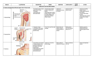
  • 1. MUSCLE ILLUSTRATION DESCRIPTION ORIGIN INSERTION NERVE SUPPLY BLOOD SUPPLY ACTION MUSCLES OF THE PECTORAL REGION A. Anterior Axioappendicular Muscles (Upper Limb to Thoracic wall) 1. Pectoralis Major -Fan-shaped muscle; Covers superior part of thorax -Convergent -Has clavicular and sternocostal heads -Clavicular head: Anterior surface of medial half of clavicle -Sternocostal head: Anterior surface of sternum, superior six costal cartilages, and aponeurosis of external oblique muscles Lateral lip of intertubercular sulcus of humerus Lateral and medial pectoral nerves -Adducts and medially rotates humerus -Draws scapula anteriorly and inferiorly -Clavicular head: flexes humerus -Sternocostal head: extends humerus (from a flexed position) 2. Pectoralis Minor -Triangular in shape -In the anterior wall, almost completely covered by the pectoralis major 3rd to 5th ribs near their costal cartilages Medial border and superior surface of coracoid process of scapula Medial pectoral nerve from brachial plexus (C8, T1) Stabilizes scapula by drawing inferiorly and anteriorly against thoracic wall 3. Subclavius -Found inferior to the clavicle -Provides protection to the subclavian vessels and to the superior trunk of the brachial plexus -Resist tendency for clavicle to be dislocated at the sternoclavicular joint Junction of 1st rib and its costal cartilage Inferior surface of middle third of clavicle Nerve to subclavius (C5, C6) Anchors and depresses clavicle
  • 2. 4. Serratus anterior -Overlies the lateral part of the thorax and forms the medial wall of the axilla -Named due to sawtooth appearance of fleshy slips or digitations -“Boxer’s muscle” as it is one of the most powerful muscles of the pectoral girdle, a strong protractor of the scapula, and is used when punching or reaching anteriorly -Multipennate External surface of lateral parts of 1st – 8th ribs Anterior surface of medial border of scapula Long thoracic nerve (C5, C6, C7) -Protracts scapula and holds in against thoracic wall -Rotates scapula B. Posterior Axioappendicular muscles (Upper limb to vertebral column) Superficial (Extrinsic shoulder) muscles 1. Trapezius -Covers the posterior aspect of the neck and superior half of the trunk -Convergent -Muscles of the 2 sides form a trapezium Medial third of superior nuchal line; External occipital protuberance; nuchal ligament; spinous process of C7 to T12 vertebrae -Upper fibers: lateral third of clavicle -Middle and lower fibers: acromion and spine of scapula -Spinal accessory nerve (CN XI) (motor fibers) -C3, C4 spinal nerves (Pain and proprioceptive fibers) -Descending/Superior/Upper – Elevate scapula -Middle fibers (all parts together) – Retracts scapula -Ascending/Inferior/Lower fibers – depress scapula
  • 3. 2. Latissimus dorsi -Large, fan-shaped muscle -Convergent -Passes from trunk to the humerus -Acts directly on the glenohumeral joint and indirectly on the pectoral girdle (Scapulothoracic joint) Spinous processes of inferior 6 thoracic vertebrae, thoracolumbar fascia, iliac crest, and inferior 3 or 4 ribs Floor of intertubercular sulcus of humerus Thoracodorsal nerve (C6, C7, C8)* -Extends, adducts, and medially rotates humerus -Rotates body towards arm Deep (Extrinsic shoulder) muscles 1. Levator Scapulae -Strap-like -Superior third lies deep to the sternocleidomastoid; interior third is deep to the trapezius Posterior tubercles of transverse processes of C1- C4 vertebrae Medial border of the scapula superior to the root of scapular spine Dorsal scapular (C4, C5) and Cervical (C3, C4) nerves -Elevates scapula medially -Rotates glenoid cavity inferiorly by rotating scapula 2. Rhomboid Major -Forms an oblique equilateral parallelogram with rhomboid minor -Thin, flat, two times wider than rhomboid minor lying superior Spinous processes of T2 to T5 vertebrae Medial border of scapula from level of spine to inferior angle Dorsal scapular nerve (C4, C5) Retracts scapula and rotates glenoid cavity inferiorly -Fix scapula to thoracic wall 3. Rhomboid Minor -Form an oblique equilateral parallelogram with rhomboid major Nuchal ligament; spinous process of C7 and T1 vertebrae Smooth triangular area at medial end of scapular spine Dorsal scapular nerve (C4, C5) -Retracts scapula and rotates its glenoid cavity inferiorly -Fix scapula to thoracic wall
  • 4. C. Posterior scapulohumeral muscles (Scapula to Humerus) 1. Deltoid -Thick, coarse-textured muscle covering the shoulder and forms its rounded contour (Shaped like the greek letter delta (triangle)) -Multipennate -Lateral third of clavicle -Acromion -Spine of scapula Deltoid tuberosity of the humerus* Axillary nerve (C5, C6) -Clavicular part (anterior): Flexion and medial rotation of the arm -Acromial part (middle): Abduction of the arm -Spinal part (posterior): Extension and lateral rotation of the arm 2. Supraspinatus Occupies the supraspinous fossa of the scapula Supraspinous fossa of the scapula Superior facet of greater tubercule of humerus Suprascapular nerve (C4, C5, C6) -Initiates and assists deltoid in arm abduction -Acts with rotator cuff muscles 3. Infraspinatus Occupies the medial three quarters of the infraspinous fossa of the scapula (Partly covered by the deltoid and trapezius) Infraspinous fossa of the scapula Medial facet of greater tubercule of the humerus Suprascapular nerve (C5, C6) -Lateral rotation of arms (acts with teres minor) -Main lateral rotator of the arm 4. Teres Major Thick rounded muscle -Latin “teres”, round -Assists the Latissimus dorsi in extending the humerus Posterior surface of the inferior angle of the scapula Medial lip of the inertubular sulcus of the humerus* Lower subscapular nerve (C5, C6) Adduction and medial rotation of arm 5. Teres Minor Narrow, elongate muscle, hidden by the deltoid -Assists the infraspinatus in lateral rotation of arm and adduction Middle part of lateral border of the scapula -Inferior facet of greater tubercule of the humerus -Glenohumeral joint capsule Axillary nerve (C5, C6) Lateral rotation of arms (acts with infraspinatus)
  • 5. Subscapularis Thick, triangular muscle lying on the costal surface of the scapula -Forms part of the posterior wall of the axilla Subscapular fossa (most of the anterior surface of the scapula) Lesser tubercle of humerus Upper and lower subscapular nerves (C5, C6, C7) -Main medial rotator of the arm -Assists in holding head of humerus in the glenoid fossa Rotator cuff muscles (SITS) Muscles involved Supraspinatus Infraspinatus Teres minor Subscapularis -Grasps and pulls humeral head to glenoid cavity -Protects and stabilizes the joint capsule Axillary region 1. Axilla -Pyramidal space inferior to glenohumeral joint and superior to axillary fascia -Contains loose connective tissues
  • 6. A. Subspacial spaces 1. Quadrangular space Borders: Teres minor (above) Teres major (below) Triceps brachii, long head (medial) Surgical neck of humerus (lateral) Axillary nerve Post humeral circumflex artery 2. Triangular space (Upper) Borders: Teres minor (above) Teres major (below) Triceps brachii, long head (lateral) Circumflex scapular artery from subscapular artery 3. Triangular space (Lower) Borders: Teres major (below) Triceps brachii, long head (medial) Humerus (lateral) Radial nerve Profunda brachial artery MUSCLES OF THE ARM A. Anterior arm: flexor muscles 1. Biceps brachii -A fusiform with 2 heads that are proximally attached -“bi” two + “caput” head -“Three-joint muscle” -Although located at the anterior compartment of the arm, it has no attachment to the humerus -Short head: tip of the coracoid process of the scapula -Long head: supraglenoid tubercle of the scapula -Radial tuberosity -Fascia of the forearm via bicipital aponeurosis *Bicipital apneurosis -A triangular membranous band that runs from the biceps tendon across the cubital fossa and merges with the antebrachial fascia -Protects the structures in the cubital fossa and helps to lessen the pressure of the biceps tendon Musculocutaneous nerve (C5, C6, C7) BRACHIAL ARTERY -Supination of the forearm (Primary supinator) -Flexion of elbow joint -Weak flexion of shoulder -also resists dislocation of shoulder
  • 7. 2. Brachialis -A flattened fusiform muscle lying posterior (deep) to the biceps -Main flexor of the forearm and is the only pure flexor of the forearm -Always contracts when the elbow is flexed Distal half of anterior surface of the humerus -Coronoid process -Ulnar tuberosity -Musculocutaneous nerve (C5, C6) -Radial nerve (C5, C7): some lateral parts are innervated by a branch of the radial nerve BRACHIAL ARTERY Flexion of the forearm 3. Coracobrachialis -Elongated muscle in the superomedial part of the arm -Landmark for locating other structures -Pierced by the musculocutaneous nerve Tip of the coracoid process of the scapula Middle third of the medial surface of the humerus Musculocutaneous nerve (C5, C6, C7) BRACHIAL ARTERY -Flexion and adduction of the arm -Resists downward dislocation of the head of the humerus (Shunt muscle) B. Posterior arm: Extensor muscles 1. Triceps Brachii -A large fusiform muscle in the posterior arm -“tri” three + “caput” head -Main extensor of the forearm -Three heads: long, lateral, and medial -Long head: Infraglenoid tubercle of the scapula -Lateral head: Posterior surface of the humerus, superior to radial groove (proximal half) Medial head: Posterior surface of the humerus, inferior to radial groove (distal 2/3) Proximal end of olecranon of ulna and fascia of forearm Radial nerve (C5, C7, C8) Profunda brachii artery Long head -least active head -Extension and adduction of the arm -Resists dislocation of the head of the humerus (Shunt muscle) Lateral head -strongest head -primarily acts as resistance Medial head -Deep -Extensor of the forearm -Active at all speeds with or without resistance 2. Arconeus -Small, triangular muscle on the posterolateral aspect of the elbow -partially blended with the triceps Posterior surface of lateral epicondyle of the humerus -Lateral surface of the olecranon fossa -Superior part of the posterior surface of the ulna Radial nerve (C7, C8, T1) Profunda brachii artery -Assists in extension of the forearm -Stabilizes the elbow joint -Abducts the ulna during pronation of the forearm
  • 8. MUSCLES OF THE FOREARM Anterior forearm: Flexor/ Pronator muscles A. Superficial group – Origin: Common flexor tendon, attached to the medial epicondyle of humerus 1. Pronator Teres -Located most laterally -Fusiform muscle Humeral head: medial epicondyle of the humerus Ulnar head: coronoid process of the Ulna Lateral aspect of shaft of radius Median nerve Pronates and flexes the forearm 2. Flexor Carpi radialis -Located on radial side; flexes the carpals only, not the fingers/metacarpals Medial epicondyle of humerus Base of 2nd metacarpal Median nerve Flexion and abduction of hand at wrist joint 3. Palmaris longus Medial epicondyle of humerus Distal half of flexor retinaculum and palmar aponeurosis Median nerve Flexes hand at wrist and tenses palmar aponeurosis Flexor Carpi Ulnaris * -Located most medially; simultaneously flexes and adductsthe hand at the wrist Humeral head: medial epicondyle of humerus Ulnar head: Olecranon and posterior border of ulna -Pisiform -Hook of hamate -Base of 5th metacarpal Ulnar nerve (C7, C8) Flexion and adduction of hand at wrist joint B. Intermediate group – Crosses the elbow *ALL anterior compartment muscles of the forearm are innervated by the MEDIAN NERVE except the flexor carpi ulnaris and the medial part of the flexor digitorum profundus which are innervated by the ulnar nerve 1. Flexor digitorum superficialis -Humeroulnar head: medial epicondyle of humerus and coronoid process of ulna -Radial head: Superior half of anterior radius -Bodies of middle phalanges of medial 4 digits -Flexes middle and proximal phalanges of medial 4 digits -Flexes hand at wrist joints
  • 9. C. Deep group 1. Flexor digitorum profundus (FDP) (Proximal) medial and anterior surface of proximal Âľ of ulna and interosseous membrane (Distal) palmar base of distal phalanges of medial 4 digits Medial part – Ulnar nerve Lateral part – Median nerve Assists in flexion of wrist Stabilizes the elbow joint Flexes distal interphalangeal joint 2. Flexor pollicis longus (FPL) Lies lateral to the FDP Long flexor of the thumb (Proximal) anterior surface of radius and interosseous membrane (Distal) palmar base of distal phalanx of the thumb Median nerve (anterior interosseous) Flexion of thumb 3. Pronator quadratus Quadrangular muscle and pronates the forearm PRIME MOVER for pronation DEEPEST muscle in the anterior aspect of the forearm (Proximal) distal 4th of anterior ulna (Distal) distal 4th of anterior radius Median nerve (anterior interosseous) Pronates forearm ANTEROIR FOREARM SUBFASCIAL SPACE BOUNDARIES CONTENTS (lateral to medial) 1. Cubital fossa Triangular area on the anterior aspect of the forearm at the area of the elbow Site of venipuncture due to large veins Lateral – brachioradialis Medial – pronator teres Floor – brachialis and supinator Roof – brachial and antebrachial fascia - Radial nerve and branches - Biceps brachii tendon - Brachial artery and vein - Median nerve
  • 10. MUSCLE ILLUSTRATION DESCRIPTION ORIGIN INSERTION NERVE SUPPLY BLOOD SUPPLY ACTION POSTERIOR FOREARM: EXTENSOR/ SUPINATOR MUSCLES In the posterior compartment of the forearm Held in place by extensor retinaculum (prevents bowstring o ftendon) Radial nerve A. SUPERFICIAL LAYER 1. Brachiradialis Forms lateral border of cubital fossa Proximal 2/3 of supra-condylar ridge of humerus Lateral surfave of distal end of radius proximal to styloid process Weak flexion of forearm; Maximal flexion if forearm is in midprone position 2. Extensor carpi radialis longus (ECRL) Fusiform muscle and is overlapped by the brachioradialis Lateral supracondylar ridge of the humerus Dorsal base of 2nd metacarpal ECRL + ECRB = abducts hand at extension ECRL + ECRB + Flexor carpi radialis = pure abduction ECRL + ECRB + Extensor carpi ulnaris = extends hand Extends and abducts hand at wrist Active during fist clenching 3. Extensor carpi radialis brevis (ECRB) Lateral epicondyle of humerus Dorsal base of 3rd metacarpal Extension and abductin of hand Acts as synergist to other muscles 4. Extensor diditorum Principal extensor of the digits; Occupying the posterior surface of the forearm Lateral epicondyle of humerus Extensor expasions of medial 4 digits Extension of medial 4 digits (proximal, middle, distal phalanges) Assist in wrist extension 5. Extensor digiti minimi (EDM) Fusiform slip of muscle Detached from extensor digitorum Lateral epicondyle of humerus Extensor expansion of 5th digit Extension of 5th digit 6. Extensor carpi ulnaris (ECU) Long fusiform muscle Located at the medial border of the forearm Lateral epicondyle of humerus Posterior border of ulna Dorsal aspect of base of 5th metacarpal Extension and adduction of hand Fist clenching Synergist to ECRB B. DEEP GROUP 1. Supinator PRIME MOVER for slow, unopposed supination Form the floor of cubital fossa Lateral epicondyle of humerus Supinator fossa Crest of ulna Lateral, posterior, anterior surfaces of proximal 3rd of radius Supination of forearm; Rotation of radius when elbow is flexed 2. Abductor pollicis longus (APL) Long fusiform belly Distal to the supinator Closely related to extensor pollicis brevis Posterior surface of ulna, radius and interosseous membrane Base of 1st metacarpal Posterior interosseus nerve (continuation of deep branch of radial nerve) Abducts thumb with Abductor pollicis brevis Extends thumb with Extensor pollicis
  • 11. 3. Extensor pollicis brevis (EPB) Lies distal to the aabductor pollicis longus Helps expends the first metacarpal and abducts the hand Posterior surface of radius and onterosseous membrane Dorsal base of proximal phalanx of thums Posterior interosseus nerve (continuation of deep branch of radial nerve) Extension of proximal phalanx and carpometacarpal joint of thumb 4. Extensor pollicis longus (EPL) Passes under extensor retinaculum Medial to the dorsal tubercle of the radius Posterior surface of middle third of ulna and interosseous membrane Dorsal base of distal phalanx of thumb Posterior interosseus nerve (continuation of deep branch of radial nerve) Extension of distal phalanx of thumb Adducts and extends the thumb Rotates the thumb laterraly 5. Extensor indicis Medial alongside to Extensor pollicis longus Confers independence of index finger Posterior surface of ulna and interosseous membrane Extensor expansion of 2nd digit Posterior interosseus nerve (continuation of deep branch of radial nerve) Extendion of index finger and assist in extension of hand FASCIAL SPACES and COMPARTMENTS Triangular skin depression on the radial aspect of wrist WALLS: Medial – EPL Lateral – APL + EPB FLOOR: Scaphoid + Trapezium Radial artery
  • 12. HAND A. DEEP FASCIA 1. Flexor retinaculum Makes up CARPAL TUNNEL – anterior/palmar side of wrist that connects the forearm to the middle compartment of the deep plane of the palm Structures that pass through this muscle: - 4 tendons of Flexor digitorum superficialis (FDS) - 4 tendons of Flexor digitorum pofundus (FDP) - 1 tendon of Flexor policis longus (FPL) - Median nerve CARPAL TUNNEL SYNDROME - Condition where there is pain, tingling and swelling of hand caused by pressure on the MEDIAN NERVE - Usually affects the thumb, index, middle and ring fingers - Relieved surgically (open or endoscope) releasing the Flexor retinaculum 2. Extensor retinaculum Keeps extensor tendons in place 3. Palmar fascia Palmar aponeurosis - Strong well-defined pat of the deep fascia of palm - Covers soft tissues and overlies long flexor tendons - Located at Thenar and Hypothenar eminences Dupuytren Contracture - Disease of palmar fascia resulting in progressive shortening, thickening and fibrosis of the palmar fascia and aponeurosis 4. Dorsal fascia Digital fibrous flexor sheath – covers the flexor digitorum
  • 13. FASCIAL COMPARTMENTS and SPACES LOCATION ADDITIONAL DESCRIPTION 1. Hypothenar compartment Space between the attachment of palmar aponeurosis to the 5th metacarpal; Middle of fibrous septum Contains hypothenar muscles 2. Thenar compartment Space between the attachment of palmar aponeurosis to the 3rd metacarpal; Side of the lateral fibrous septum Contains thenar muscles 3. Central compartment Space between the Hypothenar and Thenar compartment Contains: - Flexor tendons and sheaths - Lumbricals - Superficial palmar arterial arch - Digital vessels and nerves Adductor compartment: deepest muscular plane of the hand containing the Adductor pollicis 4. Palmar spaces Midpalmar space - Under Central compartment - Covers the flexor group of tendons Thenar space - Under Thenar compartment - Covers the thumb synovial flexor When there is swelling, this is where the fluid accumulates 5. Synovial flexor sheath Contains: Ulnar bursa – protects the Flexor digitorum superficialis and Profundus Radial bursa – protects the Flexor pollicis longus B. INTRINSIC MUSCLES OF THE HAND DESCRIPTION ORIGIN INSERTION NERVE SUPPLY BLOOD SUPPLY ACTION THENAR COMPARTMENT Median nerve (recurrent branch) Thumb movement (opposition) 1. Abductor pollicis brevis Flexor retinaculum and Tubercles of Scaphoid and Trapezium Lateral side of proximal phalanx of the thumb Abduction of thumb 2. Flexor pollicis brevis Flexor retinaculum and Tubercles of Scaphoid and Trapezium Lateral side of proximal phalanx of the thumb Median nerve – large superficial head Ulnar nerve – smaller deep head Flexes thumb 3. Opponens pollicis Flexor retinaculum and Tubercles of Scaphoid and Trapezium Lateral side of 1st metacarpal Oppose thumb by drawing the 1st metacarpal medially to the center of palm and rotates itmedially
  • 14. HYPOTHENAR COMPARTMENT DESCRIPTION ORIGIN INSERTION NERVE SUPPLY BLOOD SUPPLY ACTION Ulnar nerve (deep branch) Little finger movement 1. Abductor digiti minimi Most superficial among 3 hypothenar muscles Pisiform Medial side of proximal phalanx of 5th digit Abduction of 5th digit, assists in flexion of its proximal phalanx 2. Flexor digiti minimi Hook of hamate and Flexor retinaculum Medial side of proximal phalanx of 5th digit Flexes proximal phalanx of 5th digit 3. Opponens digiti minimi Hook of hamate and Flexor retinaculum Medial border of 5th metacarpal Draws 5th metacarpal anteriorly and rotates it to face the thumb 4. Palmaris brevis NOT in the hypothenar compartment but is part of the hypothenar eminence Wrinkles the skin that deepen the hollow of the palm, thereby aiding palmar grip CENTRAL COMPARTMENT 1. Lumbricalss Worm-like form 1st and 2nd – lateral 2 tendons of Flexor digitorum profundus; 3rd and 4th – lateral 3 tendons of Flexor digitorum Lateral sides of extensor expansions of 2nd – 5th digits 1st and 2nd median nerve; 3rd and 4th ulnar nerve (deep branch) Flex metacarpophalangeal joints Extend interphalangeal joints of 2nd – 5th digits 2. Interossei 4 Dorsal interosseous muscles (D) - located between metacarpals 3 Palmar interosseous muscles (P) - located at palmar surface of the metacarpals in the interosseous compartment of the hand Ulnar nerve (deep branch) D – Abduction (D-Ab) P – Adduction (P- Ad) 3. Adductor pollicis Located at Adductor compartment of the hand Tendon usually contains sesamoid bone Oblique head - bases of 2nd and 3rd metacarpals, capitate and adjacent bones Transverse head – anterior surface of 3rd metacarpal Medial side of base of proximal phalanx of thumb Adduction of thumb toward the lateral border of palm