

Recommended
More Related Content
What's hot
What's hot (20)
Similar to Chodatess
Similar to Chodatess (20)
More from Fidy Zegge
More from Fidy Zegge (20)
Recently uploaded
Recently uploaded (20)
Chodatess
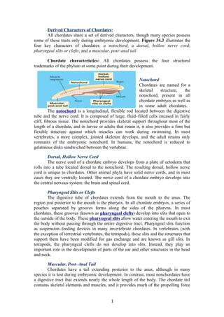
- 1. Derived Characters of Chordates: All chordates share a set of derived characters, though many species possess some of these traits only during embryonic development. Figure 34.3 illustrates the four key characters of chordates: a notochord; a dorsal, hollow nerve cord; pharyngeal slits or clefts; and a muscular, post–anal tail Chordate characteristics: All chordates possess the four structural trademarks of the phylum at some point during their development. Notochord Chordates are named for a skeletal structure, the notochord, present in all chordate embryos as well as in some adult chordates. The notochord is a longitudinal, flexible rod located between the digestive tube and the nerve cord. It is composed of large, fluid–filled cells encased in fairly stiff, fibrous tissue. The notochord provides skeletal support throughout most of the length of a chordate, and in larvae or adults that retain it, it also provides a firm but flexible structure against which muscles can work during swimming. In most vertebrates, a more complex, jointed skeleton develops, and the adult retains only remnants of the embryonic notochord. In humans, the notochord is reduced to gelatinous disks sandwiched between the vertebrae. Dorsal, Hollow Nerve Cord The nerve cord of a chordate embryo develops from a plate of ectoderm that rolls into a tube located dorsal to the notochord. The resulting dorsal, hollow nerve cord is unique to chordates. Other animal phyla have solid nerve cords, and in most cases they are ventrally located. The nerve cord of a chordate embryo develops into the central nervous system: the brain and spinal cord. Pharyngeal Slits or Clefts The digestive tube of chordates extends from the mouth to the anus. The region just posterior to the mouth is the pharynx. In all chordate embryos, a series of pouches separated by grooves forms along the sides of the pharynx. In most chordates, these grooves (known as pharyngeal clefts) develop into slits that open to the outside of the body. These pharyngeal slits allow water entering the mouth to exit the body without passing through the entire digestive tract. Pharyngeal slits function as suspension–feeding devices in many invertebrate chordates. In vertebrates (with the exception of terrestrial vertebrates, the tetrapods), these slits and the structures that support them have been modified for gas exchange and are known as gill slits. In tetrapods, the pharyngeal clefts do not develop into slits. Instead, they play an important role in the development of parts of the ear and other structures in the head and neck. Muscular, Post–Anal Tail Chordates have a tail extending posterior to the anus, although in many species it is lost during embryonic development. In contrast, most nonchordates have a digestive tract that extends nearly the whole length of the body. The chordate tail contains skeletal elements and muscles, and it provides much of the propelling force 1
- 2. in many aquatic species. Tunicates Data from various morphological and molecular studies support the hypothesis that tunicates (subphylum Urochordata) belong to the deepest–branching lineage of chordates. The tunicates most resemble other chordates during their larval stage, which may be as brief as a few minutes. In many species, the larva uses its tail muscles and notochord to swim through water in search of a suitable substrate on which it can settle, guided by cues it receives from light– and gravity–sensitive cells. Once a tunicate has settled on a substrate, it goes through a radical metamorphosis in which many of its chordate characters disappear. Its tail and notochord are resorbed; its nervous system degenerates; and its remaining organs rotate 90°. As an adult, a tunicate draws in water through an incurrent siphon; the water then passes through the pharyngeal slits into a chamber called the atrium and exits through an excurrent siphon. Food particles are filtered from the water by a mucous net and transported by cilia to the esophagus. The anus empties into the excurrent siphon. Some tunicate species shoot a jet of water through their excurrent siphon when attacked, earning them the informal name of “sea squirts. Figure: A tunicate, a urochordate. Lancelets (subphylum Cephalochordata) get their name from their bladelike shape (Figure 34.5) . As larvae, lancelets develop a notochord, a dorsal, hollow nerve cord, numerous pharyngeal slits, and a post–anal tail. They feed on plankton in the water column, alternating between upward swimming and passive sinking. As they sink, they trap plankton and other suspended matter in their pharynx. 2
- 3. Figure: The lancelet Branchiostoma, a cephalochordate. This small invertebrate displays all four main chordate characters. Water enters the mouth and passes through the pharyngeal slits into the atrium, a chamber that vents to the outside via the atriopore. Food particles trapped by mucus are swept by cilia into the digestive tract. The serially arranged segmental muscles produce the lancelet′s undulatory (wavelike) swimming movements. Adult lancelets can be up to 5 cm long. They retain key chordate traits, closely resembling the idealized chordate shown in Figure 34.3. Following metamorphosis, adult lancelets swim down to the seafloor and wriggle backward into the sand, leaving only their anterior end exposed. A mucous net secreted across the pharyngeal slits removes tiny food particles from seawater drawn into the mouth by ciliary pumping. The water passes through the slits, and the trapped food enters the intestine. The pharynx and pharyngeal slits play a minor role in gas exchange, which occurs mainly across the external body surface. A lancelet frequently leaves its burrow to swim to a new location. Though feeble swimmers, these invertebrate chordates display, in a simple form, the swimming mechanism of fishes. Coordinated contraction of muscles serially arranged like rows of chevrons (<<<<) along the sides of the notochord flexes the notochord, producing side–to–side undulations that thrust the body forward. This serial musculature is evidence of the lancelet′s segmentation. The muscle segments develop from blocks of mesoderm called somites, which are found along each side of the notochord in all chordate embryos. Globally, lancelets are rare, but in a few regions (including Tampa Bay, along the Florida coast) they occasionally reach densities in excess of 5,000 individuals per square meter. Early Chordate Evolution: Although tunicates and lancelets are relatively obscure animals, the rise of evolutionary biology has focused much attention on them. Possessing many—but not all—of the derived characters shared by vertebrates, they can provide clues about the evolutionary origin of the vertebrate body plan. As you have read, tunicates display a number of chordate characters only as larvae, whereas lancelets retain those characters as adults. Thus, an adult lancelet looks much more like a larval tunicate than like an adult tunicate. In the 1920s, based on these observations, biologist William Garstang proposed that tunicates represent an early stage in chordate evolution. He suggested that ancestral tunicate–like chordates accelerated their sexual maturity, becoming mature while still in their larval stage. Thus, they and the chordates that evolved from them retained the notochord and other features as adults. This process, which has been documented in a number of 3
- 4. evolutionary transitions, is known as paedomorphosis. While Garstang′s idea was popular for several decades, today the weight of evidence is against it in the case of tunicates. The degenerate adult stage of tunicates appears to be a derived trait that evolved only after the tunicate lineage branched off from other chordates. Even the tunicate larva appears to be highly derived, rather than a faithful reproduction of the body plan of early chordates. Studies of Hox gene expression (see Chapter 21) suggest that the tunicate larva does not develop the posterior regions of its body axis. Rather, the anterior region is elongated and contains a heart and digestive system. While Garstang′s idea was popular for several decades, today the weight of evidence is against it in the case of tunicates. The degenerate adult stage of tunicates appears to be a derived trait that evolved only after the tunicate lineage branched off from other chordates. Even the tunicate larva appears to be highly derived, rather than a faithful reproduction of the body plan of early chordates. Studies of Hox gene expression (see Chapter 21) suggest that the tunicate larva does not develop the posterior regions of its body axis. Rather, the anterior region is elongated and contains a heart and digestive system. Research on lancelets has revealed several important clues about the evolution of the chordate brain. Rather than a full–fledged brain, lancelets have only a slightly swollen tip on the anterior end of their dorsal nerve cord. But the same Hox genes that organize major regions of the forebrain, midbrain, and hindbrain of vertebrates express themselves in a corresponding pattern in this small cluster of cells in the lancelet′s nerve cord (Figure 34.6) . This suggests that the vertebrate brain apparently is an elaboration of an ancestral structure similar to the lancelet′s simple nerve cord tip. Expression of developmental genes in lancelets and vertebrates. Hox genes (including BF1, Otx, and Hox3) control the development of major regions of the vertebrate brain. These genes are expressed in the same anterior–to–posterior order in lancelets and vertebrate 4
- 5. Q1. Humans are chordates, yet they lack most of the main derived characters of chordates. Explain. A1. In humans, these characters are present only in the embryo. The notochord becomes disks between the vertebrae, the tail is almost completely lost, and the pharyngeal clefts develop into various adult structures. Q2. How do pharyngeal slits function in feeding in tunicates and lancelets? A2. As water passes through the slits, food particles are filtered from the water and transported to the digestive system Craniates are chordates that have a head: After the evolution of the basic chordate body plan, seen in tunicates and lancelets, the next major transition in chordate evolution was the appearance of a head. Chordates with a head are known as craniates (from the word cranium, skull). The origin of a head—consisting of a brain at the anterior end of the dorsal nerve cord, eyes and other sensory organs, and a skull—opened up a completely new way of feeding for chordates: active predation. (Note that heads evolved independently in other animal lineages as well, as described in Chapter 33.) Derived Characters of Craniates Living craniates share a set of derived characters that distinguish them from other chordates. On a genetic level, they possess two clusters of Hox genes (lancelets and tunicates have only one). Other important families of genes that produce signaling molecules and transcription factors are also duplicated in craniates. This additional genetic complexity made it possible for craniates to develop more complex morphologies than those of tunicates and lancelets. One feature unique to craniates is the neural crest, a collection of cells that appears near the dorsal margins of the closing neural tube in an embryo. Neural crest cells disperse throughout the body, where they give rise to a variety of structures, including teeth, some of the bones and cartilage of the skull, the inner layer of skin (dermis) of the facial region, several types of neurons, and the sensory capsules in which eyes and other sense organs develop. The neural crest, embryonic source of many unique vertebrate characters. In aquatic craniates, the pharyngeal clefts evolved into gill slits. Unlike the pharyngeal slits of lancelets, which are used primarily for suspension feeding, gill slits are associated with muscles and nerves that allow water to be pumped through the slits. This pumping can assist in sucking in food, and it facilitates gas exchange. (In terrestrial craniates, the pharyngeal clefts develop into other structures, as explained later.) Craniates, which are more active than tunicates and lancelets, also have a higher metabolism and a much more extensive muscular system. Muscles lining their digestive tract aid digestion by moving food through the tract. Craniates also have a heart with at least two chambers, red blood cells, and hemoglobin, as well as kidneys that remove waste products from the blood. 5
- 6. The Origin of Craniates: In the late 1990s, paleontologists working in China discovered a vast supply of fossils of early chordates that appear to straddle the transition to craniates. The fossils were formed during the Cambrian explosion 530 million years ago, when many groups of animals were diversifying (see Chapter 32). The most primitive of the fossils are those of the 3–cm–long Haikouella (Figure 34.8a ). In many ways, Haikouella resembled a lancelet. Its mouth structure indicates that, like lancelets, it probably was a suspension feeder. However, Haikouella also had some of the characters of craniates. For example, it had a small but well– formed brain, eyes, and muscular segments that resemble those of vertebrates. It also had hardened structures in its pharynx that may have been tooth–like “denticles.” However, Haikouella did not have a skull, suggesting that this character emerged with innovations to the chordate nervous system. Fossils of primitive chordates. The colors in the illustrations are fanciful. In other Cambrian rocks, paleontologists have found fossils of even more advanced chordates, such as Haikouichthys. About the same size as Haikouella, Haikouichthys had a skull that may have been composed of cartilage (Figure 34.8b) . Based on this and other characters, paleontologists have identified Haikouichthys as a true craniate. Hagfishes: The least derived craniate lineage that still survives is class Myxini, the hagfishes. Hagfishes have a skull made of cartilage, but they lack jaws and vertebrae. They swim in a snakelike fashion by using their segmental muscles to exert force against their notochord, which they retain in adulthood as a strong, flexible rod of cartilage. Hagfishes have a small brain, eyes, ears, and a nasal opening that connects with the pharynx. They also have tooth–like formations made of the protein keratin in their mouth. All of the 30 living species of hagfishes are marine. Measuring up to 60 cm in length, most are bottom–dwelling scavengers that feed on worms and sick or dead fish. Rows of slime glands on a hagfish′s flanks secrete a substance that absorbs water, forming a slime that may repulse other scavengers when a hagfish is feeding (see Figure 34.9). When a hagfish is attacked by a potential predator, it can produce several liters of slime in less than a minute. The slime coats the gills of an attacking 6
- 7. fish, sending it into retreat or even suffocating it. Several teams of biologists and engineers are investigating the properties of hagfish slime in hopes of producing an artificial slime that could act as a space–filling gel. Such a gel might be used, for instance, to curtail bleeding during surgery. Vertebrate systematists do not consider hagfishes to be fishes, despite their common name. Traditionally, the term fish has been applied to any craniate except tetrapods. But used in this way, it does not refer to a monophyletic group; therefore, systematists use the term fish to refer only to a specific clade of vertebrates, the actinopterygians (see Concept 34.4). We will follow that practice in this chapter. Q3. Which extinct chordate is more closely related to humans, Haikouichthys or Haikouella? Explain your answer. A3. Haikouichthys; it had a skull and thus was a craniate, as are humans. Haikouella did not have a skull. Q4. What characteristics do hagfishes have that tunicates and lancelets lack? A4. Hagfishes have a head and skull made of cartilage, plus a small brain, sensory organs, and tooth–like structures. They have a neural crest, gill slits, and more extensive organ systems. In addition, hagfishes have slime glands that ward off predators and may repel competing scavengers. 7