SlideShare a Scribd company logo
1 of 42
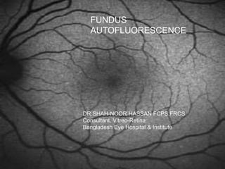
FUNDUS
AUTOFLUORESCENCE
DR.SHAH-NOOR HASSAN FCPS,FRCS
Consultant, Vitreo-Retina
Bangladesh Eye Hospital & Institute
Fluorophore
Autofluorescence
 Some materials contain a naturally
autofluorescent component that can be
visualised when excited with a light of
particular wavelength
Retinoid Fluorophores
RPE and lipofuscin
 RPE constitutes a monolayer of
polygonal cells between the
choroid and neuro-sensory
retina
 Multiple functions
 RPE dysfunction implicated in
variety of retinal diseases
 LF is a byproduct of
accumulation of sheded outer
segments of the photoreceptors
 Lipofuscin accumulates as a byproduct
of phagocytosis of photoreceptors’ outer
segment
 In advanced age: It may occupy 20% of
free cytoplasmic space of RPE cells
The older we grow the more we glow
lipofuscin
 WHY IS IT DANGEROUS ?
 A2-E (N-retinylidene-N-retinylethanol-
amine) the dominant fluorophore possess
toxic properties
 Interferes with the normal cell function
 Precursors of A2-E are also toxic
 Products of photo-oxidation of RPE
lipofuscin serves as trigger for
complement activation inflammation
Fundus Autofluorescence
 Major source of fundus
autofluorescence is the lipofuscin of the
RPE
Fundus autofluorescence
 Metabolically mapping the RPE
 Developed as a tool to evaluate the RPE
during aging and ocular disease
Andrea von Ruckmann, Fredrick W. Fitzke and Alan C. Bird- Moorfield’s
eye hospital
Scanning Laser
Ophthalmoscope
 Webb and co-workers
 55° of field in one frame
 Low power laser source
 Scan in x and y axis
 Confocality ensures that light fluorescence
and reflectance is derived from same ocular
plane
 HRA2: Excitation 488nm
Emitted light above 500nm
FAF image acquisition
ART Mean image
Live image
FAF image
OCT image
FAF image acquisition
 Align the camera using the IR
illumination.
 Spectralis HRA+OCT only: Once you
see a sharp well-focused image,
change to Redfree illumination to fine-
tune focus.
 Change to the FA illumination. The
image will now be considerably darker.
 Automatic (recommended!) or
manual (Spectralis HRA+OCT only)
sensitivity control will outline the
retinal blood vessels.
Turn
or
automatic
FAF image acquisition – Mean
function
 Activate the ART (Automatic Real
Time) Mean function to generate a ‘live
Mean’ Autofluorescence image online
and view it as it is created.
 Note: Following the injection of
fluorescein dye, it will be impossible to
perform FAF imaging.
Press
Normal Autofluorescence
distribution
 Optic nerve head
 Absence of
autofluorescent pigment
 Retinal blood vessels
 Absorption by blood
vessels
 Foveal area
 Absorption by luteal
pigment
 Parafoveal area
 Mildly decreased
intensity due to high
melanin content and
lower density of
lipofuscin granules in
central RPE
In disease state
 Excessive accumulation of lipofuscin in
the lysosomal compartment of the RPE
is the downstream process in many
hereditary and age related diseases
AF and ARMD- Basic
Considerations
 RPE is thought to play a key role in the
early and late phases of the disease
 Hallmark of aging is the accumulation of
lipofuscin granules in the cytoplasm of
the RPE cells
 Lipofuscin accumulation is the common
downstream process
 Ability to document spatial distribution of
lipofuscin and its changes over time.
 The amount of autofluorescence is signature for
previous or possible future oxidative injury
Hyperfluorescence in FAF FA 36 sec FA 69 sec
Geographic Atrophy
 Seen as hypo-
autofluorescent areas
 No RPE, NO
LIPOFUSCIN  NO
AUTOFLUORESCENCE
 Measure the atrophic
area to see for the
progression
Geographic atrophy
 Identification of peri-lesional abnormalities
 Hyperautofl signals bordering  denote sick
RPE
 The damage marches in areas of high signals
 Predictor of future trouble
Pigment Epithelial Detachment
 Serous PED:
 Increased FAF signal
corresponding to area of
detachmet
 Underlying CNVM: No
specific findings
 Surrounding area: Hypo AF
signal
Choroidal
neovascularization
 Irregular FAF in areas of CNV
 High FAF outside the edge of the
lesion
 FAF intensity decreased over the
disciform scars
 In early stages, preservation of
FAF
 Extent of abnormal areas on FAF
is more than that on FA
CNVM
CLASSIC FA
CSR
 Leaks at the level of RPE
leading to central serous
detachment
 Chronic disease associated
with atrophic changes at the
level of RPE and retina
 FA and ICG – hemodynamics
and fluid dynamics
 OCT- clues to size and
elevation of detachment
 Autofluorescence- functional
status
CSR
Bull’s eye maculopathy
Macular dystrophies-
Stargradt’s disease
 Areas of atrophy on fundus
corresponded to hypo-
autofluorescence
 Flecks seen as
depigmented lesion
appeared as areas of hypo-
autofl
 Predictive value is yet to be
determined
Macular dystrophies: Best’s
disease
 Autofl characters: central round
area of increased FAF
 Pseudohypopyon stage:
increased FAF in the lower part
 Late stages: irregular FAF
within the lesion with
disseminated spots of
increased FAF
Macular dystrophies: Best’s
disease
 Pattern of spread on FAF: centrifugal
 Atrophic regions are associated with low
levels of background FAF, lower visual
acuity, abnormal colour vision, central
scotomas and poorer
electrophysiological results
 FAF appears more striking and
widespread
 Spoke like, diffuse or combination
X-linked retinoschisis
 FAF finding reflect typical radiating
cystic changes
 The changes on FAF are most likely due
to altered passage of exciting and
emitted light from the retinal folds
Retinitis Pigmentosa
 In dominant and recessive
and rod-cone dystrophies
 Absent FAF in areas of outer
retinal atrophy
 Normal FAF in adjacent
regions of surviving retina
 High FAF in surviving areas in
some cases
 Macular oedema of more than
4 months  high FAF
Retinitis Pigmentosa
 Parafoveal ring of increased FAF
 Correlation exists between these areas
of high FAF and photopic and scotopic
sensitivity
Serpigenous Choroiditis
 In SC: Autofluorescence is
detected within 2-5 days
after the appearance of
lesion
 Provides a clear
delineation of the area of
RPE damage
 Progressive decrease in
autofluorescence was seen
during the scarring phase
APMPPE
 Affected RPE
demonstrate
increase AF in
early phase of the
disease
 More pronounced
after 3 wks
 Fade away after 1
yr.
Macular Hole
 Bright fluorescence of macular holes
similar to images on FA
 Pseudoholes: no such high
autofluorescence
 Attached operculum shows focal
decreased autofluorescence
Macular hole surgery
VKH
 Hypo AF in the areas of serous
detachments
 NIR AF: hyper AF at the macula and
hypo AF in the areas of serous
detachment
 With treatment: BL-FAF: subtle FAF
 NIR FAF: more wide spread FAF
LASER & AF
1 hour 3 hour 3 month
Applications for therapeutic
interventions
 In advanced atrophic AMD: useful to
develop and assess the therapeutic
interventions
 Fenritidine, an oral medicine shown to
reduce the production of toxic fluophores
 In retinal dystrophies: useful to assess
the functional preservation of the outer
retina
 In Leber’s: normal or slightly reduced FAF
RPE FAF & therapeutic
outcome
 The RPE-FAF of exudative AMD lesions varies
greatly.
 FAF differences have a great influence on the
chances of antivascular endothelial growth factor
(VEGF) therapy success.
 Development of visual acuity is less favorable in
eyes with initially increased central FAF.
Heimes et al. - Foveal RPE FAF as a prognostic factor for anti-VEGF therapy in exudative AMD - GraefesArch 2008
CONCLUSION
 Non invasive ,Easy to perform
 Provides a novel prognostic marker for
disease progression
 Metabolic changes and loss of RPE
integrity corresponds to visual function
 In combination with SD OCT it adds to
our understanding of retinal diseases
from a broad point of view.
THANK YOU

More Related Content

What's hot

AS OCT & UBM - Dr Shylesh B Dabke
AS OCT & UBM - Dr Shylesh B DabkeAS OCT & UBM - Dr Shylesh B Dabke
AS OCT & UBM - Dr Shylesh B DabkeShylesh Dabke
 
LASIK: COMPLICATIONS AND THEIR MANAGEMENT
LASIK: COMPLICATIONS AND THEIR MANAGEMENTLASIK: COMPLICATIONS AND THEIR MANAGEMENT
LASIK: COMPLICATIONS AND THEIR MANAGEMENTLaxmi Eye Institute
 
Macularfunctiontests seminar
Macularfunctiontests seminarMacularfunctiontests seminar
Macularfunctiontests seminarVaibhav Kanduri
 
NW2010 Epiretinal membrane
NW2010 Epiretinal membraneNW2010 Epiretinal membrane
NW2010 Epiretinal membraneNawat Watanachai
 
Tractional RD
Tractional RD Tractional RD
Tractional RD Nikhil Rp
 
Correcting presbyopia - Modern Options
Correcting presbyopia - Modern OptionsCorrecting presbyopia - Modern Options
Correcting presbyopia - Modern OptionsJason Higginbotham
 
Role of imaging in glaucoma management
Role of imaging in glaucoma management Role of imaging in glaucoma management
Role of imaging in glaucoma management gunjan chadha
 
Premium intraocular lenses The past, present and-3.pptx
Premium intraocular lenses The past, present and-3.pptxPremium intraocular lenses The past, present and-3.pptx
Premium intraocular lenses The past, present and-3.pptxMushtaq Ahmad
 
Optical coherence tomography in glaucoma - Dr Shylesh Dabke
Optical coherence tomography in glaucoma - Dr Shylesh DabkeOptical coherence tomography in glaucoma - Dr Shylesh Dabke
Optical coherence tomography in glaucoma - Dr Shylesh DabkeShylesh Dabke
 
Malignant glaucoma
Malignant glaucomaMalignant glaucoma
Malignant glaucomaNikhil Rp
 
Biometry: Iol calculation
Biometry: Iol calculation Biometry: Iol calculation
Biometry: Iol calculation Noor Munirah Aab
 
Techniques of tear film evaluation by Raju Kaiti
Techniques of tear film evaluation  by Raju KaitiTechniques of tear film evaluation  by Raju Kaiti
Techniques of tear film evaluation by Raju KaitiRaju Kaiti
 
Choroidal detachment -Nov 2017
Choroidal detachment  -Nov 2017Choroidal detachment  -Nov 2017
Choroidal detachment -Nov 2017sameerasep13
 
Phakic Intraocular lens
Phakic Intraocular lensPhakic Intraocular lens
Phakic Intraocular lensrumi das
 
WHITE DOT SYNDROMES OF THE RETINAL INFLAMATION
WHITE DOT SYNDROMES OF THE RETINAL INFLAMATIONWHITE DOT SYNDROMES OF THE RETINAL INFLAMATION
WHITE DOT SYNDROMES OF THE RETINAL INFLAMATIONAjayDudani1
 
femtosecond laser in ophthalmology by Dr. Hind Safwat (Al Azhr university)
femtosecond laser in ophthalmology by Dr. Hind Safwat (Al Azhr university) femtosecond laser in ophthalmology by Dr. Hind Safwat (Al Azhr university)
femtosecond laser in ophthalmology by Dr. Hind Safwat (Al Azhr university) Hind Safwat
 

What's hot (20)

AS OCT & UBM - Dr Shylesh B Dabke
AS OCT & UBM - Dr Shylesh B DabkeAS OCT & UBM - Dr Shylesh B Dabke
AS OCT & UBM - Dr Shylesh B Dabke
 
OCT
OCTOCT
OCT
 
LASIK: COMPLICATIONS AND THEIR MANAGEMENT
LASIK: COMPLICATIONS AND THEIR MANAGEMENTLASIK: COMPLICATIONS AND THEIR MANAGEMENT
LASIK: COMPLICATIONS AND THEIR MANAGEMENT
 
Scleral lens fitting
Scleral lens fittingScleral lens fitting
Scleral lens fitting
 
Macularfunctiontests seminar
Macularfunctiontests seminarMacularfunctiontests seminar
Macularfunctiontests seminar
 
NW2010 Epiretinal membrane
NW2010 Epiretinal membraneNW2010 Epiretinal membrane
NW2010 Epiretinal membrane
 
Tractional RD
Tractional RD Tractional RD
Tractional RD
 
Correcting presbyopia - Modern Options
Correcting presbyopia - Modern OptionsCorrecting presbyopia - Modern Options
Correcting presbyopia - Modern Options
 
Role of imaging in glaucoma management
Role of imaging in glaucoma management Role of imaging in glaucoma management
Role of imaging in glaucoma management
 
Autofluorescence
AutofluorescenceAutofluorescence
Autofluorescence
 
Premium intraocular lenses The past, present and-3.pptx
Premium intraocular lenses The past, present and-3.pptxPremium intraocular lenses The past, present and-3.pptx
Premium intraocular lenses The past, present and-3.pptx
 
Optical coherence tomography in glaucoma - Dr Shylesh Dabke
Optical coherence tomography in glaucoma - Dr Shylesh DabkeOptical coherence tomography in glaucoma - Dr Shylesh Dabke
Optical coherence tomography in glaucoma - Dr Shylesh Dabke
 
Malignant glaucoma
Malignant glaucomaMalignant glaucoma
Malignant glaucoma
 
Biometry: Iol calculation
Biometry: Iol calculation Biometry: Iol calculation
Biometry: Iol calculation
 
Techniques of tear film evaluation by Raju Kaiti
Techniques of tear film evaluation  by Raju KaitiTechniques of tear film evaluation  by Raju Kaiti
Techniques of tear film evaluation by Raju Kaiti
 
Macular hole
Macular holeMacular hole
Macular hole
 
Choroidal detachment -Nov 2017
Choroidal detachment  -Nov 2017Choroidal detachment  -Nov 2017
Choroidal detachment -Nov 2017
 
Phakic Intraocular lens
Phakic Intraocular lensPhakic Intraocular lens
Phakic Intraocular lens
 
WHITE DOT SYNDROMES OF THE RETINAL INFLAMATION
WHITE DOT SYNDROMES OF THE RETINAL INFLAMATIONWHITE DOT SYNDROMES OF THE RETINAL INFLAMATION
WHITE DOT SYNDROMES OF THE RETINAL INFLAMATION
 
femtosecond laser in ophthalmology by Dr. Hind Safwat (Al Azhr university)
femtosecond laser in ophthalmology by Dr. Hind Safwat (Al Azhr university) femtosecond laser in ophthalmology by Dr. Hind Safwat (Al Azhr university)
femtosecond laser in ophthalmology by Dr. Hind Safwat (Al Azhr university)
 

Similar to Fundus Autofluorescence Imaging: A Powerful Tool for Evaluating Retinal Disease

Fundus fluorescein angiography and B-scan by vijay
Fundus  fluorescein angiography  and B-scan by vijayFundus  fluorescein angiography  and B-scan by vijay
Fundus fluorescein angiography and B-scan by vijayDr. vijay pratap
 
Multiple evanescent white dot syndrome
Multiple evanescent white dot syndromeMultiple evanescent white dot syndrome
Multiple evanescent white dot syndromeShruti Laddha
 
98967f8c6ec465e03a7f80b29928ece1copy-230926184118-2578187b.pdf
98967f8c6ec465e03a7f80b29928ece1copy-230926184118-2578187b.pdf98967f8c6ec465e03a7f80b29928ece1copy-230926184118-2578187b.pdf
98967f8c6ec465e03a7f80b29928ece1copy-230926184118-2578187b.pdfMainaBidiyasar
 
CENTRAL SEROUS CHORIO RETINOPATHY
CENTRAL SEROUS CHORIO RETINOPATHYCENTRAL SEROUS CHORIO RETINOPATHY
CENTRAL SEROUS CHORIO RETINOPATHYSSSIHMS-PG
 
CENTRAL SEROUS CHORIOETINOPATHY
CENTRAL SEROUS CHORIOETINOPATHYCENTRAL SEROUS CHORIOETINOPATHY
CENTRAL SEROUS CHORIOETINOPATHYSivateja Challa
 
Ffa by suraj chhetri
Ffa by suraj chhetriFfa by suraj chhetri
Ffa by suraj chhetriSuraj Chhetri
 
Retinitis pigmentosa
Retinitis pigmentosaRetinitis pigmentosa
Retinitis pigmentosakamalinineha6
 
fundus fluorescein angiography V/S indocyanine green angiography
fundus fluorescein angiography V/S indocyanine green angiographyfundus fluorescein angiography V/S indocyanine green angiography
fundus fluorescein angiography V/S indocyanine green angiographyparesh nichlani
 
Central serous chorioretinopathy
Central serous chorioretinopathyCentral serous chorioretinopathy
Central serous chorioretinopathyAnjali Maheshwari
 
hereditary macular and choroidal dystrophies
hereditary macular and choroidal dystrophies hereditary macular and choroidal dystrophies
hereditary macular and choroidal dystrophies Priyanka Choudhary
 
Hereditary macular dystrophies
Hereditary macular  dystrophiesHereditary macular  dystrophies
Hereditary macular dystrophiesShruti Laddha
 
DR WANI'S TALK ON WHITE DOT SYNDROMES.pptx
DR WANI'S  TALK ON WHITE DOT SYNDROMES.pptxDR WANI'S  TALK ON WHITE DOT SYNDROMES.pptx
DR WANI'S TALK ON WHITE DOT SYNDROMES.pptxvbwani
 
Age-Related Macular Degeneration
Age-Related Macular DegenerationAge-Related Macular Degeneration
Age-Related Macular DegenerationHossein Mirzaie
 

Similar to Fundus Autofluorescence Imaging: A Powerful Tool for Evaluating Retinal Disease (20)

Fundus fluorescein angiography and B-scan by vijay
Fundus  fluorescein angiography  and B-scan by vijayFundus  fluorescein angiography  and B-scan by vijay
Fundus fluorescein angiography and B-scan by vijay
 
Multiple evanescent white dot syndrome
Multiple evanescent white dot syndromeMultiple evanescent white dot syndrome
Multiple evanescent white dot syndrome
 
Vep
VepVep
Vep
 
98967f8c6ec465e03a7f80b29928ece1copy-230926184118-2578187b.pdf
98967f8c6ec465e03a7f80b29928ece1copy-230926184118-2578187b.pdf98967f8c6ec465e03a7f80b29928ece1copy-230926184118-2578187b.pdf
98967f8c6ec465e03a7f80b29928ece1copy-230926184118-2578187b.pdf
 
FFA AND ICG
FFA AND ICGFFA AND ICG
FFA AND ICG
 
Eye Flourecein Angiography
Eye Flourecein AngiographyEye Flourecein Angiography
Eye Flourecein Angiography
 
Retinopathy of prematurity (upload for site)
Retinopathy of prematurity (upload for site)Retinopathy of prematurity (upload for site)
Retinopathy of prematurity (upload for site)
 
CENTRAL SEROUS CHORIO RETINOPATHY
CENTRAL SEROUS CHORIO RETINOPATHYCENTRAL SEROUS CHORIO RETINOPATHY
CENTRAL SEROUS CHORIO RETINOPATHY
 
CENTRAL SEROUS CHORIOETINOPATHY
CENTRAL SEROUS CHORIOETINOPATHYCENTRAL SEROUS CHORIOETINOPATHY
CENTRAL SEROUS CHORIOETINOPATHY
 
Retina physiology
Retina physiologyRetina physiology
Retina physiology
 
Ffa by suraj chhetri
Ffa by suraj chhetriFfa by suraj chhetri
Ffa by suraj chhetri
 
Retinitis pigmentosa
Retinitis pigmentosaRetinitis pigmentosa
Retinitis pigmentosa
 
fundus fluorescein angiography V/S indocyanine green angiography
fundus fluorescein angiography V/S indocyanine green angiographyfundus fluorescein angiography V/S indocyanine green angiography
fundus fluorescein angiography V/S indocyanine green angiography
 
Central serous chorioretinopathy
Central serous chorioretinopathyCentral serous chorioretinopathy
Central serous chorioretinopathy
 
hereditary macular and choroidal dystrophies
hereditary macular and choroidal dystrophies hereditary macular and choroidal dystrophies
hereditary macular and choroidal dystrophies
 
CSR
CSRCSR
CSR
 
Central Serous Retinopathy
Central Serous RetinopathyCentral Serous Retinopathy
Central Serous Retinopathy
 
Hereditary macular dystrophies
Hereditary macular  dystrophiesHereditary macular  dystrophies
Hereditary macular dystrophies
 
DR WANI'S TALK ON WHITE DOT SYNDROMES.pptx
DR WANI'S  TALK ON WHITE DOT SYNDROMES.pptxDR WANI'S  TALK ON WHITE DOT SYNDROMES.pptx
DR WANI'S TALK ON WHITE DOT SYNDROMES.pptx
 
Age-Related Macular Degeneration
Age-Related Macular DegenerationAge-Related Macular Degeneration
Age-Related Macular Degeneration
 

More from Dr. Shah Noor Hassan

Refractory Diabetic Macular Edema - A Case Report
Refractory Diabetic Macular Edema - A Case ReportRefractory Diabetic Macular Edema - A Case Report
Refractory Diabetic Macular Edema - A Case ReportDr. Shah Noor Hassan
 
Surgical management of Uveal Effusion Syndrome-A Case Report
Surgical management of Uveal Effusion Syndrome-A Case Report Surgical management of Uveal Effusion Syndrome-A Case Report
Surgical management of Uveal Effusion Syndrome-A Case Report Dr. Shah Noor Hassan
 
How To Make a Cataract Surgery Perfect - Posterior Segment Perspective
How To Make a Cataract Surgery Perfect - Posterior Segment PerspectiveHow To Make a Cataract Surgery Perfect - Posterior Segment Perspective
How To Make a Cataract Surgery Perfect - Posterior Segment PerspectiveDr. Shah Noor Hassan
 
Approach to Disc Pallor and Automated Fields in Neuro-ophthalmology
Approach to Disc Pallor and Automated Fields in Neuro-ophthalmology  Approach to Disc Pallor and Automated Fields in Neuro-ophthalmology
Approach to Disc Pallor and Automated Fields in Neuro-ophthalmology Dr. Shah Noor Hassan
 
Optical Coherence Tomography (OCT)
Optical Coherence Tomography (OCT)Optical Coherence Tomography (OCT)
Optical Coherence Tomography (OCT)Dr. Shah Noor Hassan
 

More from Dr. Shah Noor Hassan (20)

Vitrectomy Principles
Vitrectomy PrinciplesVitrectomy Principles
Vitrectomy Principles
 
Vitreous hemorrhage
Vitreous hemorrhageVitreous hemorrhage
Vitreous hemorrhage
 
Anatomy of Retina
Anatomy of RetinaAnatomy of Retina
Anatomy of Retina
 
Prophylaxis of RD
Prophylaxis of RDProphylaxis of RD
Prophylaxis of RD
 
Refractory Diabetic Macular Edema - A Case Report
Refractory Diabetic Macular Edema - A Case ReportRefractory Diabetic Macular Edema - A Case Report
Refractory Diabetic Macular Edema - A Case Report
 
Retinoblastoma (Rb)
Retinoblastoma (Rb)Retinoblastoma (Rb)
Retinoblastoma (Rb)
 
Surgical management of Uveal Effusion Syndrome-A Case Report
Surgical management of Uveal Effusion Syndrome-A Case Report Surgical management of Uveal Effusion Syndrome-A Case Report
Surgical management of Uveal Effusion Syndrome-A Case Report
 
How To Make a Cataract Surgery Perfect - Posterior Segment Perspective
How To Make a Cataract Surgery Perfect - Posterior Segment PerspectiveHow To Make a Cataract Surgery Perfect - Posterior Segment Perspective
How To Make a Cataract Surgery Perfect - Posterior Segment Perspective
 
Hypertensive Retinopathy
Hypertensive RetinopathyHypertensive Retinopathy
Hypertensive Retinopathy
 
Pars Planitis
Pars PlanitisPars Planitis
Pars Planitis
 
Fluorescein Angiography
Fluorescein AngiographyFluorescein Angiography
Fluorescein Angiography
 
Endophthalmitis
EndophthalmitisEndophthalmitis
Endophthalmitis
 
Retinal Arterial Obstructions
Retinal Arterial ObstructionsRetinal Arterial Obstructions
Retinal Arterial Obstructions
 
Coats' Disease
Coats' DiseaseCoats' Disease
Coats' Disease
 
Approach to Disc Pallor and Automated Fields in Neuro-ophthalmology
Approach to Disc Pallor and Automated Fields in Neuro-ophthalmology  Approach to Disc Pallor and Automated Fields in Neuro-ophthalmology
Approach to Disc Pallor and Automated Fields in Neuro-ophthalmology
 
Retinoblastoma
Retinoblastoma Retinoblastoma
Retinoblastoma
 
Endophthalmitis
EndophthalmitisEndophthalmitis
Endophthalmitis
 
Diabetic Maculopathy
Diabetic MaculopathyDiabetic Maculopathy
Diabetic Maculopathy
 
Optical Coherence Tomography (OCT)
Optical Coherence Tomography (OCT)Optical Coherence Tomography (OCT)
Optical Coherence Tomography (OCT)
 
Cryotherapy
CryotherapyCryotherapy
Cryotherapy
 

Recently uploaded

Call Girls In Andheri East Call 9920874524 Book Hot And Sexy Girls
Call Girls In Andheri East Call 9920874524 Book Hot And Sexy GirlsCall Girls In Andheri East Call 9920874524 Book Hot And Sexy Girls
Call Girls In Andheri East Call 9920874524 Book Hot And Sexy Girlsnehamumbai
 
Artifacts in Nuclear Medicine with Identifying and resolving artifacts.
Artifacts in Nuclear Medicine with Identifying and resolving artifacts.Artifacts in Nuclear Medicine with Identifying and resolving artifacts.
Artifacts in Nuclear Medicine with Identifying and resolving artifacts.MiadAlsulami
 
Call Girls Colaba Mumbai ❤️ 9920874524 👈 Cash on Delivery
Call Girls Colaba Mumbai ❤️ 9920874524 👈 Cash on DeliveryCall Girls Colaba Mumbai ❤️ 9920874524 👈 Cash on Delivery
Call Girls Colaba Mumbai ❤️ 9920874524 👈 Cash on Deliverynehamumbai
 
Low Rate Call Girls Pune Esha 9907093804 Short 1500 Night 6000 Best call girl...
Low Rate Call Girls Pune Esha 9907093804 Short 1500 Night 6000 Best call girl...Low Rate Call Girls Pune Esha 9907093804 Short 1500 Night 6000 Best call girl...
Low Rate Call Girls Pune Esha 9907093804 Short 1500 Night 6000 Best call girl...Miss joya
 
Call Girls Service Bellary Road Just Call 7001305949 Enjoy College Girls Service
Call Girls Service Bellary Road Just Call 7001305949 Enjoy College Girls ServiceCall Girls Service Bellary Road Just Call 7001305949 Enjoy College Girls Service
Call Girls Service Bellary Road Just Call 7001305949 Enjoy College Girls Servicenarwatsonia7
 
Call Girls Service Chennai Jiya 7001305949 Independent Escort Service Chennai
Call Girls Service Chennai Jiya 7001305949 Independent Escort Service ChennaiCall Girls Service Chennai Jiya 7001305949 Independent Escort Service Chennai
Call Girls Service Chennai Jiya 7001305949 Independent Escort Service ChennaiNehru place Escorts
 
Best Rate (Hyderabad) Call Girls Jahanuma ⟟ 8250192130 ⟟ High Class Call Girl...
Best Rate (Hyderabad) Call Girls Jahanuma ⟟ 8250192130 ⟟ High Class Call Girl...Best Rate (Hyderabad) Call Girls Jahanuma ⟟ 8250192130 ⟟ High Class Call Girl...
Best Rate (Hyderabad) Call Girls Jahanuma ⟟ 8250192130 ⟟ High Class Call Girl...astropune
 
Call Girl Bangalore Nandini 7001305949 Independent Escort Service Bangalore
Call Girl Bangalore Nandini 7001305949 Independent Escort Service BangaloreCall Girl Bangalore Nandini 7001305949 Independent Escort Service Bangalore
Call Girl Bangalore Nandini 7001305949 Independent Escort Service Bangalorenarwatsonia7
 
Call Girls Service Navi Mumbai Samaira 8617697112 Independent Escort Service ...
Call Girls Service Navi Mumbai Samaira 8617697112 Independent Escort Service ...Call Girls Service Navi Mumbai Samaira 8617697112 Independent Escort Service ...
Call Girls Service Navi Mumbai Samaira 8617697112 Independent Escort Service ...Call girls in Ahmedabad High profile
 
Russian Call Girls in Pune Riya 9907093804 Short 1500 Night 6000 Best call gi...
Russian Call Girls in Pune Riya 9907093804 Short 1500 Night 6000 Best call gi...Russian Call Girls in Pune Riya 9907093804 Short 1500 Night 6000 Best call gi...
Russian Call Girls in Pune Riya 9907093804 Short 1500 Night 6000 Best call gi...Miss joya
 
Call Girls Horamavu WhatsApp Number 7001035870 Meeting With Bangalore Escorts
Call Girls Horamavu WhatsApp Number 7001035870 Meeting With Bangalore EscortsCall Girls Horamavu WhatsApp Number 7001035870 Meeting With Bangalore Escorts
Call Girls Horamavu WhatsApp Number 7001035870 Meeting With Bangalore Escortsvidya singh
 
VIP Call Girls Tirunelveli Aaradhya 8250192130 Independent Escort Service Tir...
VIP Call Girls Tirunelveli Aaradhya 8250192130 Independent Escort Service Tir...VIP Call Girls Tirunelveli Aaradhya 8250192130 Independent Escort Service Tir...
VIP Call Girls Tirunelveli Aaradhya 8250192130 Independent Escort Service Tir...narwatsonia7
 
Aspirin presentation slides by Dr. Rewas Ali
Aspirin presentation slides by Dr. Rewas AliAspirin presentation slides by Dr. Rewas Ali
Aspirin presentation slides by Dr. Rewas AliRewAs ALI
 
(Rocky) Jaipur Call Girl - 9521753030 Escorts Service 50% Off with Cash ON De...
(Rocky) Jaipur Call Girl - 9521753030 Escorts Service 50% Off with Cash ON De...(Rocky) Jaipur Call Girl - 9521753030 Escorts Service 50% Off with Cash ON De...
(Rocky) Jaipur Call Girl - 9521753030 Escorts Service 50% Off with Cash ON De...indiancallgirl4rent
 
(👑VVIP ISHAAN ) Russian Call Girls Service Navi Mumbai🖕9920874524🖕Independent...
(👑VVIP ISHAAN ) Russian Call Girls Service Navi Mumbai🖕9920874524🖕Independent...(👑VVIP ISHAAN ) Russian Call Girls Service Navi Mumbai🖕9920874524🖕Independent...
(👑VVIP ISHAAN ) Russian Call Girls Service Navi Mumbai🖕9920874524🖕Independent...Taniya Sharma
 
Call Girls Service Pune Vaishnavi 9907093804 Short 1500 Night 6000 Best call ...
Call Girls Service Pune Vaishnavi 9907093804 Short 1500 Night 6000 Best call ...Call Girls Service Pune Vaishnavi 9907093804 Short 1500 Night 6000 Best call ...
Call Girls Service Pune Vaishnavi 9907093804 Short 1500 Night 6000 Best call ...Miss joya
 
High Profile Call Girls Coimbatore Saanvi☎️ 8250192130 Independent Escort Se...
High Profile Call Girls Coimbatore Saanvi☎️  8250192130 Independent Escort Se...High Profile Call Girls Coimbatore Saanvi☎️  8250192130 Independent Escort Se...
High Profile Call Girls Coimbatore Saanvi☎️ 8250192130 Independent Escort Se...narwatsonia7
 
Vip Call Girls Anna Salai Chennai 👉 8250192130 ❣️💯 Top Class Girls Available
Vip Call Girls Anna Salai Chennai 👉 8250192130 ❣️💯 Top Class Girls AvailableVip Call Girls Anna Salai Chennai 👉 8250192130 ❣️💯 Top Class Girls Available
Vip Call Girls Anna Salai Chennai 👉 8250192130 ❣️💯 Top Class Girls AvailableNehru place Escorts
 

Recently uploaded (20)

sauth delhi call girls in Bhajanpura 🔝 9953056974 🔝 escort Service
sauth delhi call girls in Bhajanpura 🔝 9953056974 🔝 escort Servicesauth delhi call girls in Bhajanpura 🔝 9953056974 🔝 escort Service
sauth delhi call girls in Bhajanpura 🔝 9953056974 🔝 escort Service
 
Call Girls In Andheri East Call 9920874524 Book Hot And Sexy Girls
Call Girls In Andheri East Call 9920874524 Book Hot And Sexy GirlsCall Girls In Andheri East Call 9920874524 Book Hot And Sexy Girls
Call Girls In Andheri East Call 9920874524 Book Hot And Sexy Girls
 
Artifacts in Nuclear Medicine with Identifying and resolving artifacts.
Artifacts in Nuclear Medicine with Identifying and resolving artifacts.Artifacts in Nuclear Medicine with Identifying and resolving artifacts.
Artifacts in Nuclear Medicine with Identifying and resolving artifacts.
 
Call Girls Colaba Mumbai ❤️ 9920874524 👈 Cash on Delivery
Call Girls Colaba Mumbai ❤️ 9920874524 👈 Cash on DeliveryCall Girls Colaba Mumbai ❤️ 9920874524 👈 Cash on Delivery
Call Girls Colaba Mumbai ❤️ 9920874524 👈 Cash on Delivery
 
Low Rate Call Girls Pune Esha 9907093804 Short 1500 Night 6000 Best call girl...
Low Rate Call Girls Pune Esha 9907093804 Short 1500 Night 6000 Best call girl...Low Rate Call Girls Pune Esha 9907093804 Short 1500 Night 6000 Best call girl...
Low Rate Call Girls Pune Esha 9907093804 Short 1500 Night 6000 Best call girl...
 
Russian Call Girls in Delhi Tanvi ➡️ 9711199012 💋📞 Independent Escort Service...
Russian Call Girls in Delhi Tanvi ➡️ 9711199012 💋📞 Independent Escort Service...Russian Call Girls in Delhi Tanvi ➡️ 9711199012 💋📞 Independent Escort Service...
Russian Call Girls in Delhi Tanvi ➡️ 9711199012 💋📞 Independent Escort Service...
 
Call Girls Service Bellary Road Just Call 7001305949 Enjoy College Girls Service
Call Girls Service Bellary Road Just Call 7001305949 Enjoy College Girls ServiceCall Girls Service Bellary Road Just Call 7001305949 Enjoy College Girls Service
Call Girls Service Bellary Road Just Call 7001305949 Enjoy College Girls Service
 
Call Girls Service Chennai Jiya 7001305949 Independent Escort Service Chennai
Call Girls Service Chennai Jiya 7001305949 Independent Escort Service ChennaiCall Girls Service Chennai Jiya 7001305949 Independent Escort Service Chennai
Call Girls Service Chennai Jiya 7001305949 Independent Escort Service Chennai
 
Best Rate (Hyderabad) Call Girls Jahanuma ⟟ 8250192130 ⟟ High Class Call Girl...
Best Rate (Hyderabad) Call Girls Jahanuma ⟟ 8250192130 ⟟ High Class Call Girl...Best Rate (Hyderabad) Call Girls Jahanuma ⟟ 8250192130 ⟟ High Class Call Girl...
Best Rate (Hyderabad) Call Girls Jahanuma ⟟ 8250192130 ⟟ High Class Call Girl...
 
Call Girl Bangalore Nandini 7001305949 Independent Escort Service Bangalore
Call Girl Bangalore Nandini 7001305949 Independent Escort Service BangaloreCall Girl Bangalore Nandini 7001305949 Independent Escort Service Bangalore
Call Girl Bangalore Nandini 7001305949 Independent Escort Service Bangalore
 
Call Girls Service Navi Mumbai Samaira 8617697112 Independent Escort Service ...
Call Girls Service Navi Mumbai Samaira 8617697112 Independent Escort Service ...Call Girls Service Navi Mumbai Samaira 8617697112 Independent Escort Service ...
Call Girls Service Navi Mumbai Samaira 8617697112 Independent Escort Service ...
 
Russian Call Girls in Pune Riya 9907093804 Short 1500 Night 6000 Best call gi...
Russian Call Girls in Pune Riya 9907093804 Short 1500 Night 6000 Best call gi...Russian Call Girls in Pune Riya 9907093804 Short 1500 Night 6000 Best call gi...
Russian Call Girls in Pune Riya 9907093804 Short 1500 Night 6000 Best call gi...
 
Call Girls Horamavu WhatsApp Number 7001035870 Meeting With Bangalore Escorts
Call Girls Horamavu WhatsApp Number 7001035870 Meeting With Bangalore EscortsCall Girls Horamavu WhatsApp Number 7001035870 Meeting With Bangalore Escorts
Call Girls Horamavu WhatsApp Number 7001035870 Meeting With Bangalore Escorts
 
VIP Call Girls Tirunelveli Aaradhya 8250192130 Independent Escort Service Tir...
VIP Call Girls Tirunelveli Aaradhya 8250192130 Independent Escort Service Tir...VIP Call Girls Tirunelveli Aaradhya 8250192130 Independent Escort Service Tir...
VIP Call Girls Tirunelveli Aaradhya 8250192130 Independent Escort Service Tir...
 
Aspirin presentation slides by Dr. Rewas Ali
Aspirin presentation slides by Dr. Rewas AliAspirin presentation slides by Dr. Rewas Ali
Aspirin presentation slides by Dr. Rewas Ali
 
(Rocky) Jaipur Call Girl - 9521753030 Escorts Service 50% Off with Cash ON De...
(Rocky) Jaipur Call Girl - 9521753030 Escorts Service 50% Off with Cash ON De...(Rocky) Jaipur Call Girl - 9521753030 Escorts Service 50% Off with Cash ON De...
(Rocky) Jaipur Call Girl - 9521753030 Escorts Service 50% Off with Cash ON De...
 
(👑VVIP ISHAAN ) Russian Call Girls Service Navi Mumbai🖕9920874524🖕Independent...
(👑VVIP ISHAAN ) Russian Call Girls Service Navi Mumbai🖕9920874524🖕Independent...(👑VVIP ISHAAN ) Russian Call Girls Service Navi Mumbai🖕9920874524🖕Independent...
(👑VVIP ISHAAN ) Russian Call Girls Service Navi Mumbai🖕9920874524🖕Independent...
 
Call Girls Service Pune Vaishnavi 9907093804 Short 1500 Night 6000 Best call ...
Call Girls Service Pune Vaishnavi 9907093804 Short 1500 Night 6000 Best call ...Call Girls Service Pune Vaishnavi 9907093804 Short 1500 Night 6000 Best call ...
Call Girls Service Pune Vaishnavi 9907093804 Short 1500 Night 6000 Best call ...
 
High Profile Call Girls Coimbatore Saanvi☎️ 8250192130 Independent Escort Se...
High Profile Call Girls Coimbatore Saanvi☎️  8250192130 Independent Escort Se...High Profile Call Girls Coimbatore Saanvi☎️  8250192130 Independent Escort Se...
High Profile Call Girls Coimbatore Saanvi☎️ 8250192130 Independent Escort Se...
 
Vip Call Girls Anna Salai Chennai 👉 8250192130 ❣️💯 Top Class Girls Available
Vip Call Girls Anna Salai Chennai 👉 8250192130 ❣️💯 Top Class Girls AvailableVip Call Girls Anna Salai Chennai 👉 8250192130 ❣️💯 Top Class Girls Available
Vip Call Girls Anna Salai Chennai 👉 8250192130 ❣️💯 Top Class Girls Available
 

Fundus Autofluorescence Imaging: A Powerful Tool for Evaluating Retinal Disease

  • 1. FUNDUS AUTOFLUORESCENCE DR.SHAH-NOOR HASSAN FCPS,FRCS Consultant, Vitreo-Retina Bangladesh Eye Hospital & Institute
  • 3. Autofluorescence  Some materials contain a naturally autofluorescent component that can be visualised when excited with a light of particular wavelength
  • 5. RPE and lipofuscin  RPE constitutes a monolayer of polygonal cells between the choroid and neuro-sensory retina  Multiple functions  RPE dysfunction implicated in variety of retinal diseases  LF is a byproduct of accumulation of sheded outer segments of the photoreceptors
  • 6.  Lipofuscin accumulates as a byproduct of phagocytosis of photoreceptors’ outer segment  In advanced age: It may occupy 20% of free cytoplasmic space of RPE cells The older we grow the more we glow
  • 7. lipofuscin  WHY IS IT DANGEROUS ?  A2-E (N-retinylidene-N-retinylethanol- amine) the dominant fluorophore possess toxic properties  Interferes with the normal cell function  Precursors of A2-E are also toxic  Products of photo-oxidation of RPE lipofuscin serves as trigger for complement activation inflammation
  • 8. Fundus Autofluorescence  Major source of fundus autofluorescence is the lipofuscin of the RPE
  • 9. Fundus autofluorescence  Metabolically mapping the RPE  Developed as a tool to evaluate the RPE during aging and ocular disease Andrea von Ruckmann, Fredrick W. Fitzke and Alan C. Bird- Moorfield’s eye hospital
  • 10. Scanning Laser Ophthalmoscope  Webb and co-workers  55° of field in one frame  Low power laser source  Scan in x and y axis  Confocality ensures that light fluorescence and reflectance is derived from same ocular plane  HRA2: Excitation 488nm Emitted light above 500nm
  • 11. FAF image acquisition ART Mean image Live image FAF image OCT image
  • 12. FAF image acquisition  Align the camera using the IR illumination.  Spectralis HRA+OCT only: Once you see a sharp well-focused image, change to Redfree illumination to fine- tune focus.  Change to the FA illumination. The image will now be considerably darker.  Automatic (recommended!) or manual (Spectralis HRA+OCT only) sensitivity control will outline the retinal blood vessels. Turn or automatic
  • 13. FAF image acquisition – Mean function  Activate the ART (Automatic Real Time) Mean function to generate a ‘live Mean’ Autofluorescence image online and view it as it is created.  Note: Following the injection of fluorescein dye, it will be impossible to perform FAF imaging. Press
  • 14. Normal Autofluorescence distribution  Optic nerve head  Absence of autofluorescent pigment  Retinal blood vessels  Absorption by blood vessels  Foveal area  Absorption by luteal pigment  Parafoveal area  Mildly decreased intensity due to high melanin content and lower density of lipofuscin granules in central RPE
  • 15. In disease state  Excessive accumulation of lipofuscin in the lysosomal compartment of the RPE is the downstream process in many hereditary and age related diseases
  • 16.
  • 17. AF and ARMD- Basic Considerations  RPE is thought to play a key role in the early and late phases of the disease  Hallmark of aging is the accumulation of lipofuscin granules in the cytoplasm of the RPE cells  Lipofuscin accumulation is the common downstream process
  • 18.  Ability to document spatial distribution of lipofuscin and its changes over time.  The amount of autofluorescence is signature for previous or possible future oxidative injury Hyperfluorescence in FAF FA 36 sec FA 69 sec
  • 19. Geographic Atrophy  Seen as hypo- autofluorescent areas  No RPE, NO LIPOFUSCIN  NO AUTOFLUORESCENCE  Measure the atrophic area to see for the progression
  • 20. Geographic atrophy  Identification of peri-lesional abnormalities  Hyperautofl signals bordering  denote sick RPE  The damage marches in areas of high signals  Predictor of future trouble
  • 21. Pigment Epithelial Detachment  Serous PED:  Increased FAF signal corresponding to area of detachmet  Underlying CNVM: No specific findings  Surrounding area: Hypo AF signal
  • 22. Choroidal neovascularization  Irregular FAF in areas of CNV  High FAF outside the edge of the lesion  FAF intensity decreased over the disciform scars  In early stages, preservation of FAF  Extent of abnormal areas on FAF is more than that on FA
  • 24. CSR  Leaks at the level of RPE leading to central serous detachment  Chronic disease associated with atrophic changes at the level of RPE and retina  FA and ICG – hemodynamics and fluid dynamics  OCT- clues to size and elevation of detachment  Autofluorescence- functional status
  • 25. CSR
  • 27. Macular dystrophies- Stargradt’s disease  Areas of atrophy on fundus corresponded to hypo- autofluorescence  Flecks seen as depigmented lesion appeared as areas of hypo- autofl  Predictive value is yet to be determined
  • 28. Macular dystrophies: Best’s disease  Autofl characters: central round area of increased FAF  Pseudohypopyon stage: increased FAF in the lower part  Late stages: irregular FAF within the lesion with disseminated spots of increased FAF
  • 29. Macular dystrophies: Best’s disease  Pattern of spread on FAF: centrifugal  Atrophic regions are associated with low levels of background FAF, lower visual acuity, abnormal colour vision, central scotomas and poorer electrophysiological results  FAF appears more striking and widespread  Spoke like, diffuse or combination
  • 30. X-linked retinoschisis  FAF finding reflect typical radiating cystic changes  The changes on FAF are most likely due to altered passage of exciting and emitted light from the retinal folds
  • 31. Retinitis Pigmentosa  In dominant and recessive and rod-cone dystrophies  Absent FAF in areas of outer retinal atrophy  Normal FAF in adjacent regions of surviving retina  High FAF in surviving areas in some cases  Macular oedema of more than 4 months  high FAF
  • 32. Retinitis Pigmentosa  Parafoveal ring of increased FAF  Correlation exists between these areas of high FAF and photopic and scotopic sensitivity
  • 33. Serpigenous Choroiditis  In SC: Autofluorescence is detected within 2-5 days after the appearance of lesion  Provides a clear delineation of the area of RPE damage  Progressive decrease in autofluorescence was seen during the scarring phase
  • 34. APMPPE  Affected RPE demonstrate increase AF in early phase of the disease  More pronounced after 3 wks  Fade away after 1 yr.
  • 35. Macular Hole  Bright fluorescence of macular holes similar to images on FA  Pseudoholes: no such high autofluorescence  Attached operculum shows focal decreased autofluorescence
  • 37. VKH  Hypo AF in the areas of serous detachments  NIR AF: hyper AF at the macula and hypo AF in the areas of serous detachment  With treatment: BL-FAF: subtle FAF  NIR FAF: more wide spread FAF
  • 38. LASER & AF 1 hour 3 hour 3 month
  • 39. Applications for therapeutic interventions  In advanced atrophic AMD: useful to develop and assess the therapeutic interventions  Fenritidine, an oral medicine shown to reduce the production of toxic fluophores  In retinal dystrophies: useful to assess the functional preservation of the outer retina  In Leber’s: normal or slightly reduced FAF
  • 40. RPE FAF & therapeutic outcome  The RPE-FAF of exudative AMD lesions varies greatly.  FAF differences have a great influence on the chances of antivascular endothelial growth factor (VEGF) therapy success.  Development of visual acuity is less favorable in eyes with initially increased central FAF. Heimes et al. - Foveal RPE FAF as a prognostic factor for anti-VEGF therapy in exudative AMD - GraefesArch 2008
  • 41. CONCLUSION  Non invasive ,Easy to perform  Provides a novel prognostic marker for disease progression  Metabolic changes and loss of RPE integrity corresponds to visual function  In combination with SD OCT it adds to our understanding of retinal diseases from a broad point of view.

Editor's Notes

  1. Ability to visualize the biochemistry and look into the RPE cells
  2. Classification abnormal autofluorescence patterns in early age-related macular disease with fundus photography and autofluorescence images introduced by Bindewald et al.12 Eight phenotypic patterns are differentiated: NORMAL (A, B) -- homogenous background FAF and a gradual decrease in the inner macula toward the fovea due to the masking effect of macular pigment. Only small hard drusen are visible in the corresponding fundus photograph. MINIMAL CHANGE (C, D) -- only minimal variations from normal background FAF. There is limited irregular increase or decrease in FAF intensity due to multiple small hard drusen. FOCAL (E, F ) -- several well definied spots with markedly increased FAF. Fundus photograph of the same eye with multiple including hard and soft drusen. PATCHY (G, H) -- multiple large areas (O200 mm diameter) of increased FAF corresponding to large, soft drusen and/or hyperpigmentation on the fundus photograph LINEAR (I, J ) -- characterized by the presence of at least one linear areas with markedly increased FAF. A corresponding hyperpigmented line is visible in the fundus photograph. LACE-LIKE (K, L) -- multiple branching linear structures of increased FAF. This pattern may correspond to hyperpigmentation on the fundus photograph or to no visible abnormalities. RETICULAR (M, N) -- multiple, specific small areas of decreased FAF with brighter lines in-between. The reticular pattern not only occurs in the macular area but is found more typically in a superotemporal location. There may be visible reticular drusen in the corresponding fundus photograph. SPECKLED (O, P) -- variety of FAF abnormalities in a larger area of the FAF image. There seem to be fewer pathologic areas in the corresponding fundus.
  3. The RPE is blocked by membrane in classic
  4. However, the pathobiology of many findings in central serous chorioretinopathy has remained elusive, because of our inability to image physiologic changes induced by the disease. Autofluorescence photography provides functional images of the fundus by employing the stimulated emission of light from naturally occurring fluorophores, the most significant being lipofuscin. In the case of retinal pigment epithelial cells, the buildup of lipofuscin is related in large part to the phagocytosis of photoreceptor outer segments containing damage accumulated through use, and indigestible altered molecules are retained within lysosomes and eventually become lipofuscin.
  5. NIR-Near infra red BL-Blue light
  6. but other retinal fluorophores that may occur in pathological conditions such as fluid, hemorrhages, or melanin deposition must be differentiated.