
Recommended
More Related Content
What's hot
What's hot (20)
Similar to 4.streptococcus
Similar to 4.streptococcus (20)
More from DrMrsVishwashantiVat
More from DrMrsVishwashantiVat (20)
Recently uploaded
Recently uploaded (20)
4.streptococcus
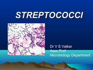
- 1. STREPTOCOCCISTREPTOCOCCI Dr V S Vatkar Asso Prof Microbiology Department
- 2. DefinitionDefinition • Gram position cocci • in chains, • non sporing, • non motile, • some capsulated, • facultatively anaerobic and fastidious in nutritional requirements.
- 3. IntroductionIntroduction • Billroth (1874): in erysipelas & calledin erysipelas & called them streptococcithem streptococci (streptos = twisted or(streptos = twisted or coiled)coiled).. • Ogston (1881) Isolated them from acuteIsolated them from acute abscesses , differs from staphylococci &abscesses , differs from staphylococci & established their pathogenicity by animalestablished their pathogenicity by animal inoculation.inoculation. • Rosenbach (1884) Isolated the cocci fromIsolated the cocci from human suppurative lesions, named themhuman suppurative lesions, named them Streptococcus pyogenesStreptococcus pyogenes..
- 4. Classification of Streptococci Aerobes & Facultative anaerobes Obligate anaerobes Peptostreptococcus Oxygen requirement hemolysis α hemolysis Streptococcus viridans Pneumococci β hemolysis β hemolytic streptococci γ hemolysis Enterococci Lancefield grouping On the basis of C Ag A-V except I & J Gr A Streptococci GRIFFITH CLASSIFICATION Based on M protein & emm gene
- 5. Based on Haemolysis on BloodBased on Haemolysis on Blood AgarAgar (i) β-haemolytic Streptococci (BHS) – complete haemolysis of the red cells around the colonies, producing clear zones around them. e.g. group A, group B etc… (ιι) α-haemolytic Streptococci – partial haemolysis with greenish discoloration of the areas surrounding the colonies. e.g.Streptococcus viridans, Streptococcus pneumoniae (iii) Non-haemolytic Streptococci e.g. Enterococcus faecalis
- 6. Lancefield GroupingLancefield Grouping • Usually done on β-haemolytic streptococci (BHS). Based on the presence of a carbohydrate component of cell wall the C carbohydrate. About 20 Lancefield groups designated as A,B,C,D, (A-H) (K-U). • Detected by reacting extract of carbohydrate C antigen with specific antisera raised against it.
- 8. M SerotypingM Serotyping • Done on only group A streptococci and based on the M protein found in Group A Streptococci. 60 such serotypes; useful for epidemiological studies.
- 9. Group A StreptococciGroup A Streptococci (Lancefield Grouping)(Lancefield Grouping) ((Streptococci PyogenesStreptococci Pyogenes)) Morphology • Spherical or oval • In chains • Nonmotile and nonsporing • It may be capsulated and the capsule is composed of hyaluronic acid.
- 10. CultureCulture • Aerobes and facultative anaerobes • 22°C - 42°C • Colony characters: virulent strains: mat finish colonies, avirulent strains:glossy colonies, capsulated colonies : mucoid
- 11. Growth & Clonial MorphologyGrowth & Clonial Morphology • Blood agar best medium with optimum temperature of 35-37°C & under aerobic conditions. • Colonies after 24 hours incubation: about 0.5 – 1mm in diameter & may/may not be surrounded by haemolysis. • They are catalase negative.
- 12. ΒΒ hemolysis on blood agarhemolysis on blood agar
- 13. Biochemical reactionsBiochemical reactions • Ferment various sugars • Produce acid but not gas • Not soluble in 10% bile • PYR
- 14. ResistanceResistance • Delicate organism • Can be stored in RCM at 4°C • More resistance to crystal violet • Sensitive to bacitracin
- 15. Antigenic structure –Antigenic structure – Virulence factorsVirulence factors • Cell wall antigens composed of 3 layers • Inner thick peptidoglycan layer: rigidity to cell wall, induces inflammatory response, thrombolytic activity • Middle layer: C or carbohydrate Ag • Outer layer: lipoteichoic acid layer: composed of M,T and R proteins • M protein : principle virulence factor, inhibits phagocytosis, inflammatory response
- 17. ToxinsToxins Streptolysin O • Oxygen labile • Soluble in oxygen so named as Sreptolysin O • Seen in deep colonies • Strongly Agenic • ASO : raised in many streptococcal infections • Increases in RF & decreases in GN and pyoderma Streptolysin S • Oxygen stable • Serum soluble so named streptolysin S • Hemolysis on surface of blood agar • Not Agenic • Not useful for serological diagnosis
- 18. Erythrogenic or pyrogenic exotoxinErythrogenic or pyrogenic exotoxin:: streptococcal pyrogenic exotoxinstreptococcal pyrogenic exotoxin (SPE)(SPE) • Scarlet fever, necrotizing fasciitis & TSS • 3 Agenic subtypes: SPE A , B & C • Subtype A & C : bacteriophage mediated , act as super Ag ----- stimulation of T-cells ------ release of cytokines ----- fever, shock, tissue damage • SPE B: chromosomally mediated • DICK test: I/D inj in susceptible children produce local erythema. Initially uesd to detect scarlet fever
- 19. EnzymesEnzymes Streptokinase: Convert plasminogen to plasmin which then lyses fibrin. Used to treat thrombotic states. e.g. Coronary thrombosis , MI Deoxyribonucleases (Streptodornase): 4 main DNAases: A B C D Antibodies produced against DNAase (anti-DNAase B) is useful for diagnosing recent Group A Streptococcal infections especially skin infections.
- 20. • Di-phospho-pyridine-nucleotidase (DPN-ase) Hyaluronidase :Degrades hyaluronic acid Spy CEP: serine protease C5a peptidase
- 21. PathogenesisPathogenesis •Causes suppurative infections and non-suppurative complications (or sequalae).
- 22. A. Suppurative (Pyogenic) Infections a) Virulence Factors (i) Principal virulence factors is the M protein Originated from the cytoplasmic membrane. Associated with pili. It is antiphagocytic. (ii) Lipotechoic Acid (LTA) For attachment to epithelial surfaces. (iii) Hyaluronic Acid An antiphagocytic capsules.
- 23. pathogenesity
- 24. PathogenicityPathogenicity Respiratory tract Acute tonsillitis Pharyngitis Scarlet fever Otitis media, Mastoiditis, Quinsy, Ludwig's angina Skin infections Wound infections, Burns, Cellulitis, Erysipelas, Impetigo or Pyoderma : An infection of the epidermis presenting as pustules. Seen most often in infants and toddlers.
- 25. Strawberry tongue in scarlet fever cellulitis erysipelas
- 27. Flesh Eating BacteriaFlesh Eating Bacteria • Cellulitis- leading toCellulitis- leading to Necrotising FasciitisNecrotising Fasciitis • Leads to necrosis ofLeads to necrosis of subcutaneoussubcutaneous issues andissues and muscular tissuesmuscular tissues and adjutant fasciaand adjutant fascia • Other complicationsOther complications Toxic shockToxic shock syndromesyndrome
- 28. INVASIVE GROUP A STREPTOCOCCI AND “FLESH-INVASIVE GROUP A STREPTOCOCCI AND “FLESH- EATING” SYNDROME OR NECROTIZING FASCIITISEATING” SYNDROME OR NECROTIZING FASCIITIS Caused by virulent strainsCaused by virulent strains ofof StreptococcusStreptococcus pyogenespyogenes
- 29. PathogenicityPathogenicity • Genital infections Both aerobic & anaerobic are normal inhabitants of female genitalia • Deep infections Bone & joint infections Lymphadenitis, Septicaemia • Non suppurative complications: Ac. rheumatic fever & Ac. Glomerulonephritis These are antigen-antibody mediated disease and occur about 1-5 weeks after the primary suppurative infection. Tend to follow either throat or skin infections or both. Streptococci are not found in the affected organ.
- 30. NON SUPPURATIVE LESIONSNON SUPPURATIVE LESIONS • Acute RheumaticAcute Rheumatic FeverFever • AcuteAcute glomerulonephritisglomerulonephritis • Carditis.Carditis. • Involvement ofInvolvement of Connective tissuesConnective tissues of the heartof the heart
- 31. NON SUPPURATIVE LESIONSNON SUPPURATIVE LESIONS
- 32. a)a) Acute Rheumatic Fever:Acute Rheumatic Fever: • Considered to be an autoimmune disease involving the myocardium and its valves, connective tissues and the big joints. • Group A Strep cell wall has some antigenic similarity with some of these human tissues. Follows after throat infections only. Tends to recur. Many serotypes are associated with acute rheumatic fever.
- 34. b)b)Acute Glomerulonephritis:Acute Glomerulonephritis: • Due to antigen-antibody complexes deposited on the basal membrane of glomeruli also can be due to similarity between group A cell components and glomerular tissue. May follow after either throat or skin. Tends not to recur. Serotypes involved are few called nephrotogenic strains.
- 35. Differences Between Glomerulonephritis &Differences Between Glomerulonephritis & Rheumatic FeverRheumatic Fever 1. Latent period between infection and first attack. 1 – 5 weeks (Average 18 days) 1 – 5 weeks (Average 10 days) 2. Preceding infection Throat only Throat or Skin 3. Pathogenesis Both Based On Immunological Reaction (Either Due to auto antibody Or due to cross reactive antigen). Similarity between organism antigens & tissue antigens Similarity between a) Organism & tissue antigens. b) Deposition of immunocomplexes in glomeruli Rheumatic Fever Glomerulonephritis
- 36. 4. Second Attacks Common Rare if any 5. Prophylactic use of penicillin. Essential Usually NOT used. 6. Serotypes (M Types) Any of the 60 serotypes Limited No. of serotypes e.g. type 12, 45 etc. 7. Serum whole complement & C3 Increased Decreased Rheumatic Fever Glomerulonephritis Differences Between Glomerulonephritis &Differences Between Glomerulonephritis & Rheumatic Fever (Continued)Rheumatic Fever (Continued)
- 37. Epidemiology ofEpidemiology of StreptococcalStreptococcal InfectionsInfections 1. Infection acquired through infected respiratory droplets. 2. Sources of Infection a) Those with active disease or convalescent carriers in throat. b) Asymptomatic carriers – the most common source. Up to 20% of school going children may carry Group A streptococci in their throats. 3. Age Group: prevalent in children especially between 3 – 8 years.
- 38. Lab. DiagnosisLab. Diagnosis A] In acute infections : by culture • Specimen : swab,pus,blood, CSF • Collection & transport • Culture
- 39. Lab. DiagnosisLab. Diagnosis • Biochem.tests . Catalase negative . Sugar fermentation with production of acid but not gas . PYR hydrolysis . Fluorescent antibody technique . Bacitracin test
- 40. Lab. DiagnosisLab. Diagnosis . Anti-deoxy-ribonuclease B (ADN-ase) . Streptozyme test . Lancefield grouping Todd hewitt broth Fullers method Rantz & Randall’s method Maxted, method . Antigen detection test : by ELISA
- 41. ProphylaxisProphylaxis Indicated only in prevention of Rh.fever to prevent reinfection and damage to the heart Long term Penicillin is given to the child who develops early stages of Rh.fever
- 42. TreatmentTreatment • Drug of choice is Penicillin • Erythromycin • Cephalexin • Tetracycline's & sulphonamides are not recommended • Chloramphenicol • Marolides
- 44. Gram stain & col.on Blood agarGram stain & col.on Blood agar
- 45. Cell surface with secreted productsCell surface with secreted products
- 47. Phagocytosis of str.pyogenes by aPhagocytosis of str.pyogenes by a macrophagemacrophage
- 48. Dividing streptococci 12000 xDividing streptococci 12000 x
- 49. Negative staining of group A streptococci viewed by TEMNegative staining of group A streptococci viewed by TEM 28,000X. The "halo" around the chain of cells (approximately28,000X. The "halo" around the chain of cells (approximately equal in thickness to the cell diameter) is the remnants of theequal in thickness to the cell diameter) is the remnants of the capsulecapsule