MCA Anatomy
-Dr Dikpal
Introduction
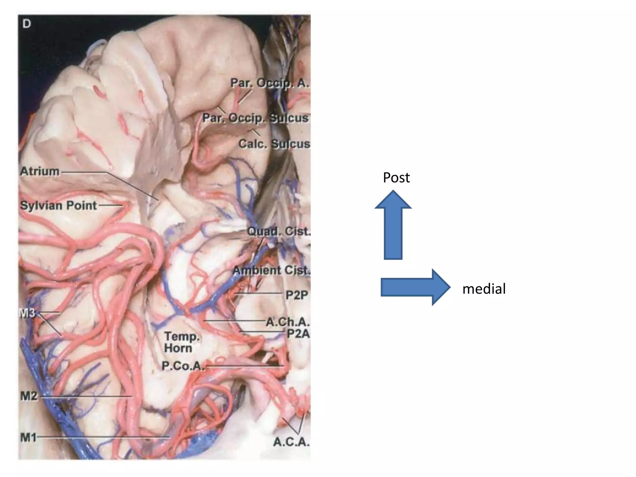
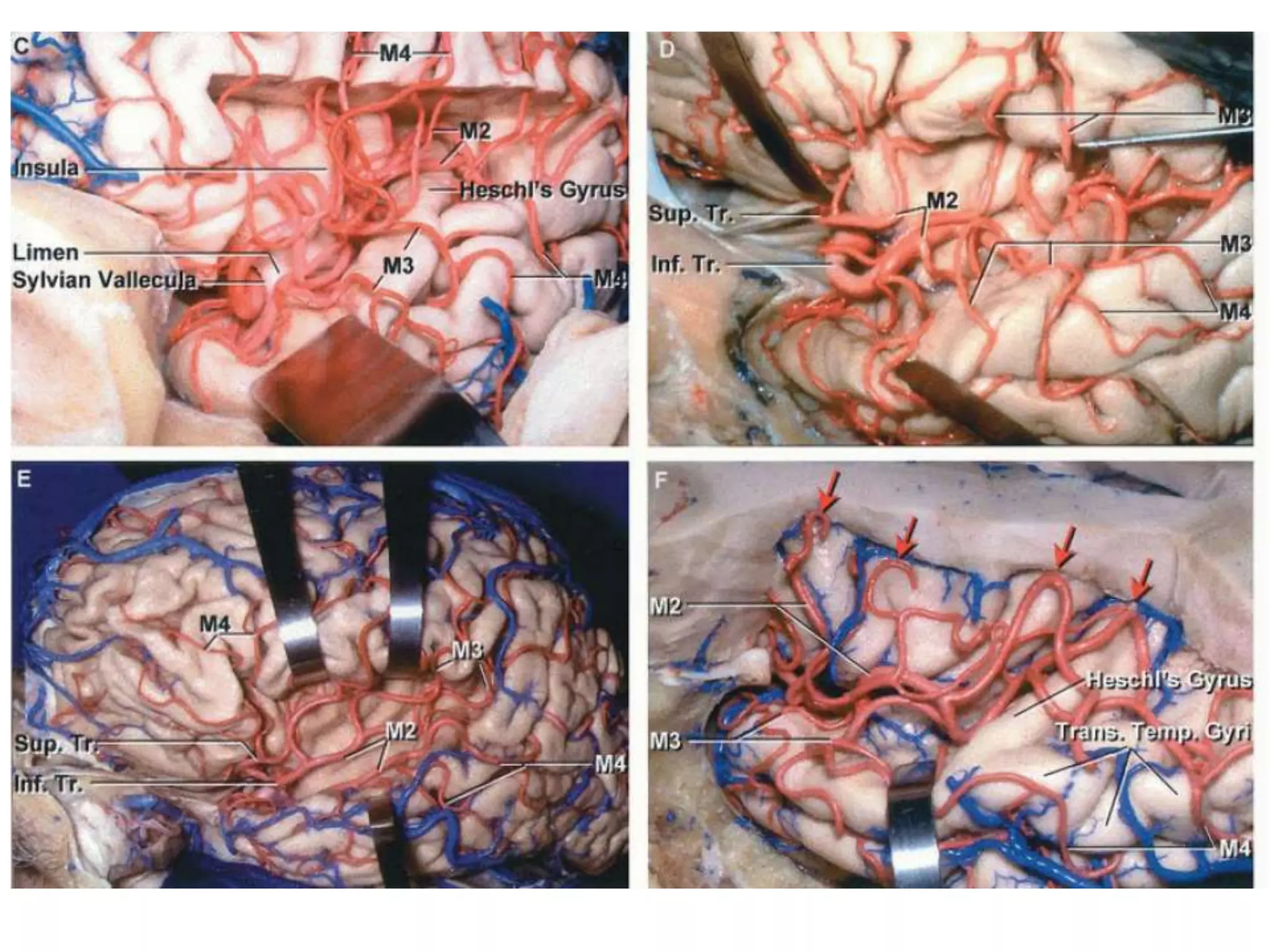
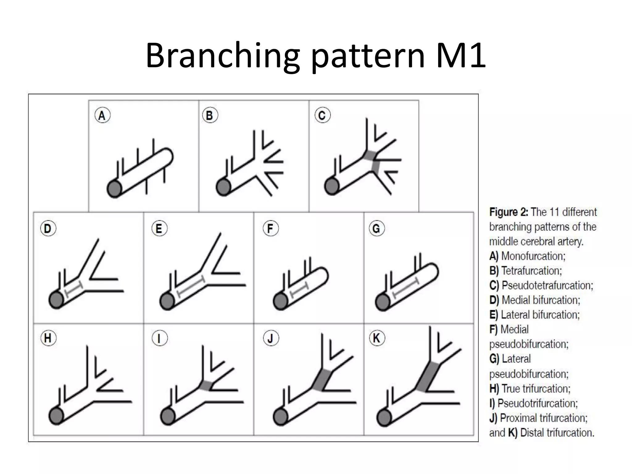
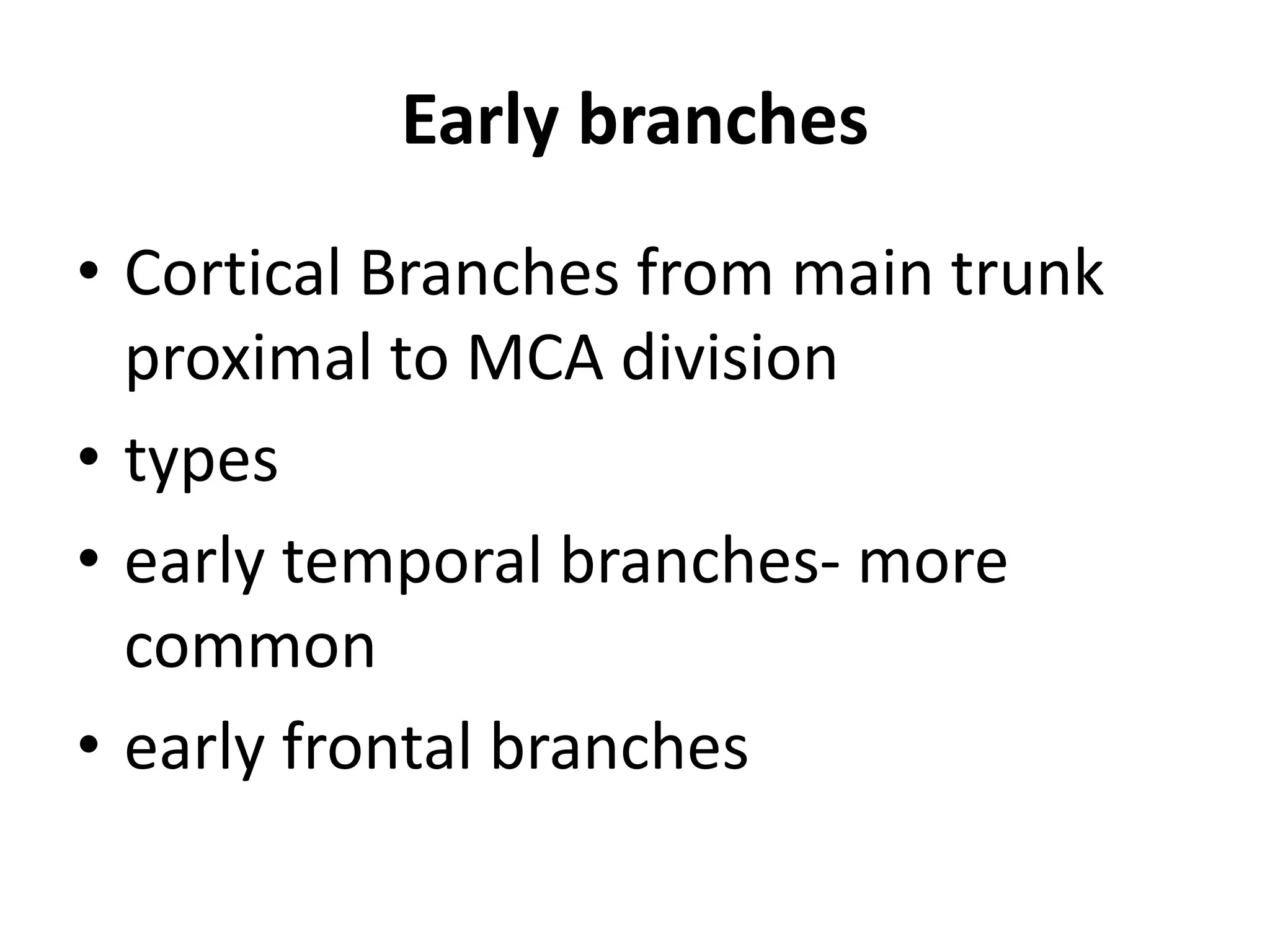
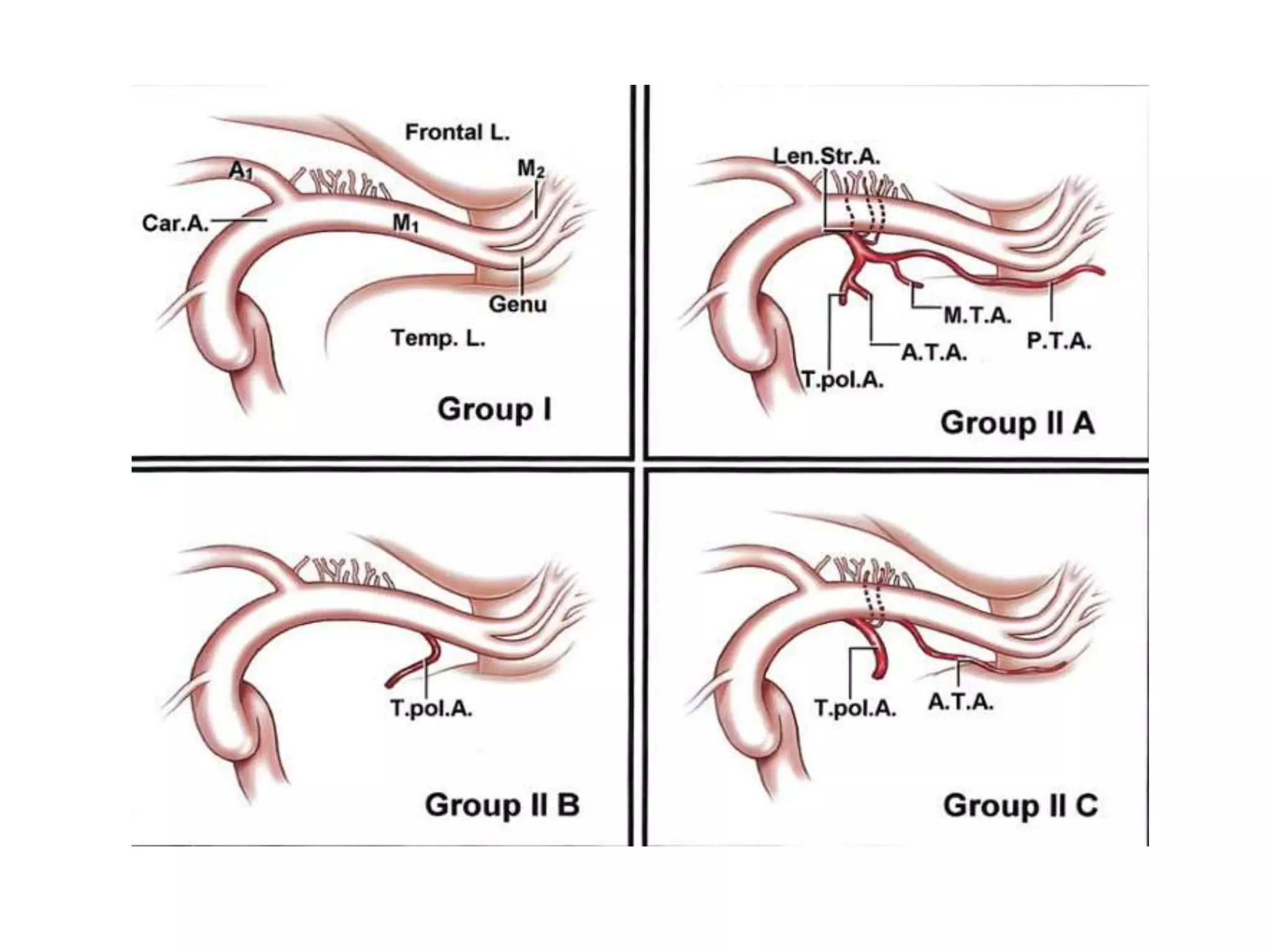
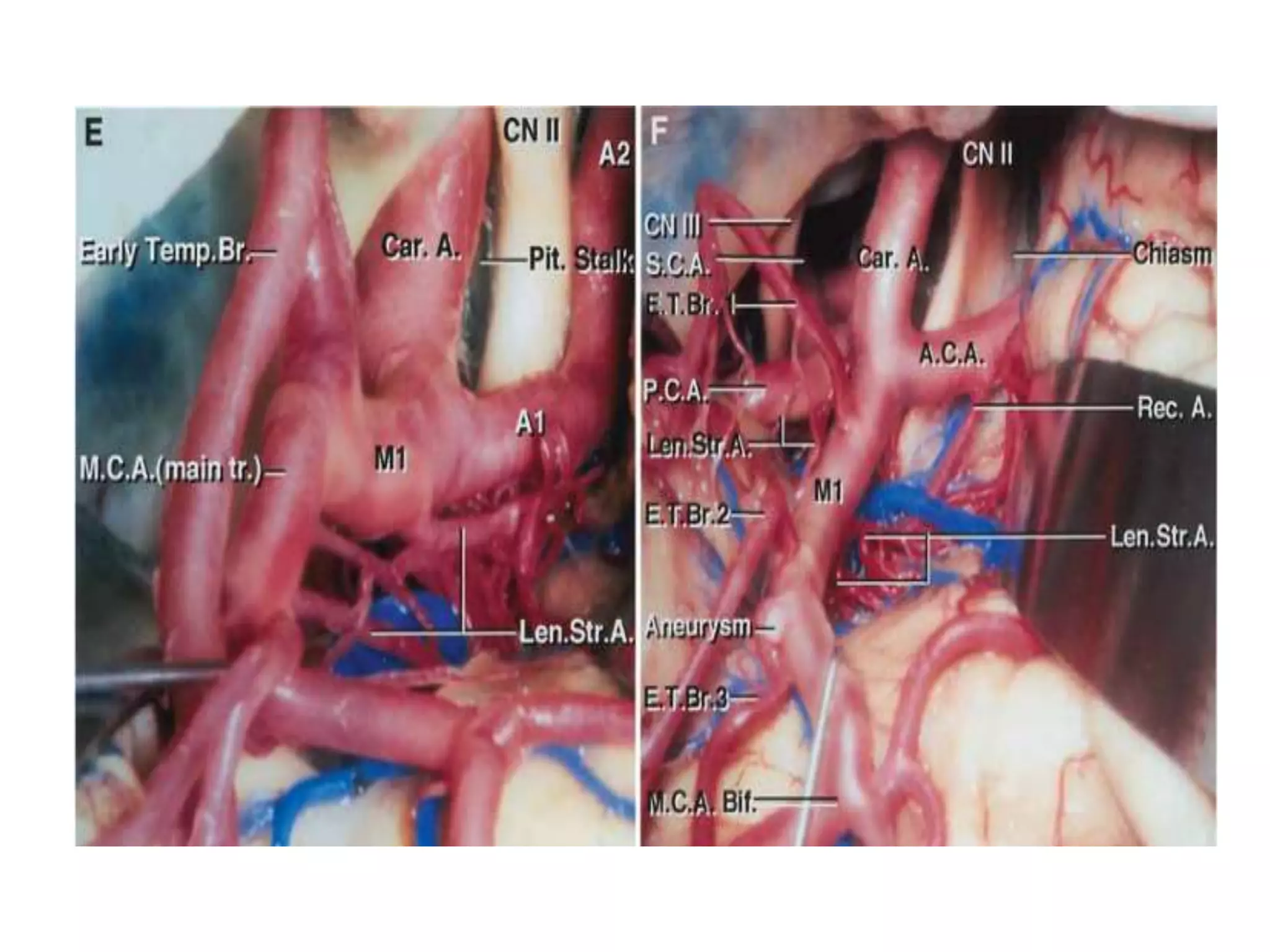
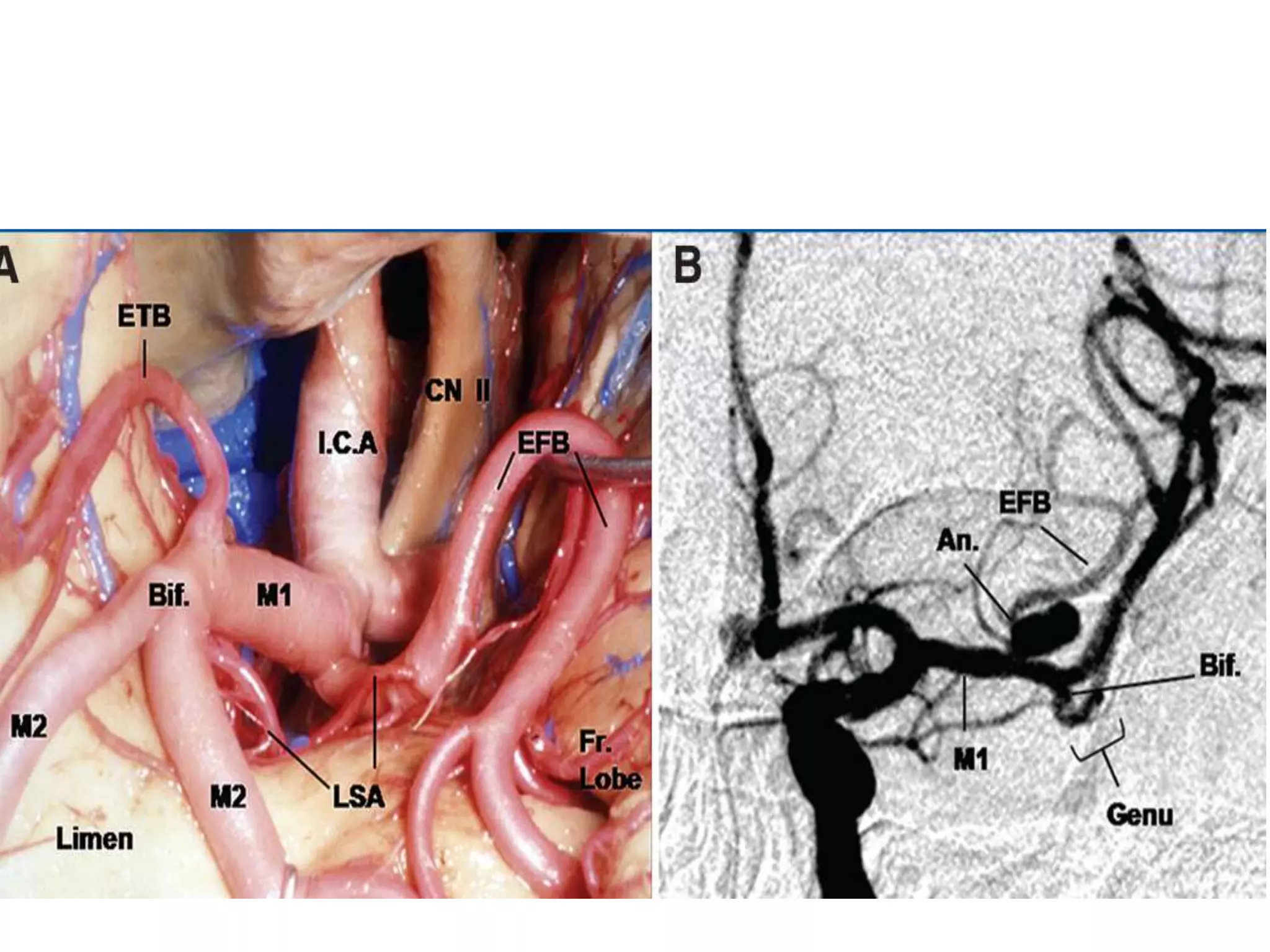
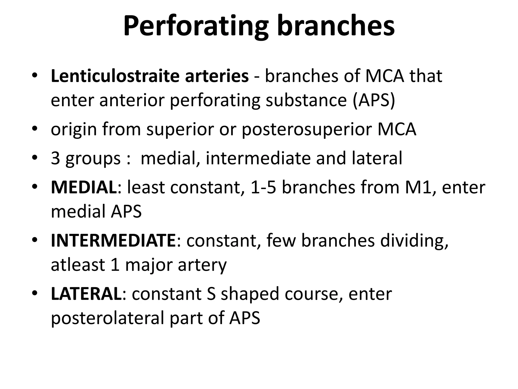
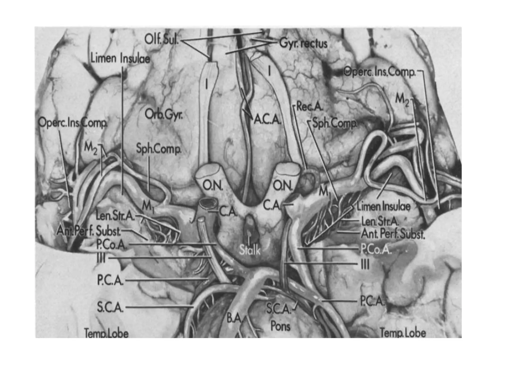
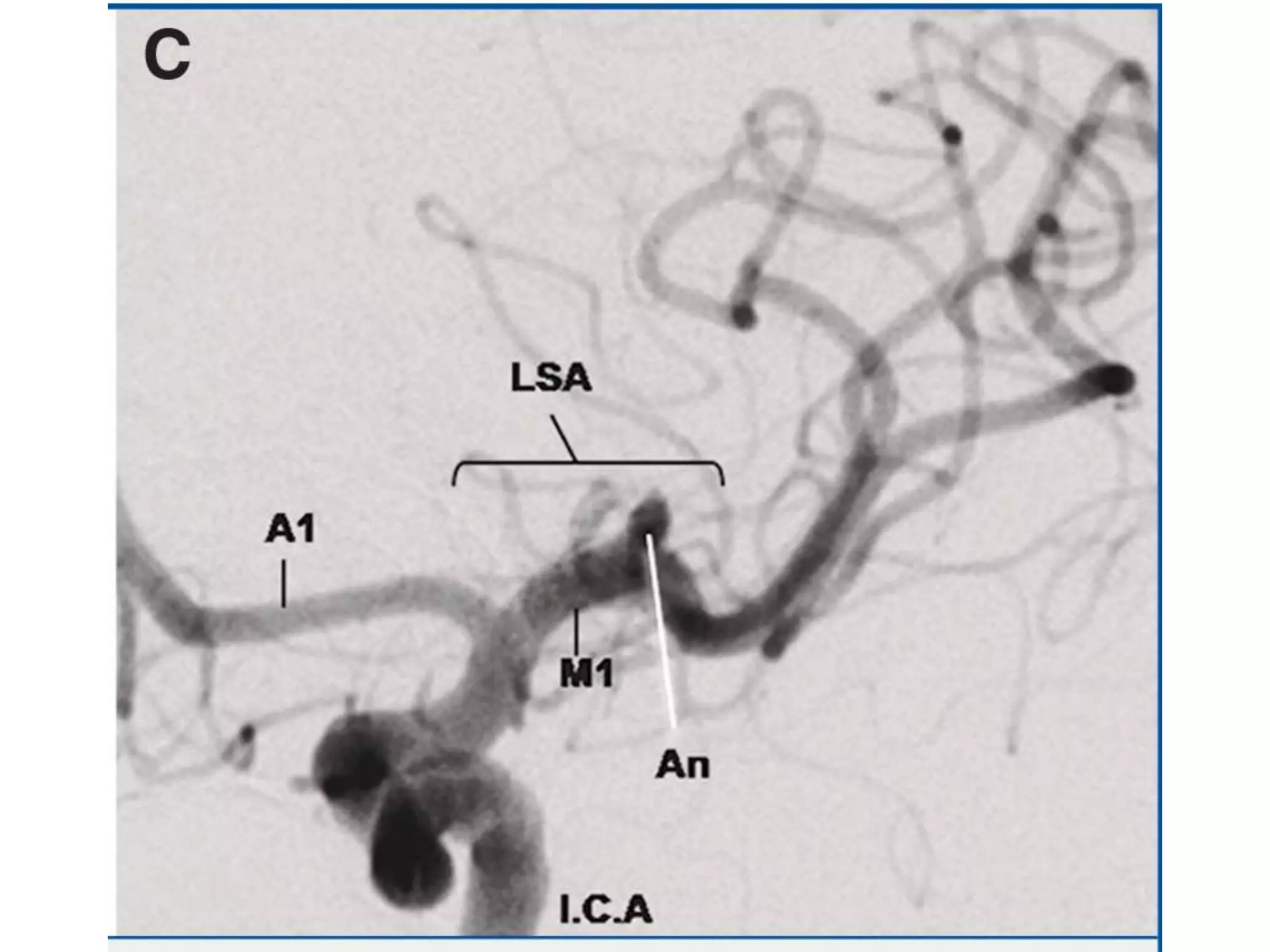
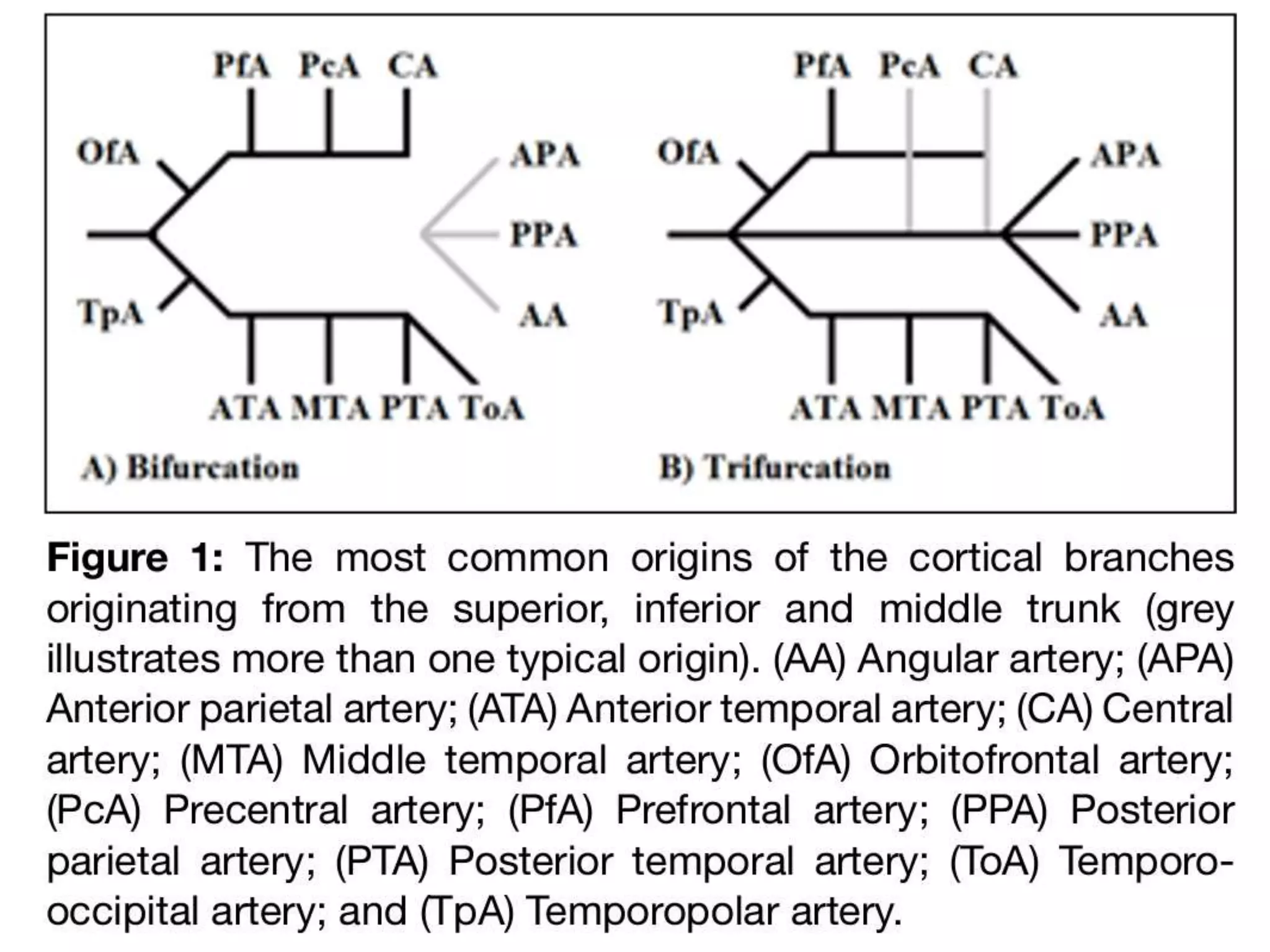
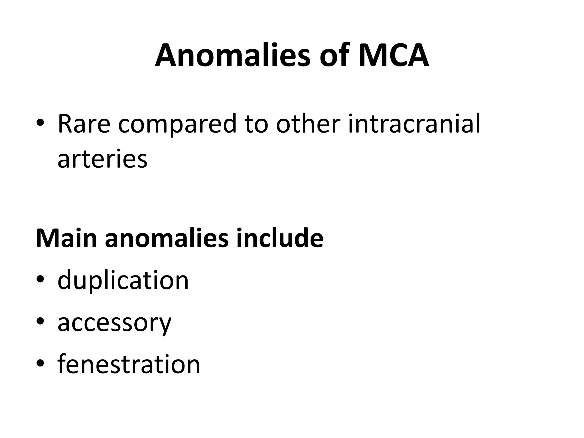
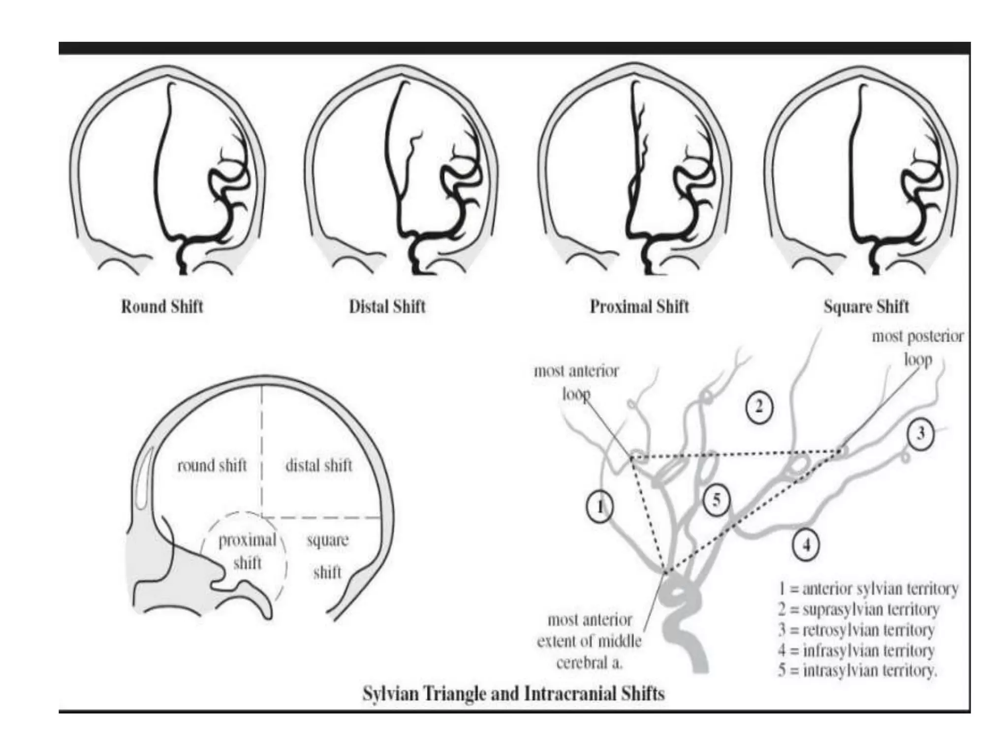
• The MCA is the largest and most complex of
the cerebral arteries
• The MCA arises as the larger of the two
terminal branches of the internal carotid
artery
• The diameter of the MCA at its origin ranges
(average, 3.9 mm), roughly twice that of the
anterior cerebral artery
Its origin and course
Post
medial
Segments
•M1 (sphenoidal),
•M2 (insular),
•M3 (opercular),
•M4 (cortical)
M1 segment
• Microneurosurgical and
angiographic
• Prebifurcation and post
bifurcation parts
• How to distinguish between early
branches and M1
Branching pattern M1
Early branches
• Cortical Branches from main trunk
proximal to MCA division
• types
• early temporal branches- more
common
• early frontal branches
Perforating branches
• Lenticulostraite arteries - branches of MCA that
enter anterior perforating substance (APS)
• origin from superior or posterosuperior MCA
• 3 groups : medial, intermediate and lateral
• MEDIAL: least constant, 1-5 branches from M1, enter
medial APS
• INTERMEDIATE: constant, few branches dividing,
atleast 1 major artery
• LATERAL: constant S shaped course, enter
posterolateral part of APS
Cortical branches
Anomalies of MCA
• Rare compared to other intracranial
arteries
Main anomalies include
• duplication
• accessory
• fenestration
Anatomy of the Middle Cerebral Artery: Cortical Branches, Branching Pattern and Anomalies; Karen CILLIERS, Benedict John PAGE; DOI:
10.5137/1019-5149.JTN.18127-16.1
Angiographic Sylvian Point (ASP)
• It is the most medial point where the last
cortical MCA branch (usually the angular
artery) turns inferiorly to exit the sylvian
fissure.(On AP view)
• This point approximates the apex of the insula
and represents the posterior limit of the
lateral cerebral sulcus.
Sylvian triangle (angiographic)
• The superior insular line ( A line tangent to the
tops of the insular loops),
• Main MCA trunk ( forms the posterior inferior
margin of the triangle)
• Most anterior branch of the ascending frontal
complex ( forms the anterior border of the
triangle)
• It is seen in lateral view and serves as angiographic
landmark for localizing supratentorial masses.
• Round shift = Frontal lesion anterior to coronal
suture
• Square shift = Lesion behind foramen of Monro
in lower half of hemisphere
• Distal shift = Posterior to coronal suture in
upper half of hemisphere
• Proximal shift = Basifrontal lesion / anterior
middle cranial fossa including anterior temporal
lobe
Exposing MCA
Sylvian fissure
Mca anatomy
Mca anatomy
Mca anatomy

Mca anatomy

  • 1.
  • 2.
    Introduction • The MCAis the largest and most complex of the cerebral arteries • The MCA arises as the larger of the two terminal branches of the internal carotid artery • The diameter of the MCA at its origin ranges (average, 3.9 mm), roughly twice that of the anterior cerebral artery
  • 3.
  • 4.
  • 7.
  • 10.
    M1 segment • Microneurosurgicaland angiographic • Prebifurcation and post bifurcation parts • How to distinguish between early branches and M1
  • 11.
  • 12.
    Early branches • CorticalBranches from main trunk proximal to MCA division • types • early temporal branches- more common • early frontal branches
  • 18.
    Perforating branches • Lenticulostraitearteries - branches of MCA that enter anterior perforating substance (APS) • origin from superior or posterosuperior MCA • 3 groups : medial, intermediate and lateral • MEDIAL: least constant, 1-5 branches from M1, enter medial APS • INTERMEDIATE: constant, few branches dividing, atleast 1 major artery • LATERAL: constant S shaped course, enter posterolateral part of APS
  • 25.
  • 28.
    Anomalies of MCA •Rare compared to other intracranial arteries Main anomalies include • duplication • accessory • fenestration
  • 30.
    Anatomy of theMiddle Cerebral Artery: Cortical Branches, Branching Pattern and Anomalies; Karen CILLIERS, Benedict John PAGE; DOI: 10.5137/1019-5149.JTN.18127-16.1
  • 31.
    Angiographic Sylvian Point(ASP) • It is the most medial point where the last cortical MCA branch (usually the angular artery) turns inferiorly to exit the sylvian fissure.(On AP view) • This point approximates the apex of the insula and represents the posterior limit of the lateral cerebral sulcus.
  • 33.
    Sylvian triangle (angiographic) •The superior insular line ( A line tangent to the tops of the insular loops), • Main MCA trunk ( forms the posterior inferior margin of the triangle) • Most anterior branch of the ascending frontal complex ( forms the anterior border of the triangle) • It is seen in lateral view and serves as angiographic landmark for localizing supratentorial masses.
  • 36.
    • Round shift= Frontal lesion anterior to coronal suture • Square shift = Lesion behind foramen of Monro in lower half of hemisphere • Distal shift = Posterior to coronal suture in upper half of hemisphere • Proximal shift = Basifrontal lesion / anterior middle cranial fossa including anterior temporal lobe
  • 37.
  • 38.

Editor's Notes

  • #4 at the medial end of the sylvian fissure, lateral to the optic chiasm, below the anterior perforated substance, and posterior to the division of the olfactory tract into the medial and lateral olfactory striae