
Recommended
Recommended
More Related Content
What's hot
What's hot (20)
Similar to Treatment of traumatised tooth
Similar to Treatment of traumatised tooth (20)
More from Deepashri Tekam
More from Deepashri Tekam (17)
Recently uploaded
Recently uploaded (20)
Treatment of traumatised tooth
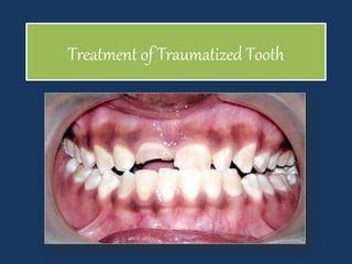
- 1. Treatment of Traumatized Tooth
- 2. Etiology of Traumatic Injuries • Automobile injury • Battered child • Child abuse • Drug abuse • Epilepsy • Fall from height • Sports related injuries
- 4. Classification by Ellis and Davey (1960) Class I Simple fracture of the crown involving little (or) no dentin. Class II Extensive fracture of the crown involving considerable dentin, but not the dental pulp. Class III Extensive fracture of the crown involving considerable dentin and exposing the dental pulp. Class IV The traumatized teeth that become nonvital with (or) without loss of crown structure. Class V Teeth lost as a result of trauma. Class VI Fracture of the root with or without a loss of crown structure. Class VII Displacement of a tooth without fracture of crown (or) root. Class VIII Fracture of crown en masse and its replacement. Class IX Injuries to primary dentition
- 5. A.Enamel fracture B.Enamel and dentine fracture C.Dentine fracture involving pulp D.Root fracture E.Extrusion F. Intrusion
- 6. WHO Classification of Traumatic Injuries 873. 60 Enamel fracture. 873.61 Crown fracture involving enamel and dentine without pulp exposure. 873.62 Crown fracture with pulp exposure. 873.63 Root fracture. 873.64 Crown root fracture 873.66 Luxation. 873.67 Intrusion or extrusion 873.68 Avulsion. 873.69 Other injuries such as soft tissue and laceration.
- 7. 873.60: Enamel fracture 873.61: Enamel and dentin fracture 873.62: Crown fracture with pulp exposure 873.63: Root fracture
- 8. 873.64: Crown root fracture 873.66: Luxation 873.67: Extrusion/intrusion 873.68: Avulsion
- 9. 873-69: Soft tissue injuries
- 10. ENAMEL FRACTURES These injuries involve the loss of the portion of coronal tooth enamel subsequent to a force directed perpendicularly or obliquely to the incisal edge of the traumatized tooth.
- 11. Diagnosis And Clinical Presentation • Enamel fracture includes superficial rough edge that may cause irritation to the tongue or lips.
- 12. Immediate treatment • Re-contouring the injured tooth, adjacent teeth and / or the opposing teeth • Eliminate the sharp enamel edges associated with minor injuries and prevents the laceration of tongue, lips or oral mucosa. When the shape and extent of the fracture precludes re-contouring a restoration is necessary • Restoration of the missing tooth structure with composite resin
- 13. DENTIN FRACTURES These injuries involve the loss of tooth substance confined to enamel and dentin but not involving the pulp.
- 14. Diagnosis And Clinical Presentation • An enamel and dentin fracture also includes a rough edge on the tooth, but sensitivity to air and hot and cold liquids and pain on mastication may be a chief complaint. • The intensity of these symptoms is related directly to the amount of exposed dentin and to the maturity of tooth.
- 15. The tooth should be tested with: a) Electric pulp tester, b) Ice, c) Ethyl chloride spray, d) Periapical radiograph.
- 16. Temporary restoration: • After the fracture, as soon as possible the exposed dentin should be protected by sedative cement such as zinc oxide eugenol held in a crown form. Permanent restoration • For uncomplicated crown fractured teeth includes the use of adhesive resin and composite resin systems.
- 17. COMPLICATED CROWN FRACTURES These injuries involve the loss of tooth substance confined to enamel and dentin but involving the pulp.
- 18. Pulp Capping and Pulpotomy • Pulp capping and pulpotomy are the measures that permit apexogenesis to take place and may avoid the need for root canal therapy. • The choice of treatment depends on the: – Size of the exposure, – The presence of hemorrhage – The length of time since the injury.
- 19. Pulp Capping • Pulp capping implies placing the dressing directly on to the pulp exposure
- 20. Indication s: • On a very recent exposure (< 24 hours) and probably on a mature, permanent tooth with a simple restorative plan. Technique : • After adequate anesthesia, a rubber dam is placed. • Crown and exposed dentinal surface is thoroughly rinsed with saline followed by disinfection with 0.12 percent chlorhexidine or betadine. • Pure calcium hydroxide mixed with anesthetic solution or saline is carefully placed over the exposed pulp and dentinal surface. • The surrounding enamel is acid etched and bonded with composite resin. Follow-up • Vitality tests, palpation tests, percussion tests and radiographs should be carried out for 3 weeks; 3, 6 and 12 months; and every twelve months subsequently. Prognosis • Prognosis is up to 80 percent
- 21. Pulpotomy • Pulpotomy refers only to coronal extirpation of vital pulp tissue. • Two Types • Partial pulpotomy • Full (cervical) pulpotomy
- 22. Partial Pulpotomy • Partial pulpotomy also termed as “Cvek Pulpotomy”, it implies removal of the coronal pulp tissue to the level of healthy pulp
- 23. Indication s: It is indicated in young permanent teeth with incomplete root formation. Technique : • After anesthetizing the area rubber dam is applied. • A 1-2 mm deep cavity is prepared into the pulp using a diamond bur. • Wet cotton pellet is used to impede hemorrhage and thereafter a thin coating of calcium hydroxide mixed with saline solution or anesthetic solution is placed over it. • The access cavity is sealed with hard setting cement like IRM. Follow-up • 1. Absence of signs or symptoms • 2. Absence of resorption either internal or external • 3. Evidence of continued root formation in developing teeth.. Prognosis • Prognosis is good (94-96%).
- 25. Cervical Pulpotomy/Deep Pulpotomy • Cervical pulpotomy involves removal of entire coronal pulp to the level of root orifices
- 26. Indication s: When the gap between traumatic exposure and the treatment provided is more than 24 hrs. • When pulp is inflamed to deeper levels of coronal pulp. Technique : • Coronal pulp is removed same as in partial pulpotomy except that it is up to level of root orifice. Follow-up • It is same as pulp capping and partial pulpotomy. • Main disadvantage of this treatment is that sensitivity tests cannot be done because of loss of coronal pulp. • Thus radiographic examination is important for follow- up. Prognosis • 80-95 percent success rate.
- 28. Apexification • If the pulp tissue is necrotic, apexification is the process which stimulates the formation of a calcified barrier across the apex. • Apexification is done to stimulate the hard tissue barrier. • Initially all canals are disinfected with sodium hypochlorite solution to remove any debris and bacteria from the canal.
- 29. Apexification • Following this calcium hydroxide is packed against the apical soft tissue and later backfilling with calcium hydroxide is done to completely obturate the canal. • When completion of hard tissue is suspected (after 3- 6 months), remove calcium hydroxide and take radiograph. • If formation of hard tissue is found satisfactory, canal is obturated using softened gutta-percha technique.
- 31. CROWN ROOT FRACTURE • Crown root fracture involves enamel, dentin and cementum with or without the involvement of pulp. • It is usually oblique in nature involving both crown and root. • This type of injury is considered as more complex type of injury because of its more severity and involvement of the pulp
- 32. Diagnosis • Coronal fragment is usually mobile. • Patient may complain of pain from mastication due to movement of the coronal portion. • Inflammatory changes in pulp and periodontal ligament are seen due to plaque accumulation in the line of fracture. • Patient may complain of sensitivity to hot and cold. • Radiographs are taken at different angles to assess the extent of fracture. • Indirect light and transillumination can also be used to diagnose this type of fracture.
- 33. Treatment • The main objective of the treatment is to: • a. Allow subgingival portion of the fracture to heal. • b. Restoration of the coronal portion. • If there is no pulp exposure, fragment can be treated by bonding alone or by removing the coronal structure and then restoring it with composites. • If pulp exposure has occurred, pulpotomy or root canal treatment is indicated depending upon condition of the tooth.
- 34. Treatment • When the fracture extends below the alveolar crest level, the surgical repositioning of tissues by gingivectomy, osteotomy, etc. should be done to expose the level of fracture and subsequently restore it. Prognosis • Long term prognosis depends on quality of coronal restoration. • Otherwise the prognosis is similar to complicated or uncomplicated fracture.
- 35. LUXATION INJURIES • Luxation injuries cause trauma to supporting structures of teeth ranging from minor crushing of periodontal ligament and neurovascular supply of pulp to total displacement of the teeth. • They are usually caused by sudden impact such as blow,
- 36. CONCUSSION • Tooth is not displaced. • Mobility is not present. • Tooth is tender to percussion because of edema and hemorrhage in the periodontal ligament. • Pulp may respond normal to testing.
- 37. SUBLUXATION • Teeth are sensitive to percussion and have some mobility. • Sulcular bleeding is seen showing damage and rupture of the periodontal ligament fibers. • Pulp responds normal to testing. • Tooth is not displaced.
- 38. Treatment of Concussion and Subluxation Rule out the root fracture by taking radiographs. Relief the occlusion by selective grinding of opposing teeth Immobilize the injured teeth. Endodontic therapy should not be carried out at first visit because both negative testing results and crown discoloration can be reversible. Follow-up is done at 3 weeks, 3, 6 and 12 months. Prognosis there is only a minimal risk of pulp necrosis and root resorption.
- 39. LATERAL LUXATION • Trauma displaces the tooth lingually, buccally, mesially or distally, in other words out of its normal position away from its long axis. • Sulcular bleeding is present indicating rupture of PDL fibers • Tooth is sensitive to percussion.
- 40. LATERAL LUXATION • Clinically, crown of laterally luxated tooth is usually displaced horizontally with tooth locked firmly in the new position. • Here percussion may elicit metallic tone indicating that root has forced into the alveolar bone.
- 41. EXTRUSIVE LUXATION In extrusive luxation • Tooth is displaced from the socket along its long axis • Tooth is very mobile • Radiograph shows the displacement of tooth.
- 42. Treatment of Lateral and Extrusive Luxation Repositioning of laterally luxated teeth require minimal force for repositioning. Before repositioning laterally luxated teeth, anesthesia should be administrated. Tooth must be dislodged from the labial cortical plate by moving it coronally and then apically. Thus tooth is first moved coronally out of the buccal plate of bone and then fitted into its original position
- 43. Treatment of Lateral and Extrusive Luxation For repositioning of extruded tooth, a slow and steady pressure is required to displace the coagulum formed between root apex and floor of the socket. After this tooth is immobilized, stabilized and splinted for approximately 2 weeks. Local anesthesia is not needed while doing this.
- 44. Follow-up: • Splint is removed 2 weeks after extrusion. • If tooth has become nonvital, inflammatory root resorption can occur, requiring immediate endodontic therapy. • Pulp testing should be performed on regular intervals. Prognosis • It depends on stage of root development at the time of injury. • Commonly seen sequel of luxation injuries are pulp necrosis,root canal obliteration and root resorption.
- 45. INTRUSIVE LUXATION • Tooth is forced into its socket in an apical direction . • Maximum damage has occurred to pulp and the supporting structures . • When examined clinically, the tooth is in infraocclusion . • Tooth presents with clinical presentation of ankylosis because of being firm in socket.
- 46. INTRUSIVE LUXATION • On percussion metallic sound is heard. • In mixed dentition, diagnosis is more difficult as intrusion can mimic a tooth undergoing eruption. • Radiographic evaluation is needed to know the position of tooth.
- 47. Treatment In immature teeth, spontaneous re-eruption is seen. If reeruption stops before normal occlusion is attained, orthodontic movement is initiated before tooth gets ankylosed If tooth is severely intruded, surgical access is made to the tooth to attach orthodontic appliances and extrude the tooth. Tooth can also be repositioned by loosening the tooth surgically and aligning it with the adjacent teeth.
- 48. ROOT FRACTURE • These are uncommon injuries but represent a complex healing pattern due to involvement of dentin, cementum, pulp and periodontal ligament. Diagnosis • Displacement of coronal segment usually reflects the location of fracture. • Radiographs at varying angles (usually at 45°, 90° and 110°) are mandatory for diagnosing root fractures.
- 49. Treatment of Root Fractures If there is no mobility of tooth and tooth is asymptomatic, only apical third fracture is suspected. In this case to facilitate pulpal and periodontal ligament healing, displaced coronal portion should be repositioned accurately. It is stabilized by splinting for 2-3 weeks
- 50. Apical segment of fractured root contains vital healing pulp whereas coronal pulp has become necrotic. Root canal therapy for both coronal and apical segment, when they are not separated. Root canal therapy of coronal segment and no treatment of apical segment, when apical segment contains vital pulp. Root canal therapy for coronal segment and surgical removal of apical third. Apexification type procedure of coronal segment, i.e. inducing hard tissue barrier at exit of coronal root canal and no treatment of apical segment. This is most commonly used procedure nowadays.
- 51. PROGNOSIS • It depends on: • Amount of dislocation and degree of mobility of coronal segment: – More is the dislocation, poorer is the prognosis. • Stage of tooth development: – More immature the tooth, better the ability of pulp to recover from trauma.
- 52. According to the Andreasen and Hjorting—Hansen, root fracture can show healing in following ways: • Healing with calcified tissue in which fractured fragments are in close contact • Healing with interproximal connective tissue in which radiographically fragments appear separated by a radiolucent line Interproximal inflammatory tissue seen in root fracture Healing of root fracture with calcified tissue
- 53. According to the Andreasen and Hjorting—Hansen, root fracture can show healing in following ways: • Healing with interproximal bone and connective tissues. Here fractured fragments are seen separated by a distinct bony bridge radiographically Healing of root fracture by interproximal bone
- 54. According to the Andreasen and Hjorting—Hansen, root fracture can show healing in following ways: • Interproximal inflammatory tissue without healing, radiographically it shows widening of fracture line Healing of root fracture by formation of connective tissue between the segments
- 55. AVULSION It is defined as complete displacement of the tooth from the alveolus. It is usually the result of trauma to an anterior tooth and is both a dental and an emotional problem.
- 56. Consequences of Trauma To Primary Teeth • Infection • Abscess • Loss of space in the dental arch • Ankylosis • Failure to continue eruption • Color changes • Injury to the permanent teeth
- 57. Consequences of Injury To Permanent Teeth • Infection • Abscess • Loss of space in the dental arch • Ankylosis • Resorption of root structure • Abnormal root development • Color changes
- 58. Storage Media For Avulsed Tooth 1. Hank’s balanced salt solution 2. Milk 3. Saline 4. Saliva 5. Visapan 6. CPP-ACP (Casein phospho-peptides–amorphous calcium phosphate) 7. Coconut water 8. Water
- 59. Hank’s Balanced Solution (Save-A-Tooth) • This pH-preserving fluid is best used with a trauma reducing suspension apparatus. • The HBSS is biocompatible with the tooth periodontal ligament cells and can keep these cells viable for 24 hours because of its ideal pH and osmolality.
- 60. Composition of HBSS is: • Sodium chloride, potassium chloride, glucose, calcium chloride, magnesium chloride, sodium bicarbonate, sodium phosphate. • Researches have shown that this fluid can rejuvenate degenerated ligament cells and maintain a success rate of over 90 percent if an avulsed tooth is soaked in it for 30 minutes prior to replantation.
- 61. Visapan • Visapan has pH of 7.4 and osmolarity of 320 mosm/L. • These properties are advantageous for cell growth. It can preserve the viability of fibroblasts for 24 hours.
- 62. Coconut Water • Studies have shown that electrolyte composition of coconut water is similar to intracellular fluid. • So it can be also used as storage media for an avulsed tooth because of its ease of availability, economical and sterile nature. • It has been shown to be equally effective as HBSS in maintaining the cell viability.
- 63. MANAGEMENT OPTIONS FOR AN AVULSED TOOTH
- 64. If the tooth has been out of its socket less than 15 minutes • Wash out the socket with the same solution, reimplant the tooth firmly, have the patient bite down firmly on a piece of gauze to help stabilize the tooth and when possible secure it to adjacent teeth with wire, arch bars, or a temporary periodontal pack. • Put the patient on a liquid diet, prescribe antibiotics preferably penicillin VK and plan next dental appointment.
- 65. If the tooth has been out 15 minutes to 2 hours, • Soak for 30 minutes to replenish nutrients. • Local anesthesia will probably be needed before reimplanting as above.
- 66. If the tooth was out over two hours, • The periodontal ligament is dead, and should be removed, along with the pulp. • The tooth should soak 30 minutes in 5 percent sodium hypochlorite and 5 minutes each in saturated citric acid, 1 percent stannous fluoride and 5 percent doxycycline before reimplanting. • The dead tooth should ankylose into the alveolar bone of the socket like a dental implant.